結論(CONCLUSIONS)
HIV 疾病表現的數量與多樣性,在皮膚上多於任何其他器官。皮膚併發症是顯著疾病負擔的來源,也可能造成污名化。雖然 ART 已降低數種 HIV 相關皮膚病症的發生率,其他疾患如 IRIS、藥物反應、代謝紊亂、HPV 感染與 SCCs 仍常被觀察到。事實上,由於 HIV 感染者存活期延長,它們的發生率甚至可能正在增加。藉由辨識 HIV 相關皮膚病症的光譜並執行適當的診斷檢查,即可及時給予治療並使結果最佳化。
我們的電子書中提供額外的圖片(存取碼請見封面內頁)。
-
Chelidze K, Thomas C, Chang AY, et al. HIV-related skin sites/default/files/media_asset/UNAIDS_FactSheet_en.pdf>.
-
UNAIDS. In Danger: UNAIDS Global AIDS Update 2022;
-
Kaushik SB, Lebwohl MG. Psoriasis: which therapy for disease in the era of antiretroviral therapy: recognition and management. Am J Clin Dermatol. 2019;20:423–442.14. Patil S, Majumdar B, Sarode SC, et al. Oropharyngeal
July 2022. <https://www.unaids.org/en/resources/ documents/2022/in-danger-global-aids-update>.
3. Rollenhagen C, Lathrop MJ, Macura SL, et al. Herpes which patient: focus on special populations and chronic infections. J Am Acad Dermatol. 2019;80:43–53.28. Menon K, Van Voorhees AS, Bebo BF Jr, et al. Psoriasis in candidosis in HIV-infected patients – an update. Front Microbiol. 2018;9:980.15. Daniel CR 3rd, Norton LA, Scher RK. The spectrum of nail simplex virus type-2 stimulates HIV-1 replication in cervical tissues: implications for HIV-1 transmission and efficacy of anti-HIV-1 microbicides. Mucosal Immunol. 2014;7:1165–1174.4. Vafai A, Berger M. Zoster in patients infected with HIV: a patients with HIV infection: from the medical board of the National Psoriasis Foundation. J Am Acad Dermatol. 2010;62:291–299.29. Adizie T, Moots RJ, Hodkinson B, et al. Inflammatory disease in patients with human immunodeficiency virus infection. J Am Acad Dermatol. 1992;27:93–97.16. Ramos-e-Silva M, Lima CMO, Schechtman RC, et al.
arthritis in HIV positive patients: a practical guide. BMC Infect Dis. 2016;16:100.30. Resnick SD, Murrell DF, Woosley JT. Pityriasis rubra pilaris,
Systemic mycoses in immunodepressed patients (AIDS). Clin Dermatol. 2012;30:616–627.17. Murakawa GJ, Harvell JD, Lubitz P, et al. Cutaneous review. Am J Med Sci. 2001;321:372–380.5. Coates SJ, Leslie KS. What’s new in HIV dermatology?
F1000Res 2019;8:F1000 Faculty Rev: 980. <https://doi. org/10.12688/f1000research.16182.1>
6. Looker KJ, Ronn MM, Brock PM, et al. Evidence of acne conglobata, and elongated follicular spines: an HIV-associated follicular syndrome? J Am Acad Dermatol. 1993;29:283.31. Eisman S. Pruritic papular eruption in HIV. Dermatol Clin.
aspergillosis and acquired immunodeficiency syndrome. Arch Dermatol. 2000;136:365–369.18. Alvar J, Aparicio P, Aseffa A, et al. The relationship synergistic relationships between HIV and human papillomavirus (HPV): systematic reviews and meta-analyses of longitudinal studies of HPV acquisition and clearance by HIV status, and of HIV acquisition by HPV status. J Int AIDS Soc. 2018;21:e25110.7. Poljak M, Sterbenc A, Lunar MM. Prevention of human
2006;24:449–457. vi.
32. Smith KJ, Skelton HG, DeRusso D, et al. Clinical and histo- between leishmaniasis and AIDS: the second 10 years. Clin Microbiol Rev. 2008;21:334–359.19. Jarvis JN, Lockwood DN. Clinical aspects of visceral pathologic features of hair loss in patients with HIV-1 infection. J Am Acad Dermatol. 1996;34:63–68.33. Woods EA, Foisy MM. Antiretroviral-related alopecia leishmaniasis in HIV infection. Curr Opin Infect Dis. 2013;26:1–9.20. Galarza C, Ramos W, Gutierrez EL, et al. Cutaneous papillomavirus (HPV)-related tumors in people living with human immunodeficiency virus (HIV). Expert Rev Anti Infect Ther. 2017;15:987–999.8. Tong WWY, Hillman RJ, Kelleher AD, et al. Anal intraepi- in HIV-infected patients. Ann Pharmacother. 2014;48:1187–1193.34. Prose NS, Abson KG, Scher RK. Disorders of the nails and acanthamebiasis infection in immunocompetent and immunocompromised patients. Int J Dermatol. 2009;48:1324–1329.21. Thomas C, Coates SJ, Engelman D, et al. Ectoparasites:
thelial neoplasia and squamous cell carcinoma in HIV-infected adults. HIV Med. 2014;15:65–76.9. Grulich AE, van Leeuwen MT, Falster MO, Vajdic CM.
hair associated with human immunodeficiency virus infection. Int J Dermatol. 1992;31:453–457.35. Cribier B, Mena ML, Rey D, et al. Nail changes in patients scabies. J Am Acad Dermatol. 2020;82:533–548.22. Aquilina C, Viraben R, Sire S. Ivermectin-responsive infected with human immunodeficiency virus. Arch Dermatol. 1998;134:1216–1220.36. Vega LE, Espinoza LR. Vasculitides in HIV infection. Curr
Demodex infestation during human immunodeficiency virus infection. A case report and literature review. Dermatol Basel Switz. 2002;205:394–397.23. Nair SP. Insect bite reaction and HIV infection. Indian
Incidence of cancers in people with HIV/AIDS compared with immunosuppressed transplant recipients: a metaanalysis. Lancet. 2007;370:59–67.10. Rigopoulos D, Paparizos V, Katsambas A. Cutaneous
Rheumatol Rep. 2020;22:60.37. Gregory N, DeLeo VA. Clinical manifestations of photo- sensitivity in patients with human immunodeficiency virus infection. Arch Dermatol. 1994;130:630–633.38. Mansourati FF, Stone VE, Mayer KH. Porphyria cutanea
J Dermatol Venereol Leprol. 2015;81:95.24. Motswaledi MH, Visser W. The spectrum of markers of HIV infection. Clin Dermatol. 2004;22:487–498.11. Sabbagh P, Riahi SM, Gamble HR, et al. The global
HIV-associated infective and inflammatory dermatoses in pigmented skin. Dermatol Clin. 2014;32:211–225.25. Mahlangeni GM, Tod BM, Jordaan HF, et al.
and regional prevalence, burden, and risk factors for methicillin-resistant Staphylococcus aureus colonization in HIV-infected people: a systematic review and metaanalysis. Am J Infect Control. 2019;47:323–333.12. Plettenberg A, Lorenzen T, Burtsche BT, et al. Bacillary tarda and HIV/AIDS: a review of pathogenesis, clinical manifestations and management. Int J STD AIDS. 1999;10:51–56.39. Wong SN, Khoo LSW. Chronic actinic dermatitis as the
Clinicopathological features of seborrheic-like dermatitis in HIV-infected adults: a single institutional descriptive cross-sectional study. Am J Dermatopathol. 2021;43:27–34.26. Alpalhao M, Borges-Costa J, Filipe P. Psoriasis in HIV angiomatosis in HIV-infected patients–an epidemiological and clinical study. Dermatol Basel Switz. 2000;201:326–331.
presenting feature of HIV infection in three Chinese males. Clin Exp Dermatol. 2003;28:265–268.40. Koethe JR, Lagathu C, Lake JE, et al. HIV and antiretro- viral therapy-related fat alterations. Nat Rev Dis Primers. 2020;6:48.
infection: an update. Int J STD AIDS. 2019;30:596–604.
在 HIV 感染者中,trimethoprim–sulfamethoxazole(TMP-SMX)是皮膚藥物反應最常見的原因。TMP-SMX 常用於 Pneumocystis jirovecii 肺炎或弓形蟲病的預防或治療,靜脈給藥時在 50%–60% 的病人中導致發疹性疹與發燒(典型於開始治療後 8–12 天)(圖 78.20)。此發生率為一般族群所觀察到的 10 倍。TMP-SMX 的其他副作用包括固定性藥物疹、SJS 與 TEN。
密切觀察往往已足夠,因為大多數皮膚藥物疹會自發消退,尤其是麻疹樣型。然而,若發生發燒等全身徵象或觀察到表皮剝離,應立即停用可疑的致病藥物,因為這可能預示更嚴重、甚至危及生命的併發症。對於某些藥物如 zidovudine、磺胺類與 dapsone,病人在發生藥物不良反應後可能成功減敏。藥物再挑戰應在受控制的情況下進行。Abacavir 禁止再挑戰,且 NNRTIs 不建議再挑戰。
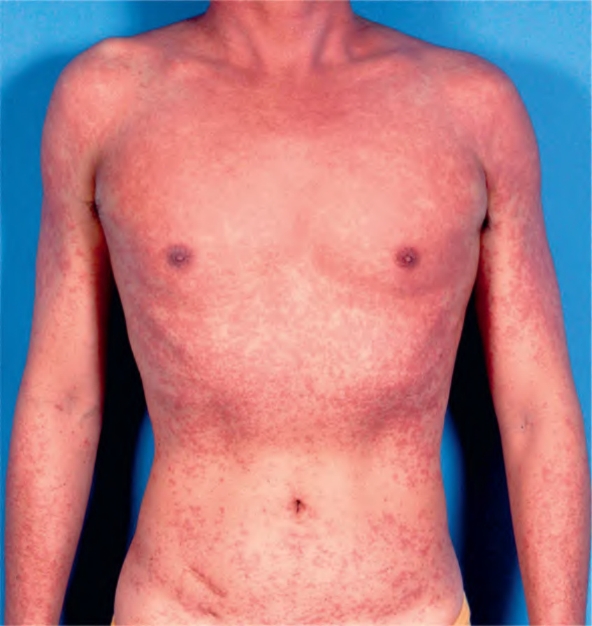
圖 78-20:由 trimethoprim–sulfamethoxazole(TMP-SMX)所致的麻疹樣藥物疹。這名 HIV 年輕男性在開始使用 TMP-SMX 後 8 天,發生廣泛的可壓退性紅斑斑與丘疹疹。注意上軀幹的融合。
Fig. 78.20 Morbilliform drug eruption due to trimethoprim–sulfamethox- azole (TMP-SMX). This young man with HIV developed a widespread eruption of blanchable erythematous macules and papules 8 days after starting TMP-SMX. Note the coalescence on the upper trunk.