CLINICAL FEATURES
Cutaneous and mucosal HPV types form two distinct groups that infect either skin or mucosa. However, viral tropism is not absolute, as DNA of skin types may be found in genital or oral warts, and in rare cases, mucosal types may be associated with skin lesions. Clinical manifestations depend on the HPV type involved, the anatomic location, and the immune status of the host (Table 79.1).
Cutaneous Infections
Cutaneous HPV types comprise a small group of viruses that infect the skin and induce common warts (Latin verrucae vulgares; singular verruca vulgaris), palmar and plantar warts (verrucae palmares et plantares), mosaic warts, flat warts (verrucae planae), and butcher’s warts (see Table 79.1). In general, classification of warts is based on morphology, histology, and anatomic location.
Common warts are hyperkeratotic, exophytic, dome-shaped papules or plaques that are typically associated with HPV-1, -2, -4, -27, or -57. These warts are most frequently located on the fingers and dorsal surfaces of the hands (Fig. 79.4 & 79.5) or in other sites prone to trauma such as the knees or elbows, but they may occur anywhere on the skin surface. Involvement of the proximal nail fold and/or ablative therapy at that location may destroy the matrix, resulting in onychodystrophy (see Fig. 79.5). Characteristic features of common warts are punctate black dots representing hemorrhage into the stratum corneum (Fig. 79.6). Autoinoculation by scratching may cause a linear arrangement of warts. Slender, exophytic filiform warts can also develop, especially in periorificial locations on the face (Fig. 79.7). Numerous verrucae can be observed in immunocompromised patients and rarely they are eruptive when the immunosuppression is drug-induced.
Palmar and plantar warts appear as thick, endophytic papules on the palms, soles, and lateral aspects of the hands and feet, with gently sloping sides and a central depression resembling an anthill (hence the term myrmecia, meaning anthill). On the soles, these are often painful from pressure when walking, due to their deep inward growth (Fig. 79.8). Plantar warts that coalesce into large plaques are referred to as mosaic warts (see Fig. 79.8). Extensive chronic verrucosis that
(HPV) types. HPV types in the genus β are in bold.
is notoriously resistant to therapy has been observed in immunocompromised individuals, and, at times, in patients with no apparent immune dysfunction (Fig. 79.9). Inclusion warts of the sole are plantar cysts from which HPV types 4, 60, 63, and 65 have been isolated. In studies from Europe, HPV-1 and the closely related types HPV-2, -27, and -57 caused the majority of palmoplantar warts, and they occurred most commonly in patients 6–10 years of age. Although a higher incidence of HPV-2-related warts was reported in atopic children, other investigators have failed to find such an association.
Flat warts are skin-colored or pinkish to brown, relatively smoothsurfaced, slightly elevated, flat-topped papules that are most commonly located on the dorsal hands, arms, or face, often in a linear array (Fig. 79.10). They are usually caused by HPV-3 or -10, and less often by HPV-28 and -29.
Butcher’s warts, which earn their name from their occurrence in meat- (or fish-) processing professionals, appear as extensive verrucous papules or cauliflower-like lesions on the dorsal, palmar, or periungual aspects of the hands and fingers. These warts are associated with HPV-7 not animal papillomaviruses.
Epidermodysplasia verruciformis (EV), first described in 1922 by Lewandowski and Lutz, is a rare genetic disease. It is characterized by a particular susceptibility to cutaneous infections with HPV types in the genus β (see above), which do not produce clinical lesions in immunocompetent individuals. The disease usually manifests in childhood with highly polymorphic, widespread lesions. About two-dozen specific HPV types have been described in EV patients, a subset of which (mainly types 5 and 8) is also detected in EV-associated skin cancers. EV typically has an autosomal recessive pattern of inheritance and is caused by truncating mutations in two genes, TMC6 (EVER1) and
The wart at the base of the distal phalanx of the hallux is painful due to deep endophytic growth; in addition, there are confluent plaques of superficial warts (mosaic warts).
TMC8 (EVER2), that encode transmembrane proteins localized to the endoplasmic reticulum. These proteins form a complex that interacts with the zinc transporter 1 (ZnT1) and affects intracellular zinc distribution, down-regulating zinc-associated transcription factors. Inhibition of ZnT1-TMC6/8 function by the HPV E5 protein is thought to facilitate viral infection; β HPV types lack E5, but they become pathogenic in EV patients with TMC6/8 defects.
Patients with EV present with widespread, discrete, or confluent papules that resemble flat warts (Fig. 79.11A); scaly, pinkish, or hypopigmented guttate macules and thin plaques that resemble pityriasis versicolor represent another characteristic finding (Figs. 79.11B & 79.12). In an individual patient with EV, warts that appear clinically distinct often contain different HPV types, and each skin lesion usually contains several HPV types. Actinic keratoses usually arise after the age of 30 years and slowly transform into invasive SCCs in approximately half of EV patients. These tumors have low metastatic potential and develop primarily in sun-exposed areas such as the forehead, ears, and hands, implicating UV irradiation as an important co-carcinogen. Patients with EV should be educated about the importance of minimizing sun exposure, and family members should be screened for the presence of similar lesions.
It may be difficult to differentiate flat warts from EV-associated lesions based on histology alone (see below), but the presence of lesional β HPV DNA (e.g. HPV-5, -8, etc.) can establish the diagnosis in an otherwise healthy individual. Beta HPV types can also induce a sporadic EV-like eruption in immunosuppressed patients, including transplant recipients and those with AIDS or a lymphoma. Of note, efflorescence of HIV-associated acquired EV has been observed following the second dose of the quadrivalent HPV vaccine.
In addition, β HPV sequences have been isolated from the normal skin of healthy individuals by highly sensitive DNA detection methods, indicating that the general population acts as the reservoir for these HPV types. There is increasing epidemiologic and biologic evidence that β HPV types may play an adjunct role in the initiation, but not maintenance, of SCCs in immunosuppressed patients.
WHIM syndrome is a rare, autosomal dominant primary immuno deficiency disorder characterized by HPV-induced warts (cutaneous and genital), hypogammaglobulinemia, recurrent bacterial infections, and neutropenia due to myelokathexis (retention of mature neutrophils in the bone marrow) (see Ch. 60). It is caused by mutations in the gene encoding the chemokine CXC receptor 4 (CXCR4), which result in impaired leukocyte trafficking and homing.
Several other primary immunodeficiencies are associated with severe HPV infection including DOCK8 deficiency, GATA2 deficiency (MonoMAC), hyper IgM syndrome, and idiopathic CD4+ lymphopenia (see Table 60.1).
Mucosal Infections
More than 40 HPV types preferentially infect the mucosa of the anogenital and upper aerodigestive tracts. Subclinical infections are much more common than visible warts. Application of 5% acetic acid (aceto-whitening) may aid in the identification of subclinical lesions as white areas.
Condylomata acuminata (singular: condyloma acuminatum), or anogenital warts, are found on the external genitalia and the perineum, perianally, or in adjacent areas such as the inguinal fold and mons pubis. Lesions may extend into the vagina, urethra, or anal canal (but rarely beyond the dentate line). Condylomata are typically discrete, sessile, smooth-surfaced exophytic papillomas or acuminate warts and may be skin-colored, brown, or whitish (especially when macerated in moist areas) (Fig. 79.13). They lack the thick cuirass of horny scale present on cutaneous warts and frequently measure one to several millimeters in diameter. Condylomata may also present as pedunculated or broad-based papillomas up to several centimeters in diameter or as large confluent plaques (Fig. 79.14). Condylomata plana, or flat cervical warts, may be difficult to recognize without aceto-whitening and the use of a (magnifying) colposcope. High-grade intraepithelial neoplasias are usually caused by high-risk types, mainly HPV-16, -18 and -31, whereas low-grade lesions may contain low-risk as well as high-risk HPV types.
Bowenoid papulosis manifests as multiple red–brown papules or confluent plaques on the external genitalia, on the perineum, or perianally (Fig. 79.15 & 79.16; see Fig. 73.13). These lesions primarily
affect young adults and may clinically resemble genital warts, but histologically represent a high-grade squamous intraepithelial lesion (HSIL) or SCC in situ. Erythroplasia of Queyrat is a distinct clinical entity, presenting as a well-demarcated, velvety erythematous plaque on the glabrous skin of the penis, vulva, or perianal region that histologically is an HSIL (Fig. 79.17). A biopsy is warranted for pigmented, erosive, bleeding, and/or therapy-resistant genital lesions to exclude malignancy. Bowenoid papulosis and erythroplasia of Queyrat contain high-risk HPV types, predominantly HPV-16, and may thus represent a precursor lesion of vulvar, penile, or perianal cancer. As noted above, these entities can represent variants of SCC in situ.
Multiple skin-colored or pink (A, B) to brown (C) smooth-surfaced, flat-topped papules. (D) Thin pink papules and plaques that are well demarcated and have more scale than the patients in A-C. These lesions are typically caused by HPV-3 or -10. B, Courtesy Kalman Watsky, MD; C, Courtesy Julie V. Schaffer, MD.
Generalized erythematous macules and plaques are seen in the same patient as shown in Fig. 79.11A. Lesions tested positive for HPV-8 and -36.
Buschke–Löwenstein tumor (giant condylomata acuminata), oral florid papillomatosis of Ackerman (oropharyngeal), epithelioma cuniculatum (plantar), and papillomatosis cutis carcinoides of Gottron (cutaneous) are types of “low-grade-malignant” verrucous carcinomas that have been associated with HPV infection. Verrucous carcinomas
are locally invasive and destructive but rarely metastasize (see Ch. 108). Buschke–Löwenstein tumor is a rare neoplasm of the anorectal area and external genitalia associated with the low-risk HPV types 6 or 11 that usually cause condylomata acuminata. The basis for the difference in biologic behavior between these two entities has not been
(VIN). Extensive red– brown or whitish papules and plaques containing high-risk mucosal HPV in an HIV-positive patient. Analogous high-grade squamous intraepithelial lesions (HSIL) were present perianally (AIN) and on the cervix (CIN).
Histology revealed high-grade anal intraepithelial neoplasia (AIN), not invasive SCC. However, continued monitoring is indicated.
established. In rare cases, pre-existing anogenital condylomata may progress into large exophytic cauliflower-like tumor masses that infiltrate deeply into underlying tissues and form fistulas and abscesses (Fig. 79.18). The histology of the Buschke–Löwenstein tumor may appear remarkably benign and resemble that of condylomata acuminata (Fig. 79.19), but its architecture is more irregular, the epithelium is more hyperplastic, and the pattern of growth is at least focally infiltrative. Focal frank malignant transformation can occur spontaneously or following X-irradiation. High-resolution imaging and a large biopsy are essential for identifying the extent of the infiltration and foci of SCC. Radical surgery may cure the disease, but recurrences are frequent, resulting in a high morbidity rate.
Oral warts appear as small, soft, pink or white, slightly elevated papules and plaques on the buccal, gingival, or labial mucosa (Fig. 79.20),
the tongue, or the hard palate. Oral condylomata are associated with HPV types 6 and 11 and may result from digital or oral–genital sexual transmissions. In HIV-positive patients, oral papillomas are frequently detected and may contain unusual HPV types such as 7, 71, 72, and 73. In focal epithelial hyperplasia, or Heck disease, multiple circumscribed papules resembling flat warts or condylomata are found on the gingival, buccal, or labial mucosa. This disorder is rare in White individuals but relatively common in children who are native South Americans, native Greenlanders, or from South African communities. It is associated exclusively with HPV-13 or -32.
HPV (especially type 16) is a causative agent in about 25% of head and neck cancers, including the majority of oropharyngeal cancers that arise from the base of tongue and palatine tonsils. HPV-positive oropharyngeal cancers can develop irrespective of exposure to tobacco. Although poorly differentiated histopathologically, they appear to have a better prognosis than HPV-negative oropharyngeal cancers whose primary risk factor is tobacco ± alcohol use.
In oral florid papillomatosis, multiple, confluent warty lesions are found in the oral cavity or the nasal sinuses. They are associated with HPV-6, -11, -16, and/or -18 in approximately half of cases. The development of these lesions is believed to be promoted by smoking,
X-irradiation, and chronic inflammation. Patients with oral papillomas need frequent examinations and repeated biopsies for early diagnosis of progression to verrucous carcinoma.
In recurrent respiratory papillomatosis (RRP), there are benign exophytic laryngeal papillomas caused by HPV-6 and -11. Patients classically present with a triad of hoarseness, stridor, and respiratory distress. Papillomas arise most often at the transition zones between the squamous and cilia-bearing epithelia of the larynx and subglottis, but they may rarely extend to the distal trachea, the bronchi, or even the bronchoalveoli. Malignant transformation of laryngeal papillomas into SCCs in patients with long-lasting disease has been reported, and exogenous factors such as X-irradiation, smoking, chemical toxins, or chemotherapy have been implicated as co-carcinogens.
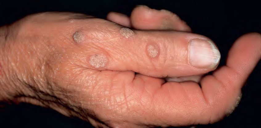
Fig. 79.4 Verrucae vulgares (common warts).Courtesy A. Geusau, MD.
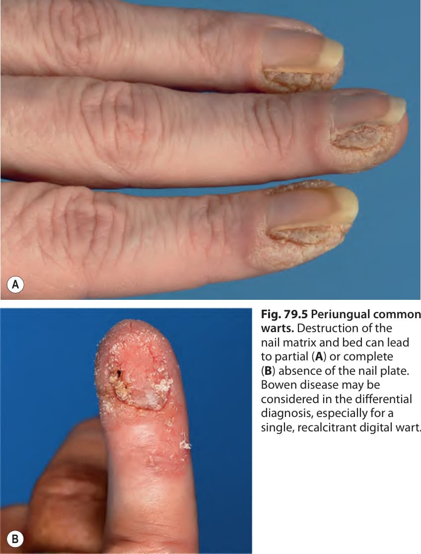
Fig. 79.5 Periungual common warts. Destruction of the nail matrix and bed can lead to partial (A) or complete (B) absence of the nail plate. Bowen disease may be considered in the differential diagnosis, especially for a single, recalcitrant digital wart.
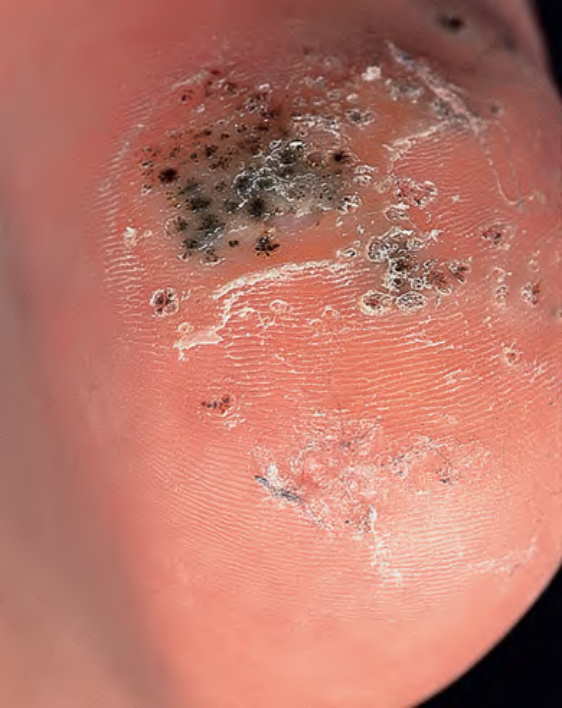
Fig. 79.6 Verrucae plantares (plantar warts). The photo was taken after shaving of the hyperkeratotic surface; the black dots represent hemorrhage into the stratum corneum.
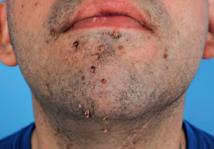
Fig. 79.7 Multiple filiform warts on the lower face.
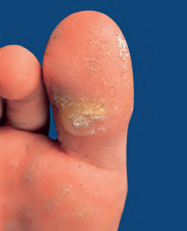
Fig. 79.8 Myrmecial wart.
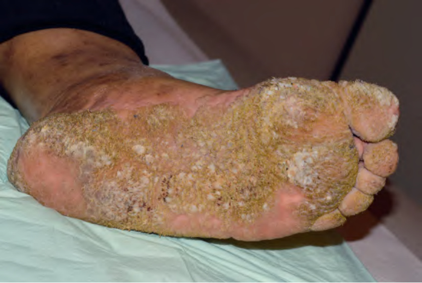
Fig. 79.9 Extensive and chronic verrucosis of the soles in a patient with hepatic cirrhosis due to alcoholism. HPV-27 DNA was isolated from the lesions.
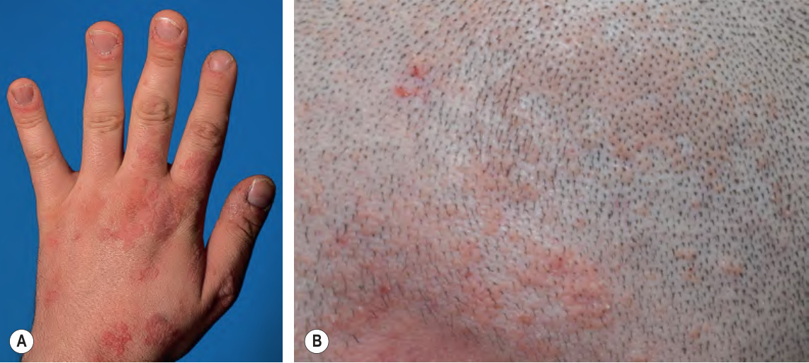
Fig. 79.10 Verrucae planae (flat warts).
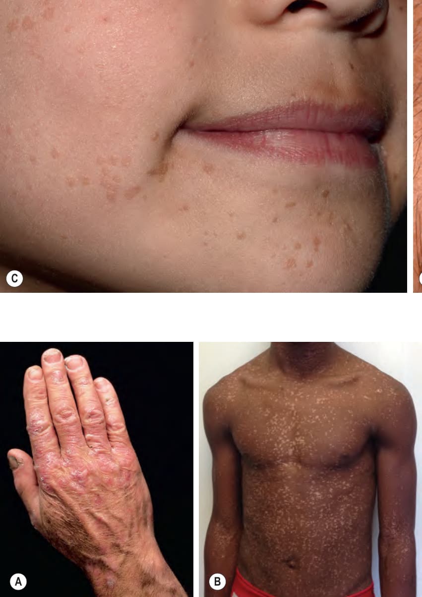
Fig. 79.11 Epidermodysplasia verruciformis (EV).A Confluent scaly papules and plaques resembling flat warts. B Numerous hypopigmented and light pink, flat-topped papules, some of which are in a linear array, in acquired HIV-associated EV. B, Courtesy Ncoza Dlova, MD.
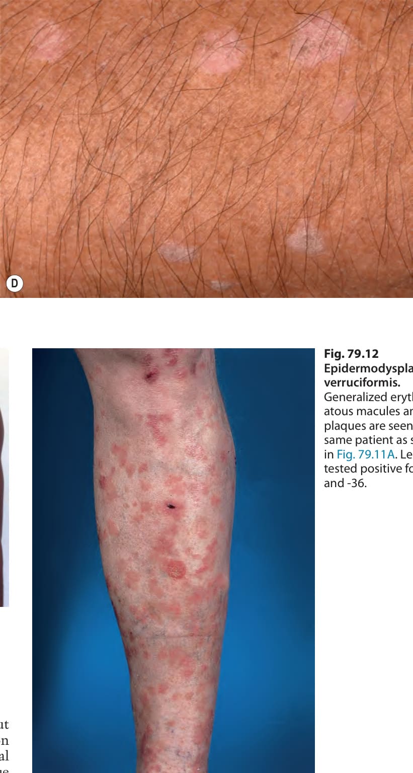
Fig. 79.12 Epidermodysplasia verruciformis.
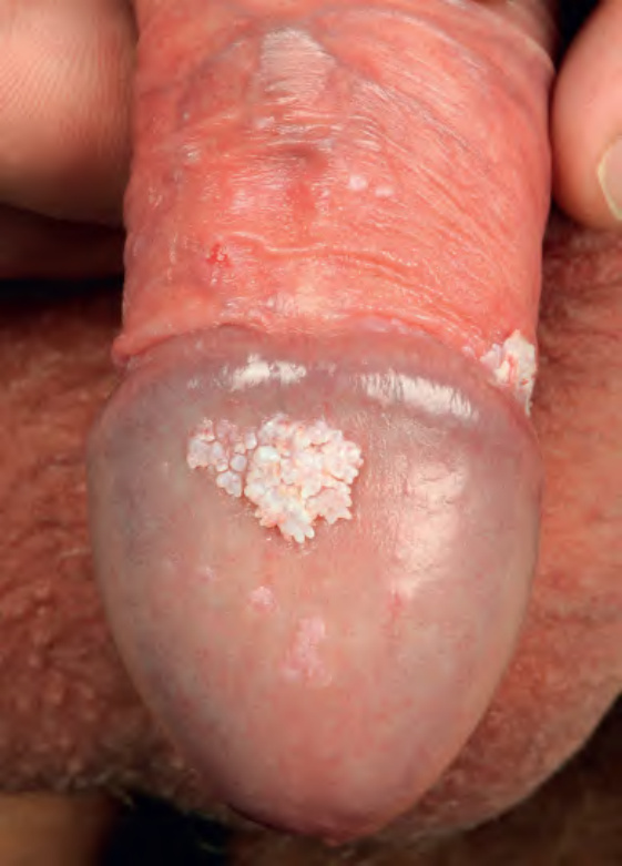
Fig. 79.13 Condylomata acuminata. Verrucous lesions on the glans and in the sulcus, with a few small papules on the shaft of the penis and distal glans. Note the acuminate topography, i.e. the tapering to a point. Courtesy Lorenzo Cerroni, MD.
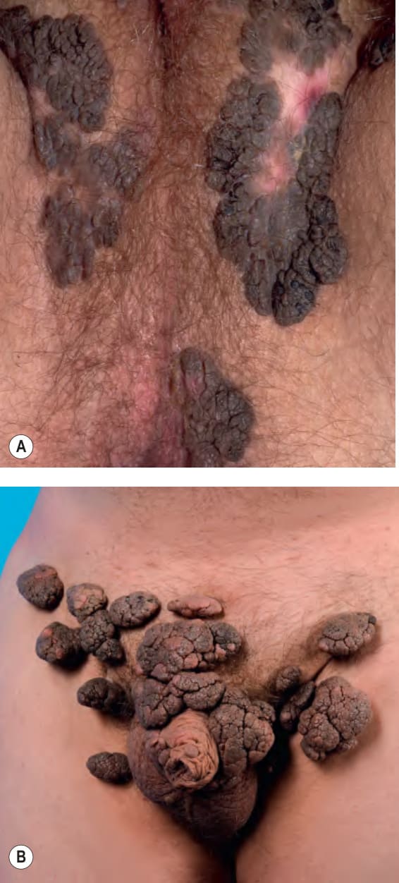
Fig. 79.14 Condylomata acuminata.A Confluent lesions forming hyperpigmented plaques in the perineal region and along the inguinal fold. A depigmented scar is seen at the site of prior treatment with liquid nitrogen. B Large, exophytic, broad-based or pedunculated papillomas in a healthy 15-year-old boy. The differential diagnosis includes Buschke- Löwenstein tumor.
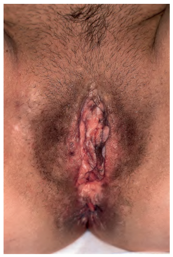
Fig. 79.15 Bowenoid papulosis of the vulva with histopathologic features of vulvar intraepithelial neoplasia
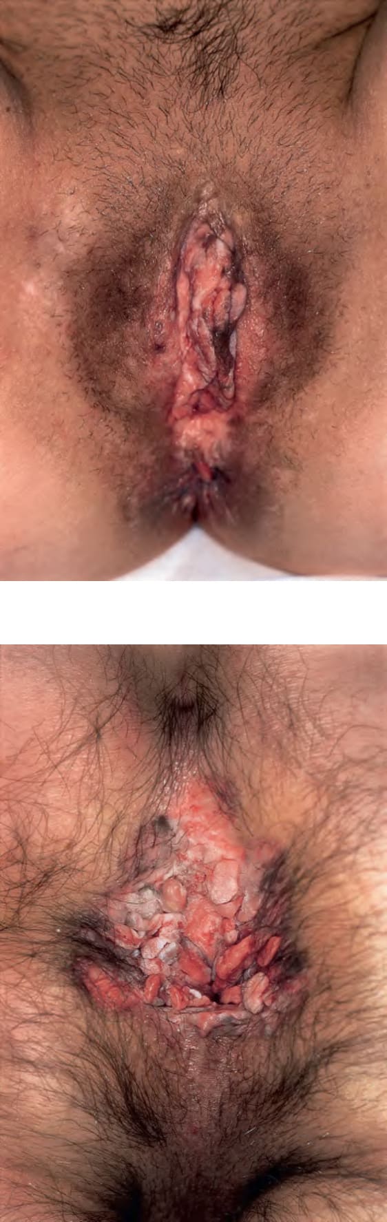
Fig. 79.16 Bowenoid papulosis of the anus positive for high-risk mucosal HPV in a man who had sex with men.
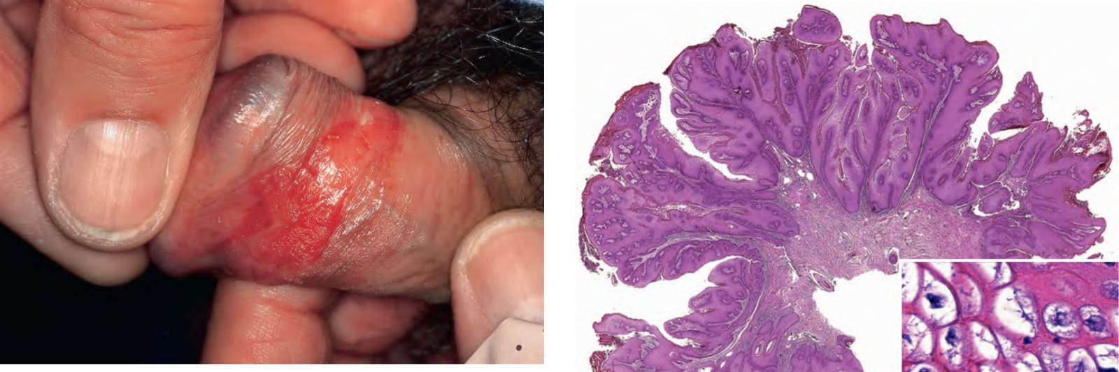
Fig. 79.17 Erythroplasia of Queyrat. A well-demarcated velvety plaque of the prepuce positive for high-risk HPV; histology revealed high-grade penile intra epithelial neoplasia (PIN).
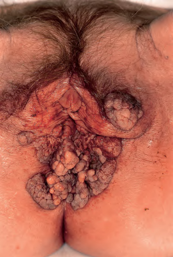
Fig. 79.18 Giant condylomata acuminata (Buschke–Löwenstein tumor). Cauliflower-like, deeply infiltrating giant condylomata acuminata in an older woman.
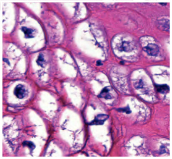
Fig. 79.19 Buschke–Löwenstein tumor – histopathologic features. Exophytic lesion with a papillomatous surface and marked, irregular epithelial hyperplasia without cellular atypia as well as the characteristic downward extension of bulbous rete ridges. Note vacuolated keratinocytes (inset). Courtesy Luis Requena, MD.
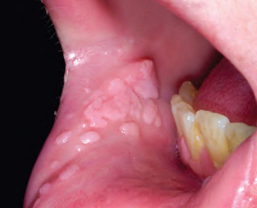
Fig. 79.20 Oral warts. Papillomas of the labial mucosa in a 6-year-old child.
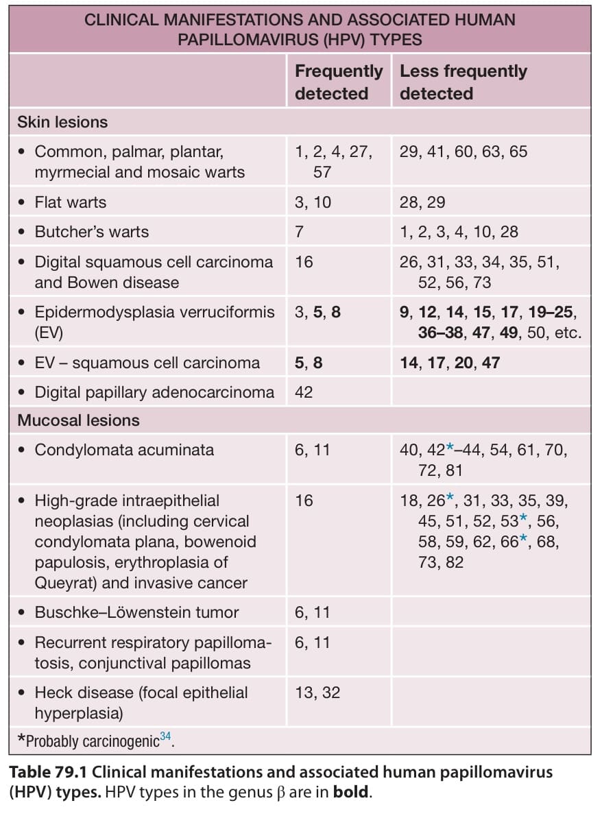
Table 79.1 Clinical manifestations and associated human papillomavirus