PATHOLOGY
The histopathologic changes induced by HPV infection are variable, reflecting the myriad clinical presentations and different anatomic sites. In productive HPV infections, a common motif in epithelial cells is the presence of cytoplasmic vacuoles isolating the nucleus from the cytoplasmic membrane. Cells with this distinctive vacuolization are referred to as koilocytotic (hollow) cells, and the presence of koilocytosis is a useful feature that distinguishes verrucae from other types of papillomas.
Common and Deep Palmoplantar Warts
Common warts are well circumscribed from the surrounding skin and characteristically have steeply sloping “church spire” papillomatosis heaped with ortho- and parakeratosis (Fig. 79.21). Parakeratosis is most commonly observed directly overlying the summits of the papillomatosis and is often accompanied by small intracorneal hemorrhages. There is also marked acanthosis and the rete ridges are elongated. In palmoplantar warts, the lateral edges of the wart bow inward to create a cup-shaped invagination below the papillomatosis. Higher-power examination reveals hypergranulosis and condensed coarse cytoplasmic keratohyalin-like granules of variable size and shape within the granular layer, along with vacuolization of cells on top of, or sometimes between, the papillae. Koilocytosis is typically observed in or immediately below the granular layer. The papillary dermis underlying the elongated rete ridges has minimal pathologic changes; increased vascularity can be observed.
Flat Warts
The characteristic features of flat warts include orthokeratosis alternating with parakeratosis, acanthosis, no or minimal papillomatosis, a uniformly thickened granular layer, and vacuolization of cells in the granular and upper spinous layers (termed “bird’s eyes”).
Some patients with flat warts develop clinically evident inflammation around these warts, which may precede their spontaneous involution. Characteristic histologic features of regressing flat warts include parakeratosis, spongiosis, exocytosis of mononuclear cells into the lower epidermis, and (occasionally) satellite cell necrosis. By immunohistochemistry, T helper cells predominate and HLA-DR antigen, a marker of immune activation, is detected on the surface of the infiltrating lymphocytes and on some keratinocytes. Many of these changes can also be observed in regressing condylomata, suggesting that wart regression is mediated, at least in part, by a delayed-type hypersensitivity-like cellular immune response directed against keratinocytes expressing wart antigens.
Epidermodysplasia Verruciformis
In patients with EV, the flat warts and pityriasis versicolor-like lesions exhibit orthokeratosis with a basket-weave appearance, parakeratosis,
and acanthosis. Under higher power, some cells within the hyperplastic spinous layer have characteristic cytopathic changes. These cells are large with perinuclear halos and a cytoplasm that has blue–gray pallor and contains keratohyaline granules of various sizes and shapes (Fig. 79.22). Keratinocytic dysplasia and actinic keratoses may be evident, especially in biopsies of lesions from sun-exposed areas. Because the histologic picture is nonspecific, it may be difficult to differentiate typical flat warts from EV-associated lesions based on histology alone.
Anogenital Warts
The most consistent histologic features seen in condylomata include epidermal hyperplasia, parakeratosis, koilocytosis, and papillomatosis. The papillomatosis is more gently rounded than is seen in common warts. The upper portions of the epithelia of mucosal surfaces normally have some degree of cytoplasmic vacuolization, so its detection is specific for condylomata acuminata only if present within the deeper portions of the spinous layer. Mitotic figures may be evident, and prior treatment with podophyllotoxin (which interferes with microtubule formation in cells undergoing mitosis) can induce the appearance of atypical mitoses that could potentially lead to misdiagnosis as SCC.
Giant Condylomata of Buschke–Löwenstein
The histology of the Buschke–Löwenstein tumor shares many features with condylomata acuminata, but there is more evident irregular epithelial hyperplasia, with marked downward extension of characteristically bulbous rete ridges, and less vacuolization of epidermal cells (see Fig. 79.19). Mitotic figures are rarely detectable, and, when present, are normal. While usually associated with extension into and destruction of local tissue, metastases to local lymph nodes from foci of SCC arising in these tumors have been reported.
Squamous Intraepithelial Neoplasias
Persistent infection with high-risk HPV types can lead to intraepithelial neoplasia that may progress to invasive carcinoma. The precancerous lesions represent a continuum of morphologic changes with indistinct boundaries. In CIN grade I, nuclear enlargement and hyperchromasia is observed in the lower epithelial layers; these changes may be accompanied by cytoplasmic halos (koilocytotic atypia). The next stage (CIN II) shows progressive atypia in all layers of the epithelium, with abnormal differentiation of the keratinizing cell layers. The atypical cells have an increased nuclear–cytoplasmic ratio, variability of nuclear size, and an increased number of mitotic figures, including abnormal mitoses and hyperchromasia. In CIN III, the epithelium is totally replaced by immature, atypical cells
Orthokeratosis alternating with parakeratosis and acanthosis are present. Note the cells with blue–gray, granular cytoplasm in the mid to upper epidermis (inset). Courtesy Lorenzo Cerroni, MD.
Note the characteristic features of abnormal keratinocyte maturation, nuclear pleomorphism, and dyskeratosis. Koilocytosis, due to cytoplasmic vacuoles that isolate the nucleus from the cytoplasmic membrane, can be observed in or immediately below the granular layer. Some pathologists refer to these intraepithelial neoplasias as SCC in situ. Courtesy R. Tyler, MD.
with a lack of surface differentiation. The analogous lesions of the vulva, vagina, penis, and anus are termed VIN (vulvar intraepithelial neoplasia; Fig. 79.23), VaIN (vaginal intraepithelial neoplasia), PIN or PeIN (penile intraepithelial neoplasia), and AIN (anal intraepithelial neoplasia), respectively. Clinically, these may appear as erythematous or whitish plaques (erythroplasia, leukoplakia) or red–brown papules (bowenoid papulosis) (see Figs. 79.15–79.17). Terminology of these lesions remains a matter of debate, and many pathologists refer to these “intraepithelial neoplasias” as “SCC in situ”.
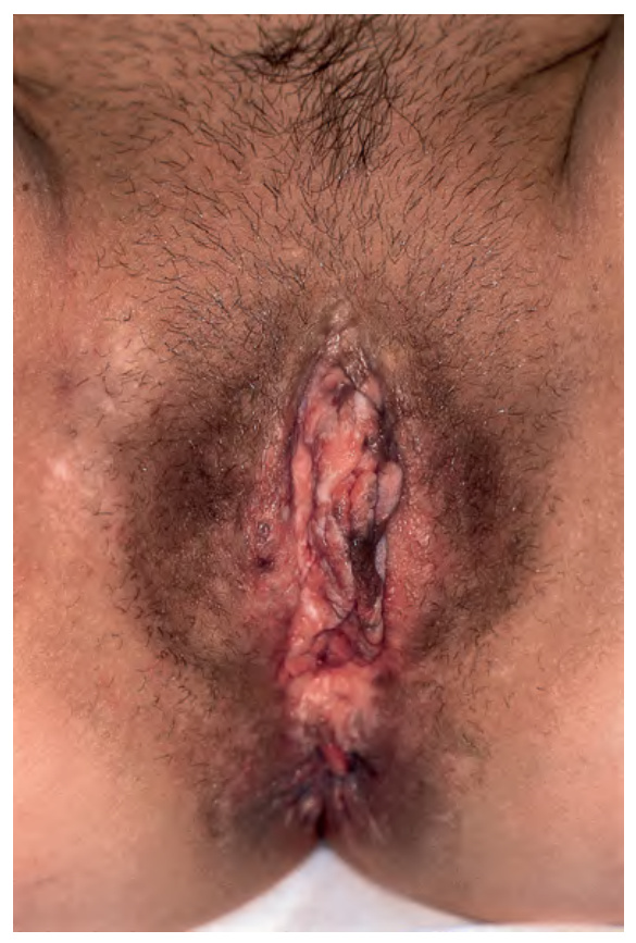
Fig. 79.15 Bowenoid papulosis of the vulva with histopathologic features of vulvar intraepithelial neoplasia
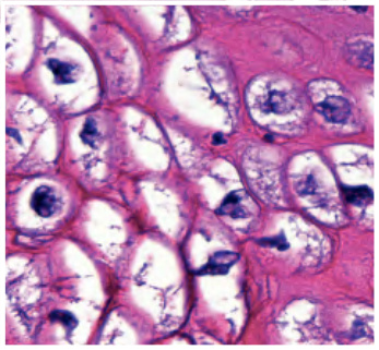
Fig. 79.19 Buschke–Löwenstein tumor – histopathologic features. Exophytic lesion with a papillomatous surface and marked, irregular epithelial hyperplasia without cellular atypia as well as the characteristic downward extension of bulbous rete ridges. Note vacuolated keratinocytes (inset). Courtesy Luis Requena, MD.
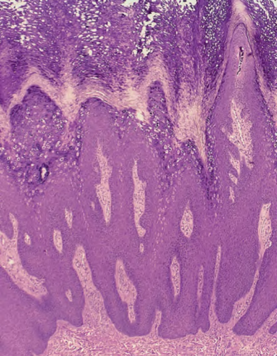
Fig. 79.21 Verruca vulgaris – histopatho- logic features. Note the characteristic features of “church spire” papillomatosis heaped with ortho- and parakeratosis, acanthosis, hypergranulosis, and koilocytosis. Courtesy R. Tyler, MD.
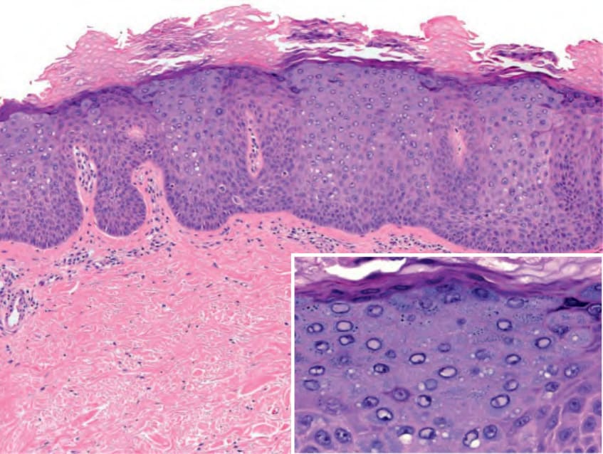
Fig. 79.22 Epidermodysplasia verruciformis – histopathologic features.
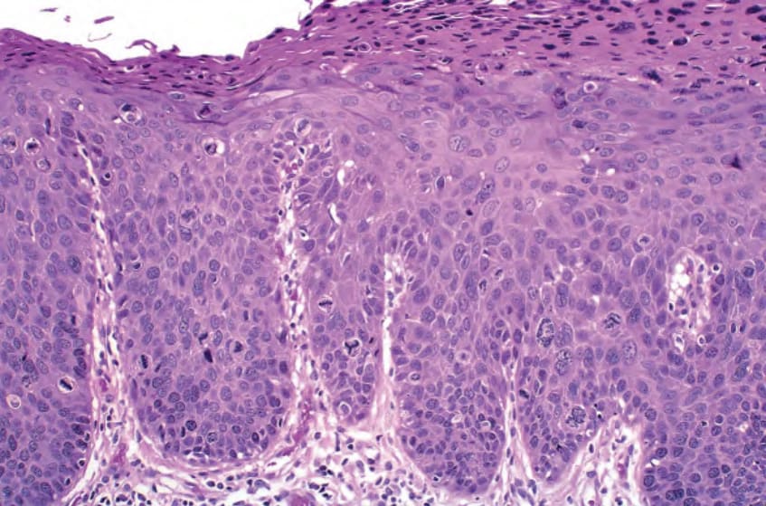
Fig. 79.23 Vulvar intraepithelial neoplasia (VIN) – histopathologic features.