DIFFERENTIAL DIAGNOSIS
The diagnosis of cutaneous and genital warts is straightforward if typical clinical features are present. Occasionally, various other diagnoses have to be considered and a biopsy may be required for confirmation and to identify dysplastic lesions. Immunohistochemistry with antibodies directed against cross-reacting epitopes of papillomavirus capsid proteins allows the detection of virions in palmoplantar warts, which are highly productive infections, but has limited sensitivity in genital warts and dysplasias. There is no routine infectivity assay available to detect virions in clinical samples, and diagnostic tests are therefore based on the molecular detection of papillomavirus DNA. Commercially available tests that utilize nucleic acid hybridization (e.g. digene Hybrid Capture® 2 [HC2] HPV DNA Test; Amplicor® HPV and Linear Array® HPV Genotyping Test, Roche [not available in the US]) can identify low-risk versus high-risk mucosal HPV types in clinical samples with high sensitivity and specificity. In routinely processed histologic sections, RNA in situ hybridization can also be performed utilizing pooled high-risk versus low-risk HPV subtypes. In addition, PCR-based techniques can detect a broad range of genital and skin HPV types. Development of circulating antibodies against the L1 major capsid protein is common following infection with genital HPV types, and ELISAs using L1 VLPs as the antigen have been useful in epidemiologic studies. However, due to low antibody titers and variable intervals between infection and seroconversion, this assay is not used for diagnosis in individual patients.
Common, Flat, and Palmoplantar Warts
Seborrheic keratoses (SKs), actinic keratoses (AKs), cutaneous horns (which can arise from warts as well as AKs, SCCs, SKs, and other neoplasms; see Ch. 109), keratoacanthomas, other SCCs, tricholemmomas, Spitz nevi, and amelanotic melanomas may resemble common warts, and even tuberculosis verrucosa cutis or a plaque of psoriasis, hypertrophic lichen planus, hypertrophic lupus erythematosus, or pseudoepitheliomatous tattoo reaction can have a warty clinical appearance. On the distal or lateral finger or periungually, a wart-like or erythematous, well-demarcated plaque may occasionally represent Bowen disease and harbor high-risk HPV types, e.g. 16 or 18. Similarly, amelanotic melanoma can masquerade as a persistent periungual or plantar wart and should be clinically suspected if erosion or subtle pigmentation is present. The differential diagnosis of flat warts may include acrokeratosis verruciformis, Gottron papules of dermatomyositis (distinguished by their localization over the knuckles), lichen nitidus, lichen planus, and lichenoid keratoses. Although lesions of both lichen planus and flat warts can have a linear array, the presence of Wickham striae points to the former diagnosis. Plantar clavi (corns) are often misdiagnosed as warts, although verruca can be differentiated by the presence of punctate black dots rather than a glassy appearance; in addition, paring of the keratotic wart surface leads to capillary bleeding from the superficially located dermal papillae. Punctate palmoplantar keratoderma, punctate porokeratosis, arsenical keratoses, poromas, and eccrine poromatosis can mimic plantar warts.
In patients with cellular immunodeficiencies, including organ transplant recipients and those with AIDS or hematologic malignancies, the number as well as incidence of cutaneous and genital warts is increased. Warts that appear at less common sites such as the face and the neck need to be differentiated from mollusca contagiosa. In immunosuppressed patients, macular and flat warts may resemble EV, but the clinical setting and absence of a family history can aid in establishing the correct diagnosis. Other heritable immunodeficiencies that predispose affected individuals to the development of warts include WHIM syndrome (see above), idiopathic CD4+ lymphopenia, DOCK8 deficiency, hyper IgM syndrome, PI3Kδ syndrome, and GATA2 deficiency (MonoMAC) (see Ch. 60 & Table 60.1).
Anogenital Lesions
Condylomata acuminata are rarely confused with condylomata lata of secondary syphilis, but serologic testing for syphilis (and, as indicated, other STIs) is recommended in patients with genital warts, and dark-field examination for spirochetes may be considered. Mollusca contagiosa preferentially involve the mons pubis and can be distinguished by their umbilication, with dermoscopy revealing a multilobular, yellow– white central structure surrounded by a “crown” of vessels. In men, pearly penile papules are normal anatomic structures consisting of several rows of discrete, monomorphic, dome-shaped, 1 to 2 mm papules circumferentially around the corona and/or sulcus of the glans penis (see Ch. 116). An analogous picture exists in women, called vestibular papillomatosis, with tiny, uniformly shaped, finger-like projections at the introitus and on the labia minora. Sebaceous glands of the prepuce and labia majora appear as white–gray to yellow, small papules with a regular array. Epidermoid cysts, steatocystomas, or angiokeratomas of the scrotum seldom pose diagnostic problems. Occasionally, verrucous herpetic infections may mimic genital warts.
Detection of subclinical genital HPV infection is facilitated by soaking with 5% acetic acid for 3 to 5 minutes; this leads to whitening (“acetowhitening”) of lesions, and the use of a colposcope for magnification will further increase diagnostic accuracy. However, aceto-whitening is not specific for HPV-induced lesions and is also observed in infectious or inflammatory conditions such as Candida vulvitis or balanoposthitis, psoriasis, lichen planus, and eczematous dermatitis.
Bowenoid papulosis consists of red–brown papules or confluent, sometimes leukoplakia-like plaques that may be difficult to distinguish from condylomata (see Fig. 79.15 & 79.16 and Ch. 73). Erythroplasia of Queyrat is a well-defined, velvety, erythematous plaque of the glans or vulva that must be differentiated from erosive lichen planus and Zoon balanitis. Bowen disease can present as a solitary patch or plaque, sometimes scaly, of the vulva or penis, most often in older individuals. Extramammary Paget disease appears as a well-defined red plaque in the groin or pubic region or on the vulva, penile root, or perianal skin, with distinctive histopathologic features (see Ch. 73).
Vulvar carcinomas are etiologically heterogeneous. Carcinomas with basaloid or warty histology are related to HPV infection, whereas keratinizing squamous carcinomas are not. Basaloid and warty vulvar carcinomas typically arise in younger women, are found adjacent to vulvar intraepithelial neoplasia (VIN; also referred to as high-grade squamous intraepithelial lesion [HSIL]), and are associated with risk factors related to sexual practices. Close to 90% of high-grade VIN/HSIL and basaloid and warty vulvar carcinomas contain high-risk HPV DNA
It developed over more than a decade from a papular lesion of the prepuce. HPV-16 and -51 DNAs were detected by RT-PCR.
(most often HPV-16, but also HPV-31 and -33), suggesting a common etiology. In contrast, the more common keratinizing vulvar carcinomas in older women, which typically arise adjacent to lichen sclerosus and differentiated VIN, infrequently contain HPV DNA and thus are regarded as a distinct entity.
Although there is heterogeneity in anal SCC, in both sexes, the vast majority of anal cancers are attributable to HPV infection. Cancers of the anal canal that are positive for high-risk HPV (mostly HPV-16, but also HPV-18, -31, -33) tend to occur in younger men (especially MSM) and women with high-risk sexual behaviors, have basaloid histology, and arise adjacent to AIN. In contrast, HPV-negative anal cancers usually affect older men, are well keratinized, and arise from perianal skin without AIN. Heterogeneous etiology has also been described for penile cancer (Fig. 79.24), analogous to that observed for vulvar and anal SCCs. Immunosuppression and a history of HPV-associated malignancy are notable risk factors for the development of a (second) anogenital cancer attributable to HPV.
Anogenital warts in children may give rise to suspicion of sexual abuse (see Ch. 90). Findings that make non-abusive transmission of HPV more likely include age <3 years, wart location somewhat distant from the anus or vulva, genital condylomata in the mother, and an absence of signs of physical abuse or of other sexually transmitted infections.
Giant condyloma acuminatum, or Buschke–Löwenstein tumor, is clinically suspected by its size and the presence of fistulas or abscesses (see Fig. 79.18). Histologic findings may be bland, and distinction from large benign genital warts can therefore be difficult at an early stage (see Fig. 79.19). However, high-resolution imaging reveals the extent of infiltration, and a large biopsy with serial sectioning may detect locally destructive growth and, rarely, transformed foci of SCC.
Oral Warts
Oral warts or condylomata may resemble the papules of focal epithelial hyperplasia (Heck disease); the latter is very rare in White individuals. A distinct oral condition with the descriptive name “proliferative verrucous leukoplakia” is clinically similar to oral florid papillomatosis but does not contain HPV DNA and has a high risk of progression to oral SCC (Fig. 79.25). Additional diagnostic considerations may include leukoedema (grayish-white translucent plaques, sometimes with a “moth-eaten” appearance, that favor the buccal mucosa and may become less evident upon stretching), white sponge nevus, and hereditary benign epithelial dyskeratosis.
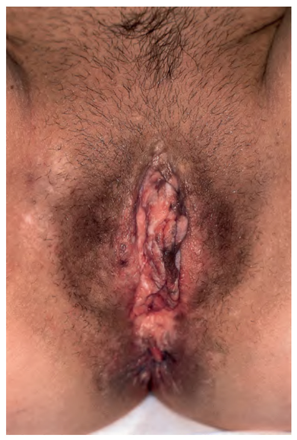
Fig. 79.15 Bowenoid papulosis of the vulva with histopathologic features of vulvar intraepithelial neoplasia
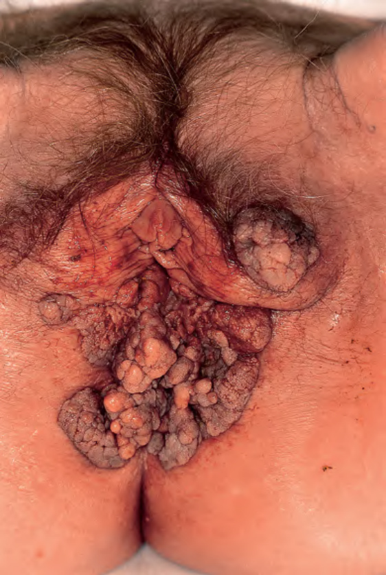
Fig. 79.18 Giant condylomata acuminata (Buschke–Löwenstein tumor). Cauliflower-like, deeply infiltrating giant condylomata acuminata in an older woman.
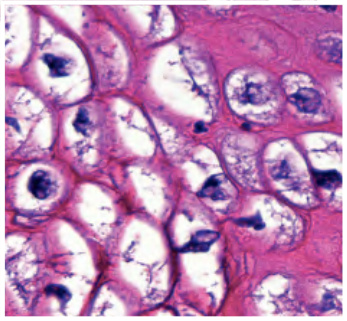
Fig. 79.19 Buschke–Löwenstein tumor – histopathologic features. Exophytic lesion with a papillomatous surface and marked, irregular epithelial hyperplasia without cellular atypia as well as the characteristic downward extension of bulbous rete ridges. Note vacuolated keratinocytes (inset). Courtesy Luis Requena, MD.
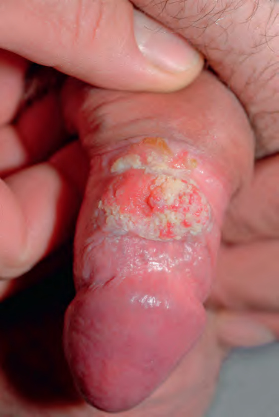
Fig. 79.24 Invasive squamous cell carcinoma of the penis.
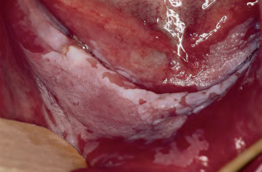
Fig. 79.25 Proliferative verrucous leukoplakia of the oral cavity. The lesions have focally progressed to invasive squamous cell carcinoma; HPV DNA was not detectable by PCR.
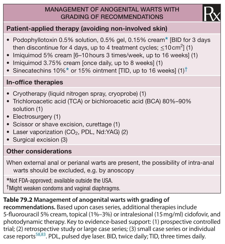
Table 79.2 Management of anogenital warts with grading of recommendations. Based upon cases series, additional therapies include 5-fluorouracil 5% cream, topical (1%–3%) or intralesional (15 mg/ml) cidofovir, and photodynamic therapy. Key to evidence-based support: (1) prospective controlled trial; (2) retrospective study or large case series; (3) small case series or individual case reports. PDL, pulsed dye laser. BID, twice daily; TID, three times daily.