HERPES SIMPLEX VIRUSES (HSV-1 AND HSV-2)
Synonyms/clinical variants: Herpes – herpes simplex Herpes labialis – cold sore, fever blister, herpes febrilis Herpes gladiatorum – scrum pox Herpetic whitlow Genital herpes – herpes progenitalis
Key features
Herpes simplex viruses produce primary and recurrent vesicular eruptions that favor the orolabial and genital regions
The use of sensitive and specific serologic markers has provided insights into the epidemiology of HSV infections
Antiviral therapy significantly influences the course and spectrum of HSV disease
Introduction
HSV-1 and HSV-2 are ubiquitous pathogens that produce primarily orolabial and genital infections. Worldwide, genital herpes is one of the most common sexually transmitted infections. The use of sensitive and specific serologic markers has enabled the epidemiology of HSV infections to be deciphered, while antiviral therapy has allowed clinicians to control the course and extent of HSV disease. Prevention of HSV infections via molecularly derived vaccines remains an important goal.
History
Herpetic lesions have been described since antiquity, including by the noted physicians Hippocrates and Galen. The word “herpes” was first defined by Greek scholars as “to creep or crawl” in reference to the spreading nature of the skin lesions. The association between orolabial herpetic lesions and genital infection was first recognized during the late eighteenth century. However, better understanding of the pathogenesis of herpes infections did not occur until the twentieth century. Lipschitz was the first to suggest antigenic differences between the two
HSV types, but it was not until 1968 that Nahmias and Dowdle demonstrated that HSV-1 was more frequently associated with orolabial and HSV-2 with genital infections. In the past several decades, there have been critical insights into the pathogenesis of HSV infections as well as advances in their diagnosis, treatment, and prevention.
Epidemiology
Herpes simplex viruses have a worldwide distribution. Their inter-action with human hosts has the following components: (1) primary infection – initial HSV infection, without pre-existing antibodies to HSV-1 or HSV-2; (2) non-primary initial infection – infection with one HSV type in an individual with pre-existing antibodies to the other HSV type; (3) latency – virus in sensory ganglia; and (4) reactivation – recurrent infection with asymptomatic viral shedding or clinical manifestations (recrudescence). Primary, non-primary initial, and recurrent infections can each be symptomatic or asymptomatic. It is estimated that approximately one-third of the world’s population has experienced a symptomatic HSV infection.
In children <10 years of age, ~80%–90% of herpetic infections are caused by HSV-1. Analyses on a worldwide basis have demonstrated HSV-1 antibodies in 50%–90% of individuals 20–40 years old. Although HSV-2 remains the major cause of recurrent genital herpes infections (70%–90% overall), the proportion due to HSV-1 has been increasing in the US, Canada, and the UK. In these countries, HSV-1 now accounts for the majority of genital herpes infections in young adults, especially college students. Recurrences of genital herpes are less common with HSV-1, which lacks HSV-2’s specific tropism for genital epithelium.
In the Centers for Disease Control and Prevention (CDC)’s 2015–2016 National Health and Nutrition Examination Survey (NHANES), the seroprevalence of HSV-2 among individuals 14–49 years of age in the US was 12% overall, with higher rates in women (16%, versus 8% in men) and Black individuals (35%). Of note, ~80% of those with positive serology never had a previous diagnosis of genital HSV infection. Other factors associated with acquisition of genital herpes include an age of 15–30 years (period of greatest sexual activity), an increased number of sexual partners, lower levels of income and education, and HIV-positivity; conversely, genital HSV-2 infection also increases the risk of acquiring and transmitting HIV infection.
Pathogenesis
Transmission of HSV can occur during asymptomatic periods of viral shedding. HSV-1 is spread primarily through direct contact with contaminated saliva or other infected secretions, while HSV-2 is spread primarily by sexual contact. Virus replicates at the mucocutaneous site of infection and then travels by retrograde axonal flow to the dorsal root ganglia, where it establishes latency until reactivation. Latency enables the virus to exist in a relatively non-infectious state for varying periods of time in its host. HSV-1 can induce intracellular accumulation of CD1d molecules in antigen-presenting cells, providing a possible explanation for how the virus evades detection and establishes latency. Normally, CD1d molecules are transported to the cell surface, where they present lipids and stimulate natural killer T cells, thus promoting an immune response. HSV’s mechanisms to evade the immune system also include downregulation of various immunologic cells and cytokines (Table 80.2).
The goals of the early host immune response against HSV are to limit viral replication and recruit other inflammatory cells. Toll-like receptors are critical to the first line of innate immune defense against HSV, and their functions include priming CD8+ T cells. HSV-specific memory CD8+ T cells, which are selectively activated and retained in
latently infected sensory ganglia, have important roles in controlling the infection and preventing symptomatic recurrences (see Table 80.2).
HSV can be reactivated either spontaneously or by a trigger such as emotional stress, ultraviolet light, fever, menstruation, immunosuppression, surgical or dental procedures, and other local tissue damage. Typically, reactivation produces vesicular lesions localized to the skin. However, viremia followed by widespread internal organ involvement can occur in immunocompromised hosts.
Clinical Features
HSV infections have a wide range of clinical presentations, and asymptomatic infection is very common. In primary infections, symptoms typically occur within 3 to 7 days after exposure. A prodrome of tender lymphadenopathy, malaise, anorexia, and fever often occurs before the onset of mucocutaneous lesions, which may be preceded by localized pain, tenderness, burning, and tingling. Painful, grouped vesicles appear on an erythematous base and may become umbilicated, followed by progression to pustules, erosions, and/or ulcerations with a characteristic scalloped border. Crusting of lesions and resolution of symptoms typically occurs within 2 to 6 weeks. A similar prodrome can precede recurrent lesions, but these are often fewer in number, with decreased severity and duration compared to those of a primary infection.
The majority of primary orolabial infections are asymptomatic. Symptomatic infections often present as gingivostomatitis in children or as pharyngitis and a mononucleosis-like syndrome in young adults. The mouth and lips are the most common sites of involvement (Fig. 80.1A,B), with oral lesions typically appearing on the buccal mucosa and gingivae. Edema and painful oropharyngeal ulcerations can lead to dysphagia and drooling.
Among individuals with latent HSV-1 infection, 20%–40% develop recurrent herpes labialis due to reactivation. Recurrent lesions appear most often on the vermilion border of the lip (Fig. 80.1C). Less common sites are perioral skin, nasal mucosa, cheek, and attached oral mucosa overlying bone (e.g. gingiva, hard palate) (Fig. 80.2). In immunocompromised hosts, recurrent herpes infections can involve intraoral movable mucosa not overlying bone (see Fig. 72.1).
Primary and non-primary initial genital herpes infections are frequently asymptomatic but (especially with the former) can also present as an excruciatingly painful, erosive balanitis, vulvitis, or vaginitis. In women, lesions often also involve the cervix, buttocks, and perineum and may be associated with inguinal adenopathy and dysuria (Fig. 80.3). Genital lesions in men typically occur on the glans or shaft of the penis, and the buttocks are occasionally affected. Systemic complaints and complications are more common in women. Extragenital lesions, urinary retention, and aseptic meningitis occur in 20%, 10%–15%, and 10% of affected women, respectively. In contrast, aseptic meningitis is a rare complication of primary genital herpes infection in men. The presence of more extensive local involvement, regional lymphadenopathy, and fever generally distinguishes primary herpes infection from recurrent disease.
Genital HSV infections can result in both subclinical viral shedding and clinically evident recurrences, which are often relatively mild; recurrences occur more frequently with HSV-2 than HSV-1. Usually, a limited number of vesicles reappear on the genitalia or buttocks (Fig. 80.4), with resolution within 7–10 days, compared to ~20 days for primary infections. Complications are uncommon. The frequency of recurrences correlates directly with the severity of the primary infection and tends to decrease over the next several years. The time interval between recurrences varies greatly, with individuals having an average of four to seven outbreaks annually. Surprisingly, although the majority of individuals with HSV-2 seropositivity report no history of genital herpes infection, symptomatic infection is eventually diagnosed in 50% of this group of patients.
Other Clinical Presentations of HSV Infection
HSV infection can occur anywhere on the skin or mucous membranes. The clinical settings and manifestations of other cutaneous and
extracutaneous presentations of HSV infection, including those in immunocompromised individuals and neonates, are summarized in Table 80.3 (Figs. 80.5–80.9).
Diagnosis and Pathology
Multiple laboratory tests are available to diagnose HSV infection, including viral culture, direct fluorescent antibody (DFA) assays, molecular techniques, and serology. Identification of HSV by viral culture usually requires 2–5 days. In addition to being the preferred method for identifying HSV in the cerebrospinal fluid, PCR is now commonly used as a more rapid, sensitive, and specific method for detecting HSV DNA in specimens from the skin and other organs.
For prevalence or incidence studies and to establish serostatus, the Western blot represents the gold standard for serologic assays; it is 99% sensitive and specific for HSV antibodies. In addition, several commercially available serologic tests can reliably identify patients who have been infected with HSV and are able to distinguish HSV-1 antibodies from HSV-2 antibodies by utilizing type-specific G-1 and G-2 glycoproteins, respectively. However, the sensitivity and specificity of these commercial assays do vary depending on the setting. The mean time to seroconversion following initial infection is 3–4 weeks.
A Tzanck smear of scrapings from early lesions, in particular the base/edges of a freshly unroofed vesicle, reveals multinucleated epithelial giant cells in ~60%–75% of HSV outbreaks (Fig. 80.10). In routinely stained biopsy specimens as well as Tzanck smears, HSV-1 cannot be differentiated from HSV-2. Enlarged, slate-gray keratinocyte nuclei with margination of chromatin represent an early histologic finding. This is followed by intraepidermal vesiculation associated with ballooning degeneration of keratinocytes, which is most prominent at the vesicle base (Fig. 80.11). These swollen, pale keratinocytes often fuse to form multinucleated giant cells and may contain eosinophilic intranuclear inclusion bodies surrounded by an artifactual cleft (Cowdry type A inclusions). Ballooning degeneration can affect adnexal structures as well as the interfollicular epidermis. A variably dense dermal infiltrate of lymphocytes, neutrophils, and eosinophils is observed, with a tendency to be milder in recurrent disease. Vascular changes may include areas of hemorrhagic necrosis and perivascular cuffing. Epidermal necrosis is sometimes extensive, especially in the setting of disseminated skin lesions.
Differential Diagnosis
Orolabial herpes can sometimes be confused with aphthous stomatitis, erythema multiforme major or Stevens–Johnson syndrome, herpangina
and other coxsackievirus infections, pharyngitis (e.g. due to EBV), oral candidiasis, Behçet disease, and chemotherapy-induced mucositis (see Table 80.1). Oral aphthae typically present as a single or few lesion(s) on the buccal mucosa without associated vesicles. Erythema multiforme
major (most often triggered by HSV) and Stevens–Johnson syndrome can usually be distinguished from HSV by the presence of characteristic cutaneous as well as other mucosal lesions. In herpangina, the posterior pharynx is more commonly involved, unlike in HSV infection, which favors the anterior portion of the mouth. Oral lesions of hand-foot-andmouth disease are generally accompanied by vesicles that favor acral and (especially with coxsackievirus A6) perioral areas. Exudative tonsillitis, diffuse hyperemia of the oropharynx, and petechiae on the hard and soft palate are common manifestations of EBV-induced mononucleosis. Although a white, curd-like coating of the tongue, buccal mucosa, and pharynx is the most common presentation of oral candidiasis, there is also an erythematous form of the disease. In Behçet disease, oral aphthae are recurrent and patients have additional manifestations such as genital aphthae, uveitis, and papulopustular or erythema nodosumlike skin lesions. In immunocompromised hosts, the possibility of coexisting infections must be considered.
Trauma, syphilitic chancres, chancroid, mpox, and lymphogranuloma venereum are considerations in the differential diagnosis of genital herpes (see Ch. 82). The syphilitic chancre is usually a single lesion that is neither painful nor recurrent. Chancroid often manifests as several tender ulcers with a yellowish-gray exudate overlying a base of granulation tissue, while lymphogranuloma venereum presents with a transient papule, herpetiform vesicle, erosion, or ulcer; both conditions typically result in tender inguinal lymphadenopathy. Reactive non-sexually related acute genital ulcers (e.g. associated with EBV, CMV, or Mycoplasma infection) and other genital aphthae can be difficult to distinguish from herpetic lesions without viral culture, DFA, or PCR. Additional entities in the differential diagnosis of genital erosions are listed in Table 73.6.
Treatment and Prevention
FDA-approved antiviral agents for treatment of recurrent episodes of orolabial herpes in immunocompetent persons include valacyclovir 2 g twice daily for 1 day, a single 1.5-g dose of famciclovir, and the topical treatments listed in Table 80.4. Randomized controlled trials
A–C Intact grouped vesicles and/or vesiculopustules with an erythematous base on the penis (A), medial buttock (B), and above the gluteal cleft (C). The buttocks represent a common location in women. D Ulcers with subtly scalloped borders on the penis. A, C, Courtesy Kalman Watsky, MD; B, Courtesy Louis A. Fragola, Jr, MD; D, Courtesy Edward Cowen, MD.
have shown that these therapies have modest benefits, decreasing the duration of mucocutaneous lesions, viral shedding, and pain; efficacy is maximized when treatment is started at the first sign or symptom of a recurrence. Both valacyclovir and famciclovir have also been shown to reduce the frequency of orofacial herpes outbreaks after laser resurfacing.
For treatment of primary and recurrent genital herpes, oral antivirals are the agents of choice (see Table 80.4). With initiation within 24–48 hours of onset, acyclovir, famciclovir, and valacyclovir reduce the duration of viral shedding, pain, and time to healing for first-episode and recurrent genital herpes. Intravenous acyclovir is indicated for neonatal HSV infection, severe infections in immunocompromised hosts, severe eczema herpeticum, and patients with systemic complications. In immunocompromised patients, it is important to treat with oral or intravenous antivirals until the cutaneous lesions are completely healed. Systemic antiviral agents need to be dose-adjusted for patients with impaired renal function (Table 80.5).
Chronic suppressive therapy with oral antiviral agents is usually reserved for patients with six or more outbreaks per year (see Table 80.4), although clinicians often exercise more lenient criteria for initiation of therapy for individuals with severe or problematic outbreaks, an insufficient prodrome to benefit from episodic therapy, or immunosuppression. In addition to decreasing the frequency of symptomatic outbreaks or even eliminating them, suppressive therapy decreases asymptomatic viral shedding by 95% and can thereby reduce transmission of genital herpes to a susceptible partner. Daily suppressive therapy, together with consistent condom use and avoidance of sexual activity during recurrences, is therefore also recommended for individuals with genital herpes who have a seronegative partner.
The emergence of acyclovir-resistant HSV is an increasing concern for immunocompromised individuals. Foscarnet is the only antiviral drug approved by the FDA for treatment of acyclovir-resistant HSV (see Fig. 127.11). Cidofovir is another antiviral agent that has shown efficacy in the treatment of acyclovir-resistant HSV. The use of foscarnet or systemic cidofovir is limited by potentially severe renal toxicity and the requirement for intravenous administration. Although not FDA-approved, compounded topical cidofovir has been advocated by the CDC as a “user-friendly”, albeit expensive, alternative treatment.
Individuals co-infected with HIV and HSV have more severe outbreaks and more frequent viral shedding than those without HIV infection. When added to the medical regimen of HIV-infected patients, antiherpetic suppressive therapy appears to allow the co-infected person to respond better to antiretroviral therapy and to reduce genital and plasma HIV-1 RNA levels, but it does not seem to decrease the risk of HIV-1 transmission. Oral acyclovir, famciclovir, and valacyclovir can be used for genital and orolabial HSV in the setting of HIV infection if there is no evidence of acyclovir resistance.
There is significant interest in prevention of HSV disease. Between 70% and 80% of HSV is transmitted during periods of
asymptomatic viral shedding. Sexual abstinence is the only method for absolute prevention of genital herpes, which can be transmitted even with the use of condoms. In addition to antiviral therapy, patient education regarding prevention of genital herpes transmission is essential.
There is currently no licensed vaccine available for HSV, although several vaccines are under development and evaluation for prevention of HSV infection and recurrences. GEN-003, a therapeutic vaccine under investigation for genital HSV-2 infection, was found in phase I/ II clinical trials to reduce the rates of outbreaks and asymptomatic viral shedding. In randomized controlled studies, adults with a history of genital herpes due to HSV-2 who received the helicase-primase inhibitor pritelivir had significantly decreased HSV shedding and fewer days with genital lesions than those who received placebo or valacyclovir. There is currently an ongoing trial of the use of oral pritelivir for acyclovir-resistant mucocutaneous HSV infections in immunocompromised hosts.
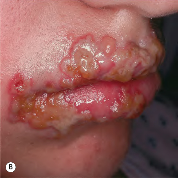
Fig. 80.1 Orolabial herpes simplex virus (HSV) infections.A Primary herpes gingivostomatitis due to HSV-1 in a child. Note the coalescing lesions with scalloped borders. B Primary versus non-primary initial HSV-2 infection in a teenager. There are grouped vesicles on an erythematous base; note the scalloped borders. C Recurrent herpes labialis (cold sore, fever blister). D Extensive HSV gingivostomatitis in a woman who was not immunosuppressed. A, Courtesy Julie V. Schaffer, MD; B, Courtesy Jean L. Bolognia, MD; D, Courtesy Kalman Watsky, MD.
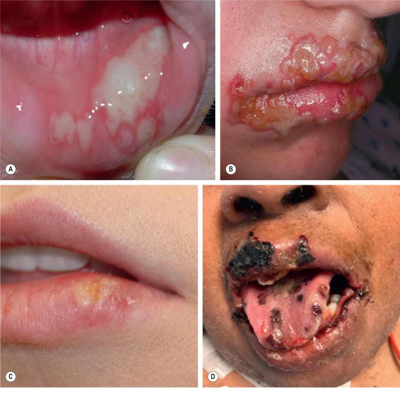
Fig. 80.2 Recurrent herpes simplex virus type 1 infection on the cheek. Occasionally, such lesions are misdiagnosed as cellulitis or bullous impetigo. Courtesy Kalman Watsky, MD.
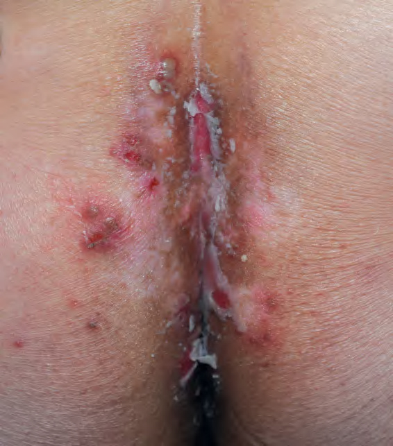
Fig. 80.3 Primary genital herpes. Grouped vesicles and erosions in the gluteal cleft. Courtesy Kalman Watsky, MD.
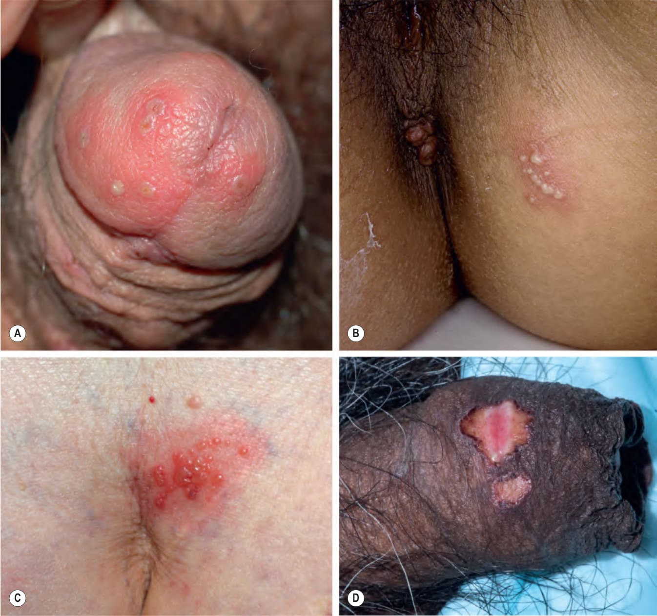
Fig. 80.4 Recurrent genital herpes.
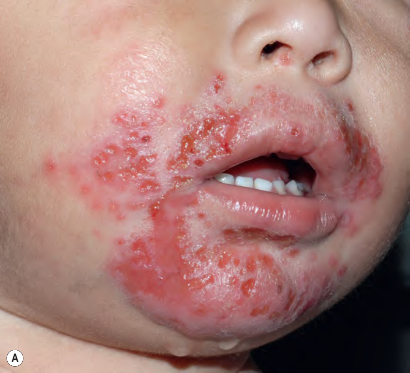
Fig. 80.5 Eczema herpeticum.A Monomorphic, punched-out erosions with a scalloped border in this infant with a history of facial atopic dermatitis. B Monomorphic, small hemorrhagic crusts and erosions coalescing in the popliteal fossae, an area of pre-existing atopic dermatitis. A, Courtesy Julie V. Schaffer, MD; B, Courtesy Kalman Watsky, MD.
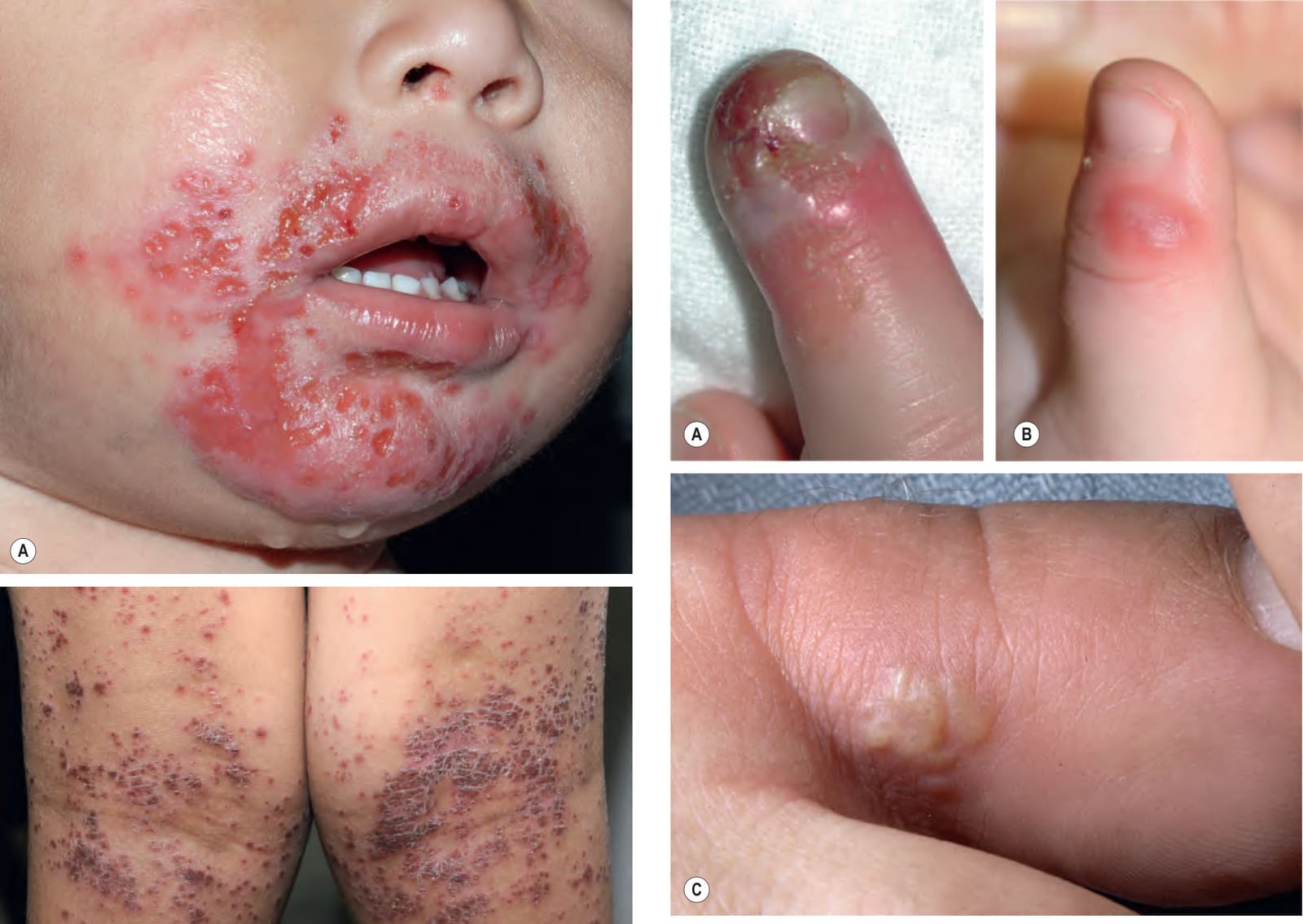
Fig. 80.6 Herpetic whitlow.A Coalescing vesicles and erosions on the distal finger of a child. B An edematous erythematous plaque with relatively subtle central vesicle formation on the thumb of a child. C Grouped vesicles on the toe of an adult. Herpetic whitlow is sometimes misdiagnosed as cellulitis or blistering distal dactylitis, or, depending on the distribution, paronychia. C, Courtesy Louis A. Fragola, Jr, MD.
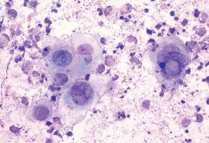
Fig. 80.10 Tzanck smear. Note the multinucleated epithelial giant cells from a patient with herpes simplex viral infection. Courtesy Louis A. Fragola, Jr, MD.
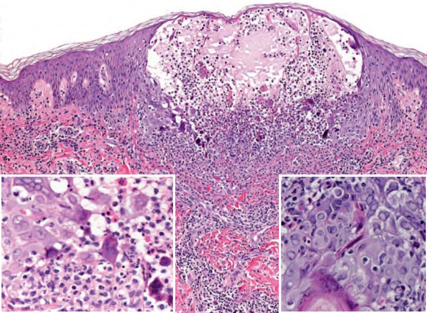
Fig. 80.11 Histology of herpes simplex viral infection. Intraepidermal vesicle with ballooning degeneration of keratinocytes and multinucleated giant cells; the latter arise from the fusion of infected keratinocytes. Note the steel-gray nuclei with margination of chromatin and inclusions (insets). Courtesy Lorenzo Cerroni, MD.
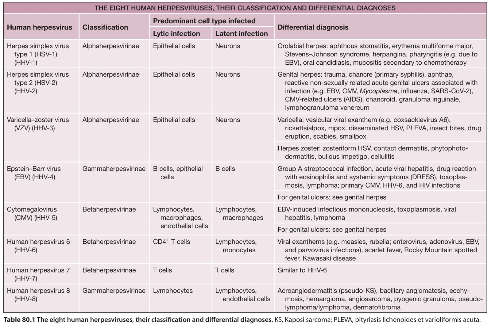
Table 80.1 The eight human herpesviruses, their classification and differential diagnoses. KS, Kaposi sarcoma; PLEVA, pityriasis lichenoides et varioliformis acuta.
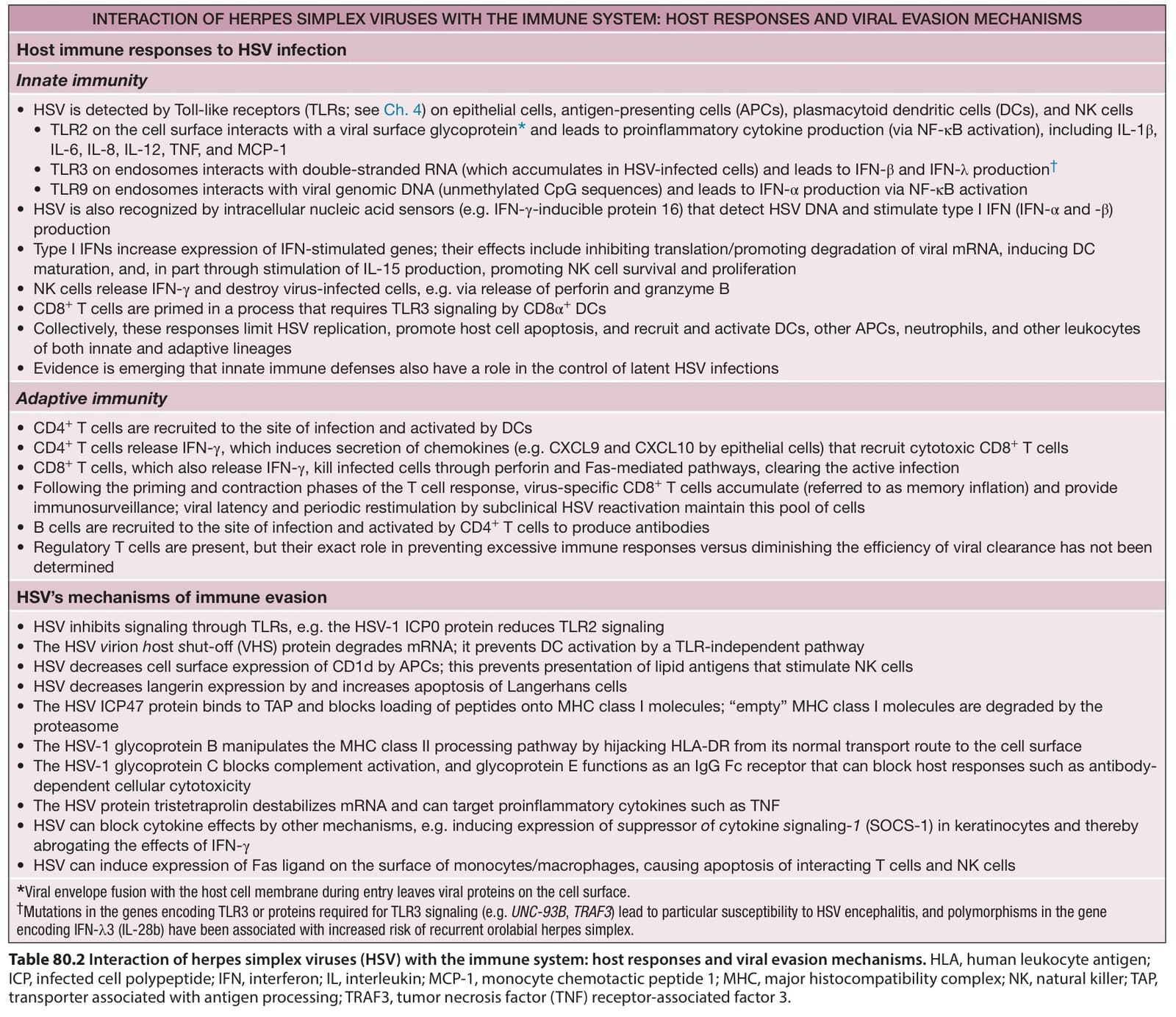
Table 80.2 Interaction of herpes simplex viruses (HSV) with the immune system: host responses and viral evasion mechanisms. HLA, human leukocyte antigen; ICP, infected cell polypeptide; IFN, interferon; IL, interleukin; MCP-1, monocyte chemotactic peptide 1; MHC, major histocompatibility complex; NK, natural killer; TAP, transporter associated with antigen processing; TRAF3, tumor necrosis factor (TNF) receptor-associated factor 3.

Table 80.3 Other clinical presentations of herpes simplex virus (HSV) infections.
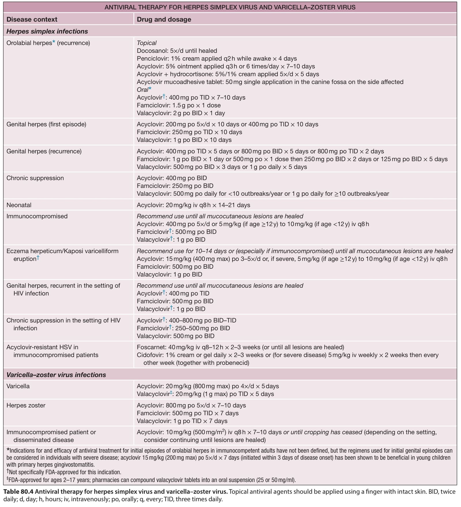
Table 80.4 Antiviral therapy for herpes simplex virus and varicella–zoster virus. Topical antiviral agents should be applied using a finger with intact skin. BID, twice daily; d, day; h, hours; iv, intravenously; po, orally; q, every; TID, three times daily.
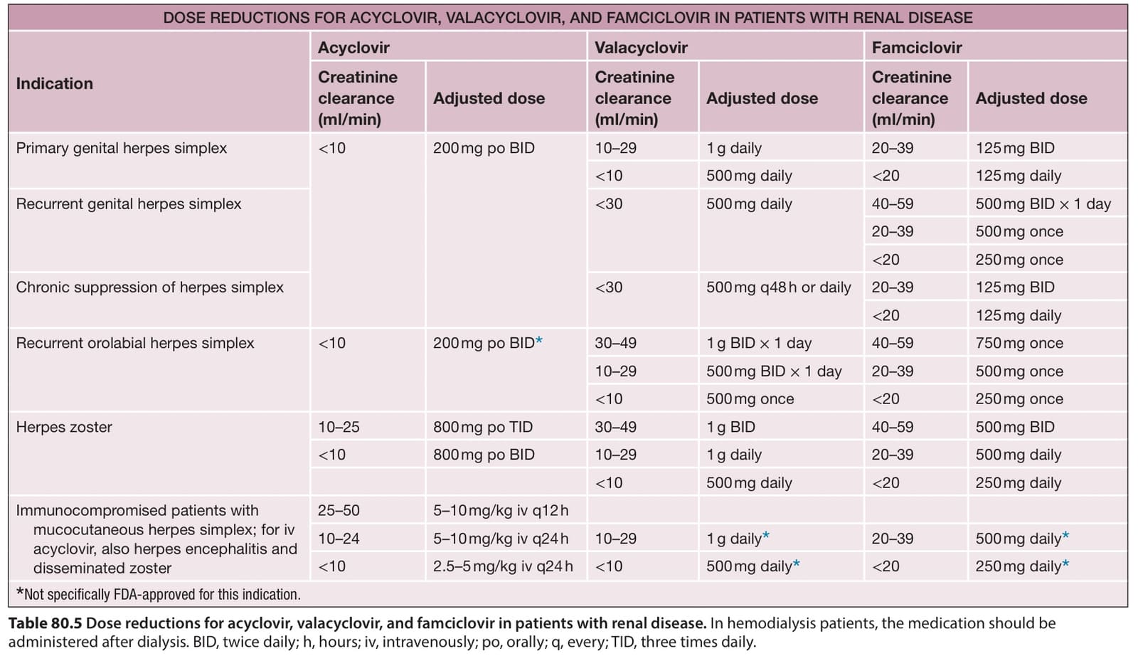
Table 80.5 Dose reductions for acyclovir, valacyclovir, and famciclovir in patients with renal disease. In hemodialysis patients, the medication should be administered after dialysis. BID, twice daily; h, hours; iv, intravenously; po, orally; q, every; TID, three times daily.