VARICELLA–ZOSTER VIRUS (VZV; HHV-3)
Synonyms/clinical forms: Varicella – chickenpox Herpes zoster – shingles
Key features
VZV is the etiologic agent of varicella (chickenpox) and herpes zoster (shingles)
VZV has a high morbidity and mortality rate in immunocompro- mised hosts
Antiviral treatment as well as vaccination can reduce or eliminate serious disease-associated sequelae and decrease the incidence of VZV infection
Introduction
VZV is the etiology of varicella (chickenpox) and herpes zoster (shingles). Varicella is usually symptomatic, and before the advent of the varicella vaccine, it occurred in 90% of children in the US by the time they reached 10 years of age. Herpes zoster represents reactivation of latent VZV infection and develops in ~20% of healthy adults and 50% of immunocompromised persons, with substantial morbidity and mortality in the latter group. Early initiation of antiviral treatment can reduce or eliminate serious sequelae of VZV infections.
Enlarging ulcerations in a child with acute lymphocytic leukemia who was presumed to have a Rhizopus infection (A) and in a young man with AIDS (B). C Coalescence of eroded, yellow–white papules and plaques on the tongue.
History
Heberden first distinguished chickenpox from smallpox in 1767. The term “chickenpox” is thought to come from either the French word “chiche-pois” for chickpea (referring to the size of the vesicles) or the Old English word “gigan” meaning “to itch”. The relationship between varicella and herpes zoster was first recognized in 1888, when von Bokay described the development of varicella in children following exposure to those with herpes zoster infection. Kundratitz (1922) and Bruusgaard (1932) more convincingly demonstrated the association between the two diseases by the development of varicella in susceptible children who had been inoculated with vesicle fluid from patients with herpes zoster. This was followed by the identification of the same virus in both diseases, confirming that their etiologies were identical.
Epidemiology
VZV has a worldwide distribution and 98% of the adult population is seropositive. In the pre-vaccine era, 90% of children <10 years of age developed varicella and ~4 million cases occurred annually in the US, with large outbreaks during the winter and spring.
Since the introduction of the varicella vaccine in 1995, the overall incidence of varicella has decreased by ~85%, with evidence of herd immunity. The age of peak incidence also shifted from 5–9 years to 10–14 years, and a higher rate of breakthrough varicella among immunized children in the latter age group led to the addition of a second varicella vaccine dose to the routine childhood immunization schedule in 2006.
Although the incidence and severity of herpes zoster increase substantially in middle to late adulthood, it can occur at any age and is more common in younger persons who had a varicella infection within the first year of life. Overall, individuals with a history of varicella have a 20% lifetime chance of developing zoster. The annual incidence of herpes zoster in the US and Europe is classically 2.5/1000 persons between ages 20 and 50 years, 5/1000 between ages 51 and 79 years, and 10/1000 in those >80 years of age. However, there is evidence that exposure to varicella can protect seropositive adults from the development of zoster, and it has been postulated that widespread vaccination against varicella could reduce this immune boosting effect and increase the incidence of zoster. Whether this hypothetical increase will be counterbalanced by zoster vaccination remains to be determined. Other risk factors for herpes zoster include mental and physical stress, a family history of zoster, use of JAK inhibitors or proteasome inhibitors (e.g. bortezomib, carfilzomib), and an immunocompromised state, especially when due to HIV infection and allogeneic hematopoietic stem cell transplantation (HSCT).
Pathogenesis
Airborne droplets are the usual route of transmission of varicella, although direct contact with vesicular fluid is another mode of spread, and the incubation period is 11–20 days. Varicella is extremely contagious, and 80%–90% of susceptible household contacts develop a clinically evident infection. The affected individual is infectious from 1–2 days before skin lesions appear until all the vesicles have crusted.
During varicella infection, primary viremia occurs after an initial 2–4 days of viral replication within regional lymph nodes. A cycle of viral replication in the liver, spleen, and other organs is then followed by a secondary viremia, which seeds the entire body 14–16 days postexposure. During this period, the virus enters the epidermis by invading capillary endothelial cells. VZV subsequently travels from mucocutaneous lesions to dorsal root ganglion cells, where it remains latent until reactivation at a later time.
Herpes zoster appears upon reactivation of latent VZV, which may occur spontaneously or be induced by stress, fever, radiation therapy, local trauma, or immunosuppression. During a herpes zoster outbreak, the virus continues to replicate in the affected dorsal root ganglion and produces a painful ganglionitis. Neuronal inflammation and necrosis can result in a severe neuralgia that intensifies as the virus spreads down the sensory nerve. Fluid from herpes zoster vesicles can transmit VZV to seronegative individuals, leading to varicella but not herpes zoster. The transmission rate to susceptible household contacts is ~15% for zoster, compared to 80%–90% for varicella.
Clinical Features
Varicella
A prodrome of mild fever, malaise, and myalgia may occur, especially in adults. This is followed by an eruption of pruritic, erythematous macules and papules, which starts on the scalp and face, and then spreads to the trunk and extremities (Fig. 80.12). Lesions rapidly evolve over ~12 hours into 1–3 mm clear vesicles surrounded by narrow red halos (“dew drops on a rose petal”). The number of vesicles varies from only a few to several hundred, and there is often involvement of the oral mucosa (see Fig. 80.12D). Sparing of the distal and lower extremities is common. Older vesicles evolve to form pustules and crusts, with individual lesions healing within 7–10 days. The presence of lesions in all stages of development is a hallmark of varicella.
The disease course is usually self-limited and benign in healthy children. However, prior to introduction of the varicella vaccine, complications resulted in 11 000 hospitalizations annually in the US. Secondary bacterial infection of the skin with subsequent scarring is the most common complication. CNS sequelae are uncommon (<1 per 1000 cases) and may include encephalitis and acute cerebellar ataxia; Reye syndrome which consists of encephalitis plus fatty liver is now
rare due to avoidance of aspirin in children with varicella. Varicella in adolescents and adults is often more severe than in children, with an increased number of skin lesions (see Fig. 80.12A) and more frequent development of pneumonia, which has a 10%–30% mortality rate if untreated. Occasional complications include thrombocytopenia, hepatitis, glomerulonephritis, optic neuritis, keratitis, arthritis, myocarditis, pancreatitis, orchitis, and vasculitis.
Maternal varicella during the first 20 weeks of pregnancy is associated with an ~2% risk of congenital varicella syndrome (varicella embryopathy). Possible congenital defects include a low birth weight, cutaneous scarring, ocular abnormalities, cortical atrophy, psychomotor retardation, and hypoplastic limbs. Children whose mothers had varicella during pregnancy may also develop zoster early in life without ever having extrauterine varicella. Severe neonatal varicella, in which amelioration by maternal antibodies is lacking, can occur when maternal varicella develops between 5 days before and 2 days after delivery.
In immunocompromised individuals, varicella may lead to severe complications and even death. These patients frequently have a more extensive and atypical cutaneous eruption, often with hemorrhagic or purpuric lesions. Involvement of the lungs, liver, and CNS is common.
Herpes zoster
Reactivation of VZV may occur at any time after the development of varicella. In >90% of patients, herpes zoster begins with a prodrome of pruritus, tingling, tenderness, hyperesthesia, and/or intense pain, which can mimic a myocardial infarction, surgical abdomen, or toothache. These symptoms occasionally occur without subsequent skin lesions, a phenomenon referred to as “zoster sine herpete”. However, most patients develop a painful eruption of grouped vesicles on an erythematous base in a dermatomal distribution (Figs. 80.13– 80.15); the lesions can involve more than one contiguous dermatome and occasionally cross the midline. Edematous papules and plaques may precede the appearance of vesicles, and progression to pustules or bullae can occur (see Fig. 80.14). Any cutaneous site may be affected, with the trunk representing the most common location, followed by the face. VZV DNA can often be detected in the peripheral blood and saliva of patients with herpes zoster, even after initiation of antiviral treatment.
Herpes zoster usually resolves without sequelae in immunocompetent children and young adults. However, the pain, cutaneous lesions, and complications of herpes zoster become more severe with increasing age and immune compromise. The most common complication is postherpetic neuralgia, which is characterized by dysesthetic pain (e.g. burning or stabbing sensation, allodynia) that persists after the skin lesions have healed. This affects 10%–20% of all herpes zoster patients and increases in both incidence and severity with age. Complications of herpes zoster related to the dermatome that is affected are presented in Table 80.6. Other complications can include secondary bacterial infection, scarring, meningoencephalitis, pneumonitis, and hepatitis. Cutaneous disorders that can develop within sites of previous herpes zoster, including granulomatous (Fig. 80.16) and pseudolymphomatous lesions, are listed in Table 80.7.
In immunocompromised individuals, herpes zoster may be more severe and can have unusual clinical presentations, e.g. persistent crusted, verrucous lesions in HIV-infected patients (Fig. 80.17) or postherpetic hyperhidrosis. Disseminated cutaneous disease, defined as more than 20 vesicles outside the area of primary or adjacent dermatomes, and/or visceral involvement occurs in ~10% of immunocompromised persons with zoster (Fig. 80.18).
Diagnosis and Pathology
A clinical diagnosis can usually be made based upon the history, including previous varicella or vaccine administration, and physical examination. A Tzanck smear, PCR, or DFA can assist in quickly confirming the diagnosis (see above and Fig. 80.10). The latter two (but not the Tzanck smear) can differentiate between HSV and VZV. Varicella and herpes zoster have the same histologic findings as HSV infections (see above and Fig. 80.11), but immunohistochemical staining can distinguish between the two viruses. Exclusive involvement of the hair follicles may be observed in herpes zoster, but not in HSV infection.
Additional laboratory tests include viral culture and serology. Viral culture is a very specific test; however, it is not very sensitive and results may not be available for >1 week. Serologic assays are diagnostic of VZV if the convalescent serum has at least a fourfold increase in the VZV titer relative to the acute serum. As a result, serology is only useful in retrospect.
Differential Diagnosis
The differential diagnosis of varicella may include vesicular viral exanthems (e.g. due to coxsackieviruses), disseminated HSV infection, pityriasis lichenoides et varioliformis acuta (PLEVA), rickettsialpox, drug eruptions, bullous insect bite reactions, and even scabies. Although no longer naturally occurring, smallpox also presents with a widespread vesicular eruption; its distinct clinical features are reviewed in Chapter 81. For herpes zoster, diagnostic considerations
may include zosteriform HSV infections (which often recur in the same site), bacterial skin infections (e.g. cellulitis, bullous impetigo), contact dermatitis, and phytophotodermatitis. The entities listed above can usually be differentiated from VZV infections based on the history and clinical presentation, but laboratory studies and histologic evaluation are sometimes needed.
Treatment and Prevention
Varicella
Varicella in immunocompetent children can be treated symptomatically with antipyretics (e.g. acetaminophen), antihistamines, calamine lotion, and tepid baths. If started within 24–72 hours after the onset of the cutaneous eruption, acyclovir has been shown to decrease the duration and severity of varicella. Oral acyclovir and valacyclovir are FDA-approved for the treatment of varicella in children (2–17 years of age) while acyclovir is approved for adults (see Table 80.4). These antiviral agents are recommended for varicella in healthy adolescents and adults as well as in children with chronic cutaneous or pulmonary disorders and in those receiving chronic salicylate therapy, inhaled corticosteroids, or intermittent oral corticosteroids. However, routine antiviral therapy is not recommended for otherwise healthy children with varicella due to the self-limited disease course and modest benefits of treatment. Intravenous acyclovir is indicated for varicella in immunocompromised patients, including those receiving chronic systemic corticosteroids, due to their increased risk of more severe disease and complications.
Erythematous, edematous plaques with early vesicle formation. Note the perifollicular accentuation (B). D, E Later stages of evolution with prominent pustule formation (D) and a dusky purple color associated with older vesicles (E). F Bullous variant on the flexor arm. B, Courtesy Jean L. Bolognia, MD; D, Courtesy Louis A. Fragola, Jr, MD.
Administration of varicella zoster immune globulin (125 U/10 kg, 625 U maximum) intramuscularly within 96 hours of varicella exposure is recommended to provide passive prophylaxis to nonimmune immunocompromised individuals and pregnant women as well as high-risk neonates (see above). Protection lasts for ~3 weeks. Another option for postexposure varicella prophylaxis in these patient groups is administration of intravenous immunoglobulin (IVIg; ≥400 mg/kg), which contains high levels of varicella-specific IgG. Prophylactic oral acyclovir administration with usual varicella dosing (see Table 80.4) for 1 week, starting 7–10 days after exposure, can also be considered. Lastly, postexposure varicella vaccination (within 72–120 hours) may prevent or modify disease in nonimmune individuals who are ≥12 months of age, immunocompetent, and eligible to receive this live attenuated vaccine (see below).
Approved by the FDA in 1995, the live attenuated VZV vaccine (Oka strain; Varivax®) is highly efficacious, with seroprotection rates of ~85% after one dose and ≥99% after two doses in healthy children. Two doses of the vaccine, routinely given at ages 12–15 months and 4–6 years, are recommended to improve protection and counteract waning vaccine-induced immunity (see below). Studies indicate that one dose of the vaccine is 70%–90% effective in preventing infection and 95%–100% effective in preventing severe disease, and recipients of two doses are more than threefold less likely to develop breakthrough varicella than those who received one dose. The incidence and severity of both varicella (usually due to wild-type VZV) and zoster (most often due to the attenuated Oka strain) (Fig. 80.19) are decreased in vaccine recipients compared to unimmunized children. However, in a report based on surveillance of 350 000 subjects who received one vaccine
dose, loss of vaccine-induced immunity over time was observed, e.g. within 1 year of vaccination, there were 1.6 cases of varicella per 1000 person-years versus 58.2 per 1000 person-years at 9 years post-vaccination. Because this is a live viral vaccine, administration is contraindicated during pregnancy and in individuals with immunosuppression due to hematologic malignancies, HIV infection (if CD4+ T cells are <15% or <200 cells/mcl), other T cell immunodeficiencies, or systemic immunosuppressive therapy such as TNF inhibitors or prednisone (≥20 mg daily for ≥2 weeks, or ≥2 mg/kg daily when weight <10 kg).
A Clustered vesicles in a linear array within the distribution of V. B In the distribution of V, grouped pustules on the left side of the chin and hemorrhagic crusting of the lower lip, with one lesion extending past the midline. This could be mistaken for an acneiform eruption or impetigo. C In the same patient, erosions are present on the left side of the tongue. The anterior two-thirds of the tongue is innervated by the facial nerve (VII; taste) as well as V (sensory); see Table 80.6 for details of Ramsay Hunt syndrome. D Focal hemorrhagic crusting with scalloped borders arising in an erythematous patch on the forehead within the V distribution. Note the periorbital edema. Courtesy Kalman Watsky, MD.
Herpes zoster
For herpes zoster, beginning antiviral treatment within 72 hours of the development of skin lesions is optimal, but initiation up to 7 days after onset also appears to be beneficial. Acyclovir, famciclovir, and valacyclovir are all FDA-approved for the treatment of herpes zoster in immunocompetent individuals and result in decreased severity and duration of both skin lesions and pain (see Table 80.4). In controlled studies of immunocompetent individuals >50 years of age with acute herpes zoster, famciclovir and valacyclovir were equally effective (and
Pink papules and plaques developed several weeks after an episode of herpes zoster in the same dermatomes. A sharp midline demarcation was observed on the back. Histologically, a granulomatous dermatitis was present within the dermis and focally a perineural distribution pattern was seen. Courtesy Rebecca Vaughn, MD.
similar or superior to acyclovir) at reducing the frequency and duration of postherpetic neuralgia. Intravenous acyclovir is indicated for the treatment of zoster in immunocompromised patients as well as those with serious complications. Amenamevir, a helicase-primase inhibitor that is approved in Japan, was found to be non-inferior to valacyclovir in treatment of adults with zoster.
Multiple treatments are available for patients who develop postherpetic neuralgia. Low-dose tricyclic antidepressants have demonstrated considerable efficacy and appear to act by a mechanism independent from their antidepressant effects. In a randomized, controlled, multi-center trial, gabapentin (up to 3600 mg/day) was found to be effective in the treatment of pain and sleep interference associated with
postherpetic neuralgia. Combined treatment with nortriptyline and gabapentin reduced pain more than either agent alone in another randomized controlled trial. In one open study, the use of gabapentin together with valacyclovir during acute herpes zoster was more effective in preventing postherpetic neuralgia than treatment with valacyclovir alone in historical controls; this regimen is recommended for patients with acute herpes zoster presenting with moderate to severe pain. Although treatment with prednisone in addition to acyclovir can reduce acute pain in patients with herpes zoster, several controlled trials have failed to demonstrate a difference in the incidence or severity of postherpetic neuralgia with this combination compared to acyclovir monotherapy. An 8% capsaicin patch was found to reduce the pain of postherpetic neuralgia for up to 12 weeks after a 1-hour application. Other treatments used for postherpetic neuralgia include analgesics, EMLA cream, lidocaine patches, narcotic analgesics, pregabalin, nerve blocks, and biofeedback. Although some evidence suggests that patients receiving TNF inhibitors for various immune-mediated diseases may have a slightly higher incidence of herpes zoster, development of postherpetic neuralgia in these patients appears to be uncommon.
Because reactivation of VZV is linked with states of decreased cellmediated immunity, the varicella vaccine was studied as a method of prophylaxis for herpes zoster. A large trial utilizing a 14-fold more concentrated version of the varicella vaccine in almost 40 000 adults ≥60 years of age showed a 51% reduction in the incidence of herpes zoster and 67% decrease in the likelihood of postherpetic neuralgia in vaccine recipients. The FDA approved this live attenuated vaccine (Zostavax®) for immunocompetent adults ≥50 years of age in 2006, and the Advisory Committee on Immunization Practices (ACIP) recommended a single dose of Zostavax® for the prevention of herpes zoster in immunocompetent individuals ≥60 years of age. However, multiple subsequent studies found that immunity induced against zoster by this vaccine was relatively short-lived, with a decrease in efficacy to <35% by year 6 after administration. Considering its limited effectiveness as well as reports of necrotizing retinitis in immunocompromised vaccine recipients, Zostavax® is no longer available in the US.
In phase III studies, a recombinant subunit vaccine containing VZV glycoprotein E and the AS01B adjuvant system (HZ/su) administered in two doses two months apart reduced the likelihood of herpes zoster
~97% and postherpetic neuralgia ~90% in adults ≥50 years of age. Efficacy of this vaccine was unaffected by advancing age and persisted for >3 years. In 2017, the FDA approved this recombinant zoster vaccine (Shingrix®) as a two-dose regimen, with the ACIP also recommending its use for the prevention of herpes zoster in immunocompetent adults ≥50 years of age (2–6 months between doses) and those ≥18 years of age who are or will be immunocompromised (1–6 months between doses). Vaccination is recommended regardless of whether or not the individual has a history of varicella, herpes zoster, or administration of a live attenuated VZV vaccine.
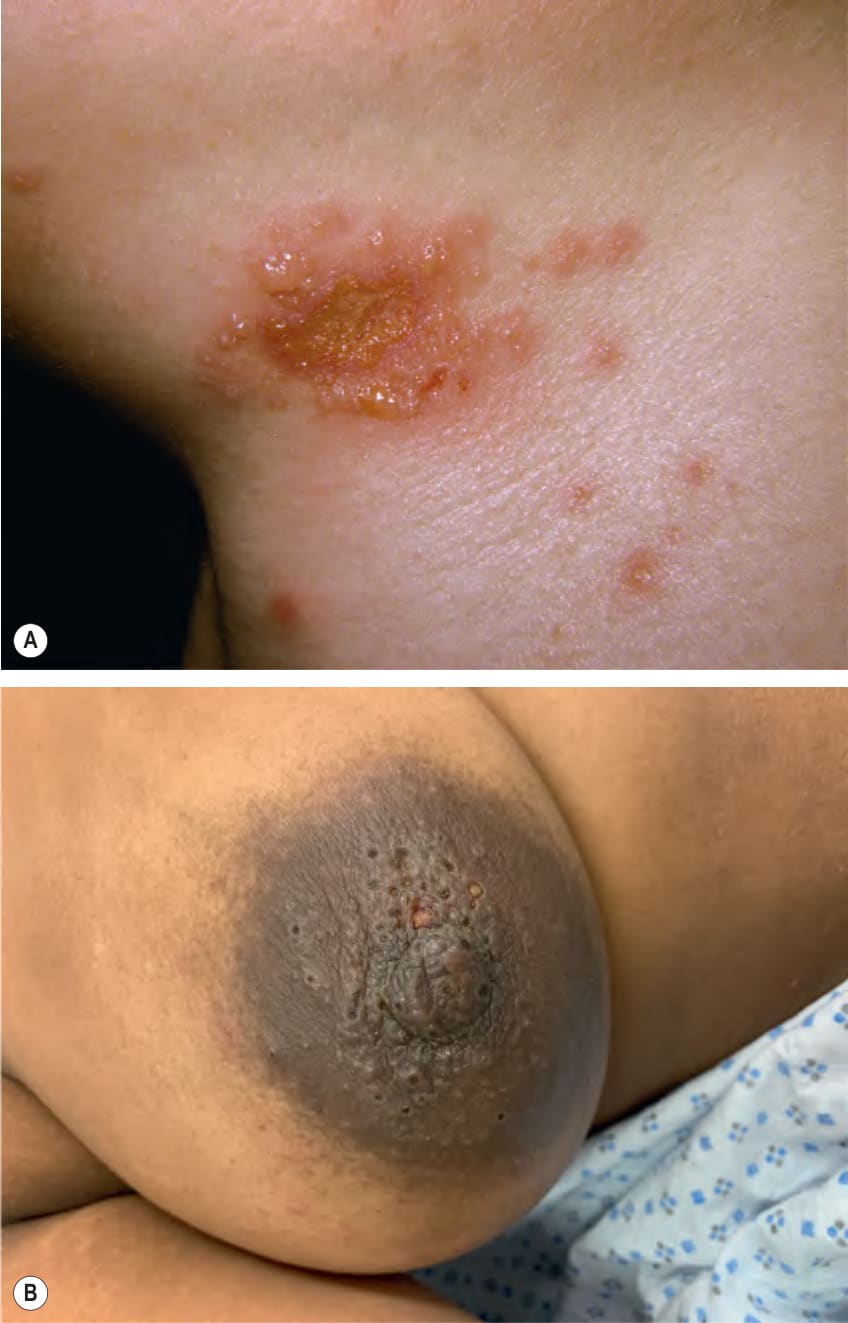
Fig. 80.7 Herpes simplex infections in less common locations.A Herpes gladiatorum presenting as grouped vesicles and erosions on the neck of a high-school wrestler. B Small vesicles and punched-out erosions clustered on the nipple and areola. A, Courtesy Louis A. Fragola, Jr, MD; B, Courtesy Kalman Watsky, MD.
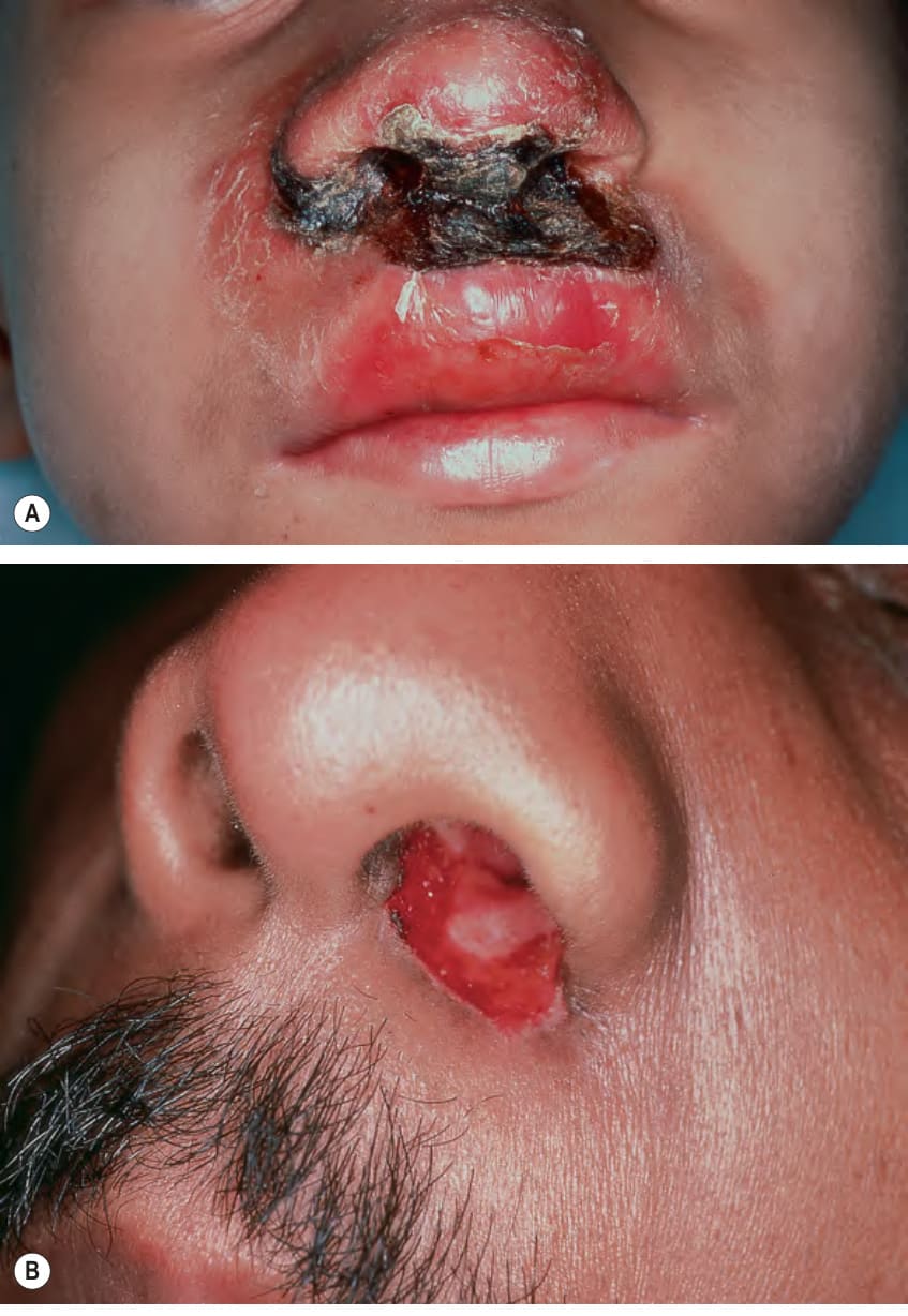
Fig. 80.8 Herpes simplex virus infections in immunocompromised hosts.A, B
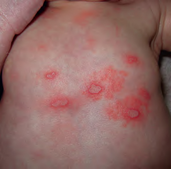
Fig. 80.9 Neonatal herpes. Grouped papulovesicles with an erythematous base on the chest. Note the scalloped borders in areas of coalescence. Courtesy Frank Samarin, MD.
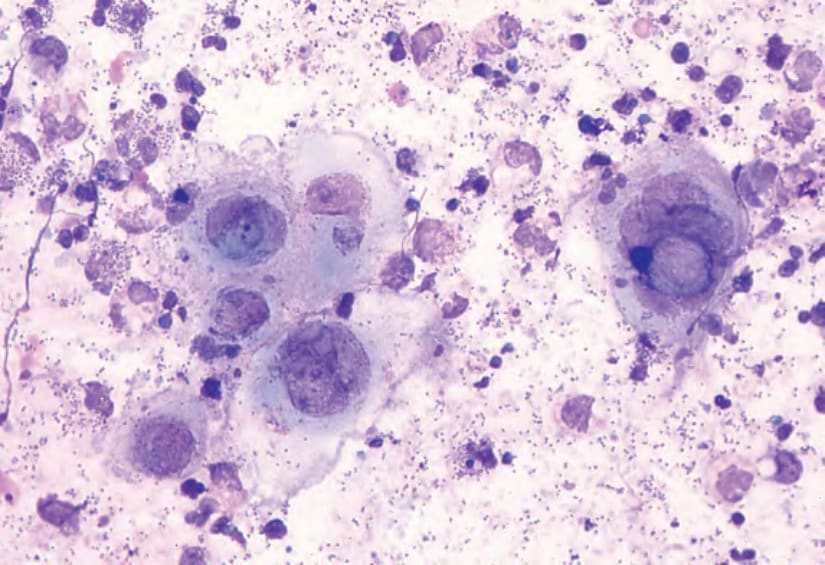
Fig. 80.10 Tzanck smear. Note the multinucleated epithelial giant cells from a patient with herpes simplex viral infection. Courtesy Louis A. Fragola, Jr, MD.
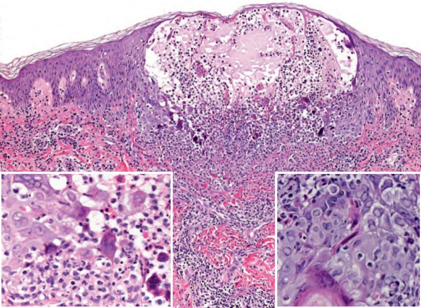
Fig. 80.11 Histology of herpes simplex viral infection. Intraepidermal vesicle with ballooning degeneration of keratinocytes and multinucleated giant cells; the latter arise from the fusion of infected keratinocytes. Note the steel-gray nuclei with margination of chromatin and inclusions (insets). Courtesy Lorenzo Cerroni, MD.
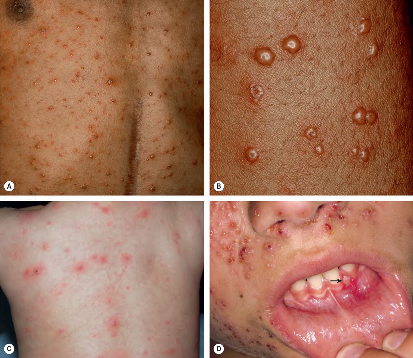
Fig. 80.12 Varicella.A–C Lesions in different stages of evolution, including vesicles, pustules, and hemorrhagic crusts. Vesicles often develop central umbilication. D Oral lesions can also occur (arrow). A, B, Courtesy Robert Hartman, MD; C, Courtesy Julie V. Schaffer, MD; D, Courtesy Judit Stenn, MD.
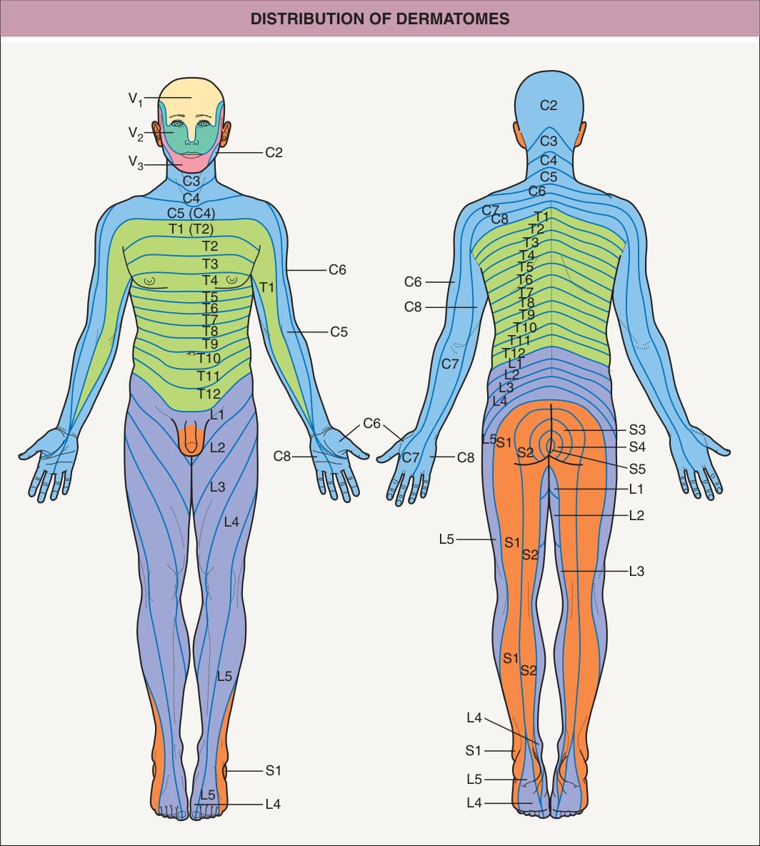
Fig. 80.13 Distribution of dermatomes.
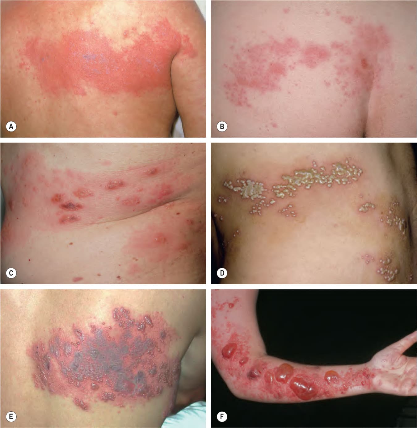
Fig. 80.14 Herpes zoster.A–C
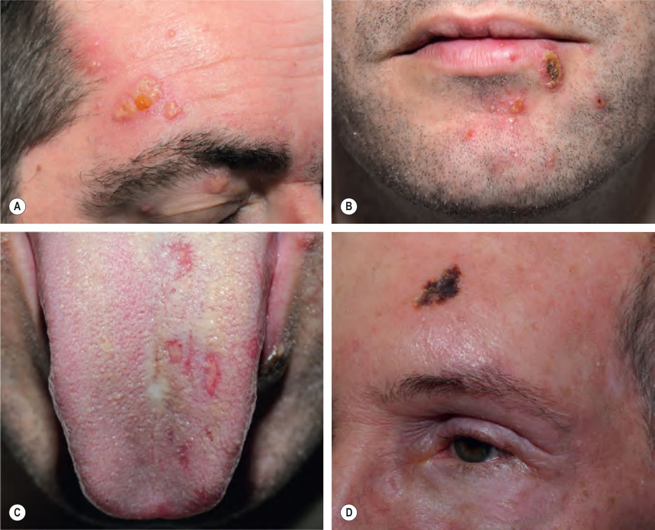
Fig. 80.15 Facial herpes zoster.
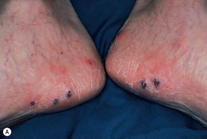
Fig. 80.16 Granulomatous dermatitis in the site of previous herpes zoster.
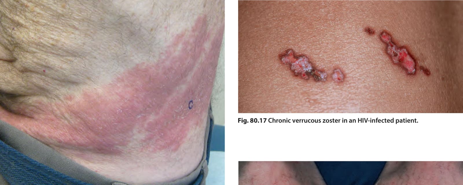
Fig. 80.17 Chronic verrucous zoster in an HIV-infected patient.
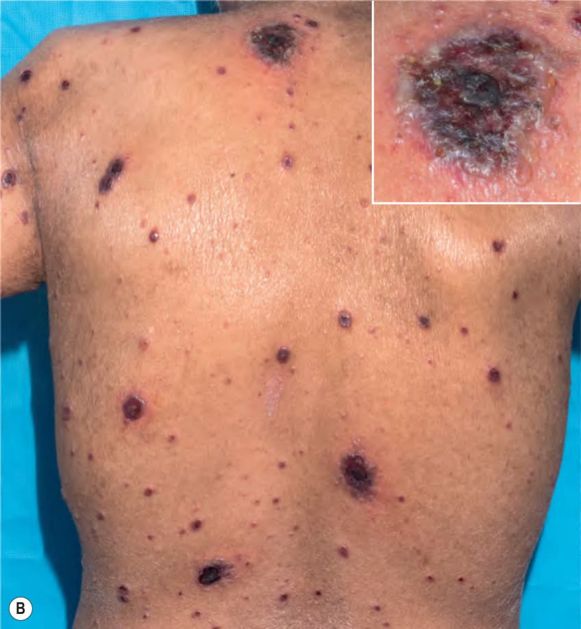
Fig. 80.18 Disseminated varicella zoster viral infection.A Multiple violet–black papules on the feet. B Widely distributed necrotizing vesicles and bullae, some of which have formed large clusters, in a patient with lymphoma. Patients with disseminated cutaneous skin lesions should be evaluated for possible visceral involvement (e.g. hepatic, pulmonary, CNS), especially if they are immunocompromised. B, Courtesy Edward Cowen, MD.
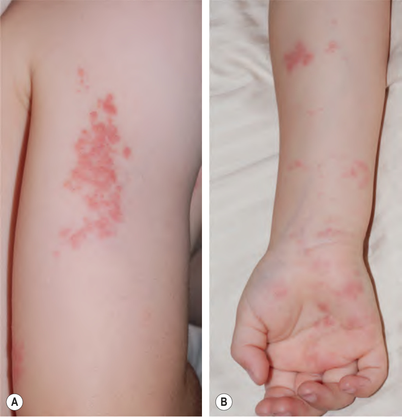
Fig. 80.19 Herpes zoster following varicella vaccination. This healthy 3-year-old boy developed an eruption of erythematous papulovesicles in the C5 and C6 dermatomes of the right arm. He had received the varicella vaccine in the right shoulder at age 12 months, and several erythematous, edematous papulonodules had appeared in the region of the injection 2 weeks later. Courtesy Julie V. Schaffer, MD.
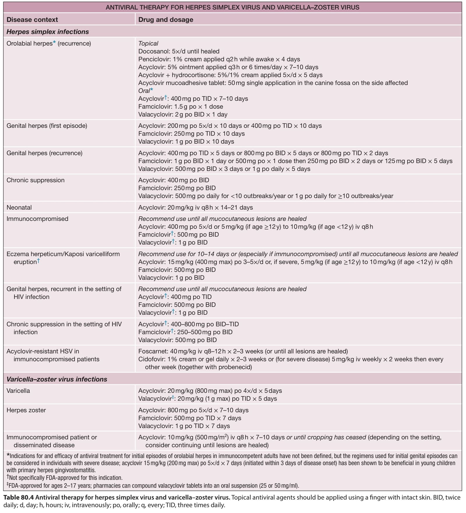
Table 80.4 Antiviral therapy for herpes simplex virus and varicella–zoster virus. Topical antiviral agents should be applied using a finger with intact skin. BID, twice daily; d, day; h, hours; iv, intravenously; po, orally; q, every; TID, three times daily.
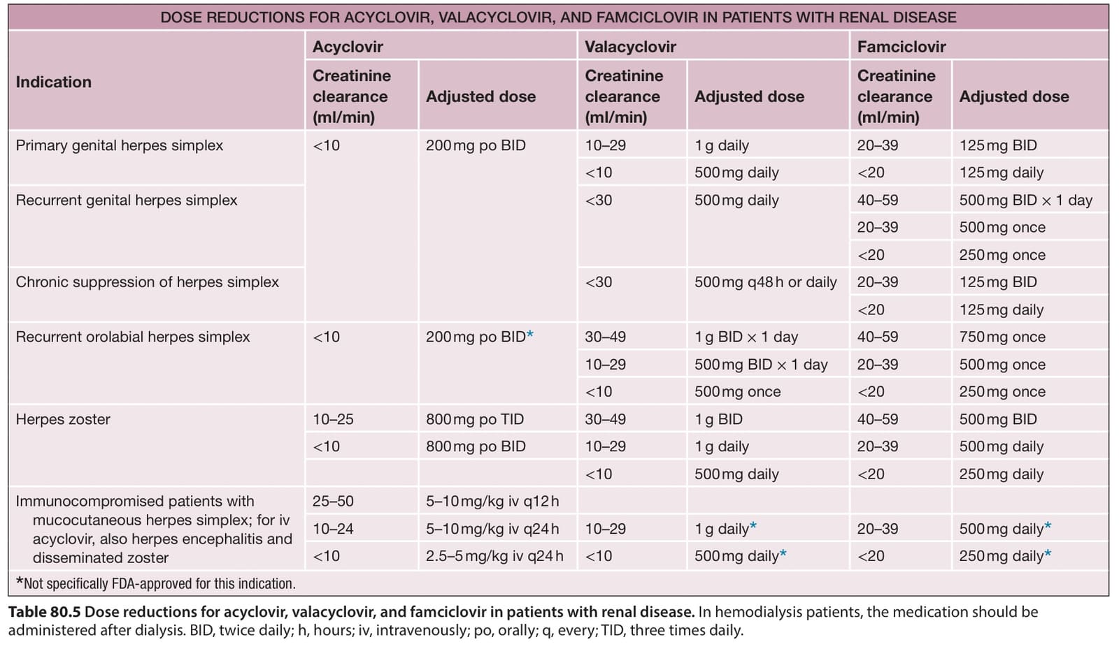
Table 80.5 Dose reductions for acyclovir, valacyclovir, and famciclovir in patients with renal disease. In hemodialysis patients, the medication should be administered after dialysis. BID, twice daily; h, hours; iv, intravenously; po, orally; q, every; TID, three times daily.
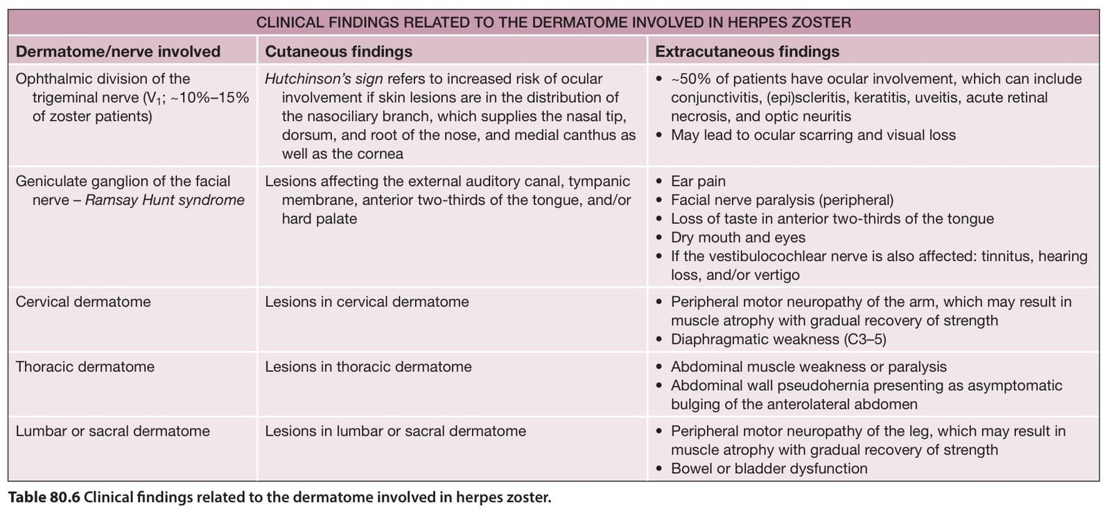
Table 80.6 Clinical findings related to the dermatome involved in herpes zoster.
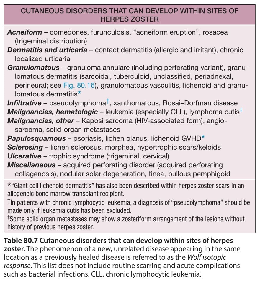
Table 80.7 Cutaneous disorders that can develop within sites of herpes zoster. The phenomenon of a new, unrelated disease appearing in the same location as a previously healed disease is referred to as the Wolf isotopic response. This list does not include routine scarring and acute complications such as bacterial infections. CLL, chronic lymphocytic leukemia.