EPSTEIN–BARR VIRUS (HHV-4)
Synonym: Infectious mononucleosis – glandular fever
Key features
EBV is responsible for a variety of clinical diseases, most notably infectious mononucleosis; it is also associated with lymphoproliferative disorders and lymphomas
Knowledge of EBV-specific serologies and other laboratory tests for
EBV is essential for the clinician
Introduction and History
EBV is a ubiquitous human herpesvirus (HHV-4) that is responsible for a broad spectrum of clinical diseases, including infectious mononucleosis, oral hairy leukoplakia (see Ch. 78), hydroa vacciniforme (see Ch. 87), and a variety of lymphoproliferative disorders, lymphomas (B and T cell), and other malignancies (Table 80.8). It replicates well in B lymphocytes and can lead to their immortalization. EBV was named after its two discoverers, who found this herpesvirus in an African Burkitt lymphoma in 1964. In 1968, a link between infectious mononucleosis and EBV was established. Since then, various clinical disorders have been associated with EBV and are classified based on the type of cell infected and state of viral infection.
Epidemiology
In high-income countries, the prevalence of EBV infection in the general population approaches 95% by early adulthood. Peaks of incidence occur in individuals 1 to 6 and 14 to 20 years of age. Approximately 60%–80% of young children in low-income countries are seropositive for EBV, compared to ≤50% in the US. Most children with primary EBV infection have either no symptoms or a mild, nonspecific febrile illness. In contrast, ~50% of adolescents and young adults with primary EBV infection develop infectious mononucleosis. In the US, EBV is commonly associated with B cell lymphomas (~20%) and lympho proliferative disorders (polyclonal and monoclonal) in immunocompromised patients. Worldwide, EBV is also implicated in Burkitt lymphoma (particularly the African-endemic form) and nasopharyngeal carcinoma (primarily in Asia) (see Table 80.8).
Pathogenesis
EBV is transmitted primarily through infectious saliva and occasionally via blood transfusion, although its presence in genital secretions and breast milk has been reported. The virus preferentially infects human mucosal epithelial cells and B lymphocytes, with primary infection originating within the oropharyngeal epithelium. Infectious mononucleosis results from active viral replication, which is subsequently terminated by the host immune response.
After infectious mononucleosis resolves, circulating B lymphocytes can become latently infected with EBV as the virus enters cells by specifically binding to cell surface complement receptors CR2 or CD21. As the B lymphocyte divides, expression of the EBV nuclear antigen-1 gene (EBNA-1) is essential for replication of EBV DNA. Furthermore, the EBNA-1 protein and the virally encoded latent membrane protein-2 (LMP-2) are important in establishment of EBV latency within B lymphocytes and evasion from the cellular immune system. In the presence
of a normally functioning immune system, cell-mediated immune responses toward EBV antigens generally prevent the transformation and immortalization of B lymphocytes. However, in patients with deficient cell-mediated immunity, B lymphocytes can become immortalized and give rise to EBV-induced lymphoproliferative disorders.
Cell-mediated immunity to EBV infection is persistent and protects against developing infectious mononucleosis upon re-exposure to different types and strains of EBV later in life. The humoral immune response, although it generates antibodies to a variety of EBV antigens, does not appear to be protective. Latent virus in EBV-infected B lymphocytes can subsequently reinfect the oropharyngeal epithelium, resulting in periodic EBV reactivation and viral shedding for the lifetime of the host. However, reactivation of EBV is less common compared to other herpesviruses.
Clinical Features
EBV-induced infectious mononucleosis most commonly occurs in adolescents and young adults 15–25 years of age. After an incubation period of 30–50 days, the clinical triad of pharyngitis, fever lasting up to 10 days, and lymphadenopathy (especially cervical) occurs in >80% of patients. An initial prodrome of headache, malaise, and fatigue often precedes onset of the triad. The absence of cervical lymphadenopathy and fatigue makes the diagnosis unlikely. Splenomegaly is found in >50% of patients and can be accompanied by hepatomegaly. Other
EBV-related genital ulcers, which are most common in adolescent girls, are often misdiagnosed as genital herpes simplex infection and in some patients represent a variant of aphthosis (see Table 80.8).
Erythematous macules and papules have become confluent on the upper trunk.
clinical findings may include exudative pharyngitis (often gray–white), anorexia, nausea, vomiting, cough, and arthralgias.
EBV infection can have a variety of dermatologic manifestations. A faint, nonspecific erythematous exanthem has been reported in up to 70% of hospitalized patients and ~10% of all patients with infectious mononucleosis. The exanthem typically develops on day 4–6 of the illness and may last up to 1 week. It can be morbilliform, urticarial, scarlatiniform, vesicular, erythema multiforme-like, purpuric, or petechial in nature, usually beginning on the trunk and proximal upper extremities with later extension to the face and forearms. Eyelid petechiae, periorbital edema, and petechiae at the junction of the hard and soft palate may also be observed. Resolution of the illness usually occurs after 2 to 3 weeks, although symptoms of fatigue and malaise may last longer.
Painful genital ulcers, often measuring >1 cm, occasionally develop in patients (especially adolescent girls) with primary EBV infection (Fig. 80.20); PCR or in situ hybridization has confirmed the presence of lesional EBV in some cases. Cutaneous conditions that are occasionally associated with primary EBV infection include Gianotti– Crosti syndrome (see Ch. 81), erythema nodosum, urticaria, acrocyanosis, erythema multiforme, erythema annulare centrifugum, and pityriasis lichenoides.
Administration of ampicillin or amoxicillin, and less commonly penicillin or cephalosporins, to patients with infectious mononucleosis, frequently (up to 90% with the former agents) leads to the development of a “hypersensitivity” skin reaction. This usually develops 7–10 days following institution of the antibiotic. Typically, a pruritic, pink to copper-colored, scarlatiniform or morbilliform eruption develops on extensor surfaces and pressure points and then becomes confluent as it spreads onto the trunk and extremities (Fig. 80.21). Desquamation occurs 1 week later. The mechanism of this enhanced immune response to aminopenicillins in the setting of EBV infection is not well under-stood. In the absence of a pre-existing allergy, such reactions do not generally preclude the use of this class of antibiotics after the infectious mononucleosis resolves.
EBV-induced infectious mononucleosis can have serious complications. Splenic rupture occurs in 0.2% of affected adults, usually after trauma. Significant enlargement of oropharyngeal lymphoid tissue can lead to airway compromise. Other potential complications include hepatitis, thrombocytopenia, hemolytic anemia, glomerulonephritis, and CNS disturbances.
Diagnosis and Pathology
Laboratory findings in patients with infectious mononucleosis include mild to moderately elevated hepatic transaminase levels, mild thrombocytopenia, and an absolute and relative lymphocytosis (up to 50 000 cells/dl) with 20%–40% atypical lymphocytes. Atypical lymphocytosis is not pathognomonic for EBV infection, as a variety of infectious agents and drugs can also cause this phenomenon. Diagnosis is usually made by a positive monospot test, a simple slide test that detects IgM heterophile antibodies, or by increased titers of heterophile antibodies; the latter reach a titer of >1 : 40 in ~90% of young adults infected with EBV. Heterophile antibodies are capable of agglutinating sheep, horse, or bovine erythrocytes, and they typically become detectable 1 week to 1 month after the onset of symptoms and persist for 3 to 18 months. Because heterophile antibody titers are positive in only a minority of young children with a primary EBV infection, EBV-specific serologies are often necessary to establish the diagnosis in this age group. Titers of at least three different antibodies should be assessed to determine the stage of EBV infection, allowing differentiation between an acute primary, past/latent, and reactivated infection (Table 80.9).
PCR-based assays to detect EBV DNA in peripheral blood or tissue samples are also available. Markedly elevated peripheral blood levels are found in patients with infectious mononucleosis and in immunodeficient individuals with EBV-associated lymphoproliferative disorders. Of note, lower levels of EBV DNA can also be detected in the peripheral blood of healthy individuals with a latent EBV infection. In situ hybridization for EBV-encoded small RNA (EBER) represents a widely available, sensitive, and specific method to detect EBV-infected cells in the skin and other tissues. This assay can be performed on paraffin-embedded specimens, and EBER-positive cells are prominent in EBV-related dermatoses (e.g. T cells in hydroa vacciniforme; see Fig. 87.11) and malignancies, but absent in inflammatory dermatoses and lymphomas that are not related to EBV infection.
Primary EBV infection is not associated with any pathognomonic histologic findings. Features are similar to those observed with other viral exanthems, i.e. slight spongiosis, parakeratosis, and a perivascular lymphocytic infiltrate. Additional findings may include focal keratinocyte necrosis, mild ballooning degeneration, and, rarely, multi-nucleated keratinocytes, but less prominent than with HSV and VZV infection. Aminopenicillin-induced eruptions in the setting of infectious mononucleosis often demonstrate vacuolar interface changes and spongiosis in association with a denser dermal inflammatory infiltrate. Biopsies of involved lymph nodes typically show reactive lymphoid hyperplasia.
Differential Diagnosis
The differential diagnosis of EBV-induced infectious mononucleosis includes drug reaction with eosinophilia and systemic symptoms (DRESS, see Ch. 21 and HHV-6 section below), which is often associated by reactivation of herpesviruses including EBV as well as HHV-6. DRESS is distinguished by exposure to an implicated drug in addition to features such as eosinophilia and marked facial edema. Group A streptococcal infection, acute viral hepatitis, toxoplasmosis, lymphoma, and primary CMV, HHV-6, and HIV infections may represent additional diagnostic considerations. A combination of history, physical examination, and laboratory studies can differentiate among these diseases.
Treatment
In most cases of infectious mononucleosis, the disease is self-limited and treatment is supportive. Acyclovir can inhibit EBV replication; however, in controlled studies where primary EBV infection was treated with acyclovir or acyclovir combined with systemic corticosteroids, there was no effect on clinical outcome. In one study of healthy individuals with a past/latent EBV infection, the administration of valacyclovir for 1 year reduced the number of EBV-infected B cells in the peripheral blood compared to an untreated control group. Due to their multiple side effects, corticosteroids are reserved for only complicated cases of infectious mononucleosis associated with hemolytic anemia, severe thrombocytopenia, liver failure, or lymphadenopathy resulting in airway compromise. In patients with EBV-associated lymphoproliferative disorders, reduction of immunosuppression can lead to spontaneous resolution, especially when the disorder is polyclonal.
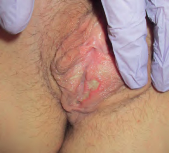
Fig. 80.20 Reactive non-sexually related acute genital ulceration associated with primary EBV infection.
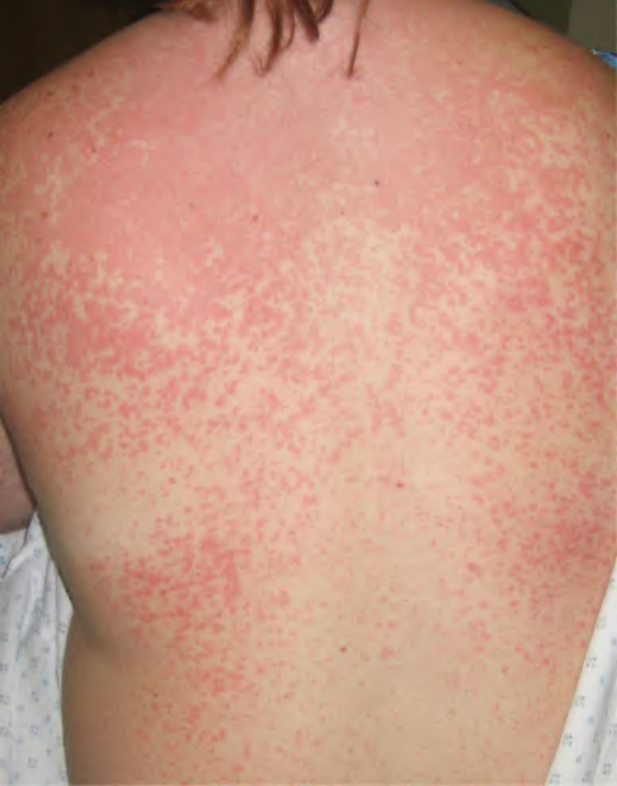
Fig. 80.21 Ampicillin-induced eruption in a patient with infectious mononucleosis due to EBV infection.
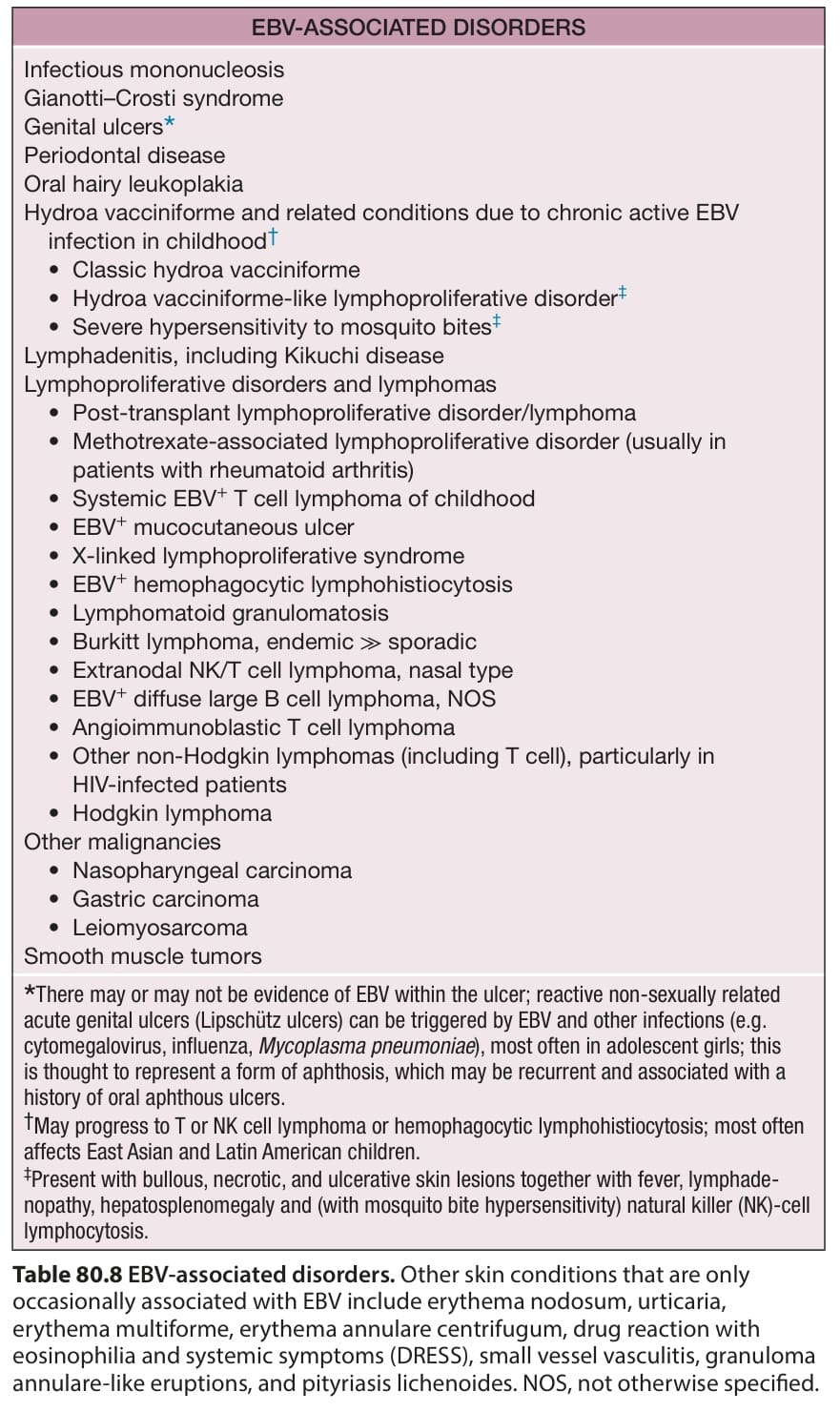
Table 80.8 EBV-associated disorders. Other skin conditions that are only occasionally associated with EBV include erythema nodosum, urticaria, erythema multiforme, erythema annulare centrifugum, drug reaction with eosinophilia and systemic symptoms (DRESS), small vessel vasculitis, granuloma annulare-like eruptions, and pityriasis lichenoides. NOS, not otherwise specified.
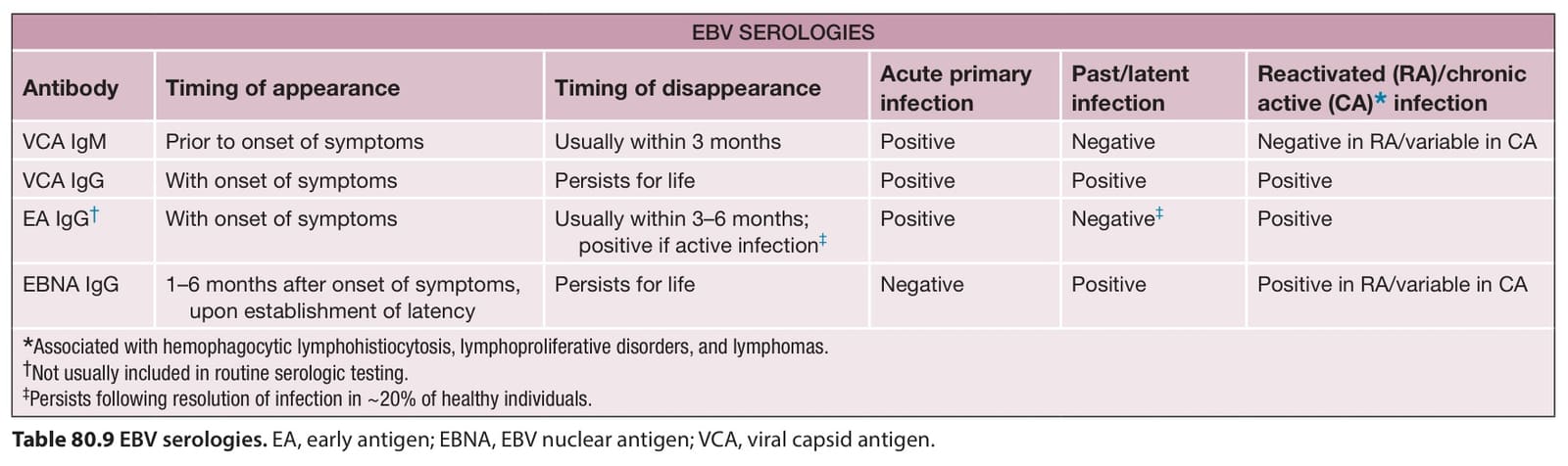
Table 80.9 EBV serologies. EA, early antigen; EBNA, EBV nuclear antigen; VCA, viral capsid antigen.