CYTOMEGALOVIRUS (HHV-5)
Key features
CMV is the leading infectious cause of congenital deafness and intellectual disability (TORCH syndrome) as well as the leading cause of blindness (due to retinitis) in AIDS patients
Neonates and immunocompromised hosts are at risk for severe clinical infections
Introduction
CMV is endemic in all parts of the world, and it is the most common cause of intrauterine infection in humans. In immunocompetent hosts, 95% of infections are asymptomatic or subclinical. Neonates and immunocompromised individuals, especially individuals with AIDS, allogeneic HSCT recipients, and patients receiving alemtuzumab, are at risk for severe clinical manifestations of CMV infection.
History
In 1881, Ribbert first observed “protozoan-like cells” in the organs of a stillborn baby with presumed congenital syphilis. Before the discovery of a viral etiology, the disease was initially called “cytomegalic inclusion
disease” based on the characteristic enlarged cells (cytomegalia) and intranuclear inclusions. In 1956, human CMV was independently isolated in vitro by three groups of investigators led by Weller, Rowe, and Smith. Originally known as the “salivary gland virus” or “salivary inclusion disease virus”, CMV was given its current name by Weller and his colleagues in 1960.
Epidemiology
The prevalence of CMV infection is inversely proportional to socioeconomic status. For example, 80%–100% of the African population is CMV-seropositive compared with 60%–70% of the US and Western European populations. The increased seroprevalence in low-income countries is thought to be due to crowded living conditions, poor hygiene, and greater numbers of individuals caring for an infant. Age-related prevalence studies in the US have shown highest risks of CMV acquisition during early childhood, adolescence, and the reproductive years.
CMV seropositivity is especially high among toddlers in day care centers, and transmission from young children to their mothers has been described. In adults, CMV infections are often contracted through sexual contact. Blood transfusions carry a 3%–4% risk of CMV infection per transfusion unit. To decrease this risk, leukocyte-depleted blood can be used in immunosuppressed patients.
Solid organ transplant and HSCT recipients, other individuals receiving immunosuppressive agents, and those infected with HIV are at increased risk of disseminated CMV infection. More recently introduced treatments such as CAR-T cellular therapy and bispecific T cell engager therapy also put patients at risk for CMV infections. CMV represents a source of significant morbidity (e.g. retinitis, colitis) and mortality in patients with AIDS. In the US, congenital CMV infection occurs in ~0.5% of all neonates.
Pathogenesis
Transmission of CMV is via body fluids, including saliva, blood, urine, semen, breast milk, and cervical and vaginal secretions, as well as in transplanted organs and rarely hematopoietic stem cells. CMV can also be spread indirectly by contaminated fomites, such as toys. Transplacental transmission of CMV to the fetus is more likely in the setting of a primary infection in the mother, with 40% of fetuses becoming infected, compared to <1% in recurrent cases.
CMV has an estimated incubation period of 4–8 weeks, followed by viremia via infected blood leukocytes and spread to various organs. It is a cytotoxic virus that causes cell enlargement (cytomegaly) and nuclear condensation (inclusion). In immunocompetent hosts, the virus induces high levels of specific anti-CMV CD4+ and CD8+ T cells. CMV infection may be either primary or recurrent, and the latter can result from either viral reactivation or reinfection with a different antigenic type.
Following primary infection, CMV persists in a latent state for the life of the host and rarely causes disease. In immunocompromised individuals, however, reactivation of latent virus can lead to recurrent infection characterized by persistent viral replication, viremia, and dissemination to distant organs. Cell injury and tissue destruction contribute to organ damage by directly altering cellular function and producing an inflammatory response.
The organ infected depends on the age of the host. Infection in healthy children and adults often involves the lymphoid tissues, while fetal and neonatal infection commonly affects the salivary glands and neurons. Infection in immunocompromised individuals frequently involves the retina and lungs as well as the liver and gastrointestinal tract.
Clinical Features
More than 90% of primary CMV infections are subclinical, although a “mononucleosis-like syndrome” similar to that seen with EBV occasionally develops in immunocompetent persons (Table 80.10). This syndrome has also been described in both immunocompetent and immunocompromised individuals after a blood transfusion. Infected patients develop non-exudative pharyngitis in addition to fever, malaise, myalgias, lymphadenopathy, and hepatosplenomegaly. Atypical lymphocytosis and elevated liver enzymes may also occur. An
exanthem, which can be morbilliform, urticarial, petechial or purpuric, develops in a small percentage of patients. As with infectious mononucleosis, the administration of ampicillin or related drugs during this symptomatic period leads to a cutaneous eruption in 80%–100% of individuals. Although the clinical course of CMV-induced mononucleosis is self-limited, rare complications include hemolytic anemia, thrombocytopenia, granulomatous hepatitis, Guillain–Barré syndrome, meningoencephalitis, myocarditis, interstitial pneumonia, arthritis, and gastrointestinal and genitourinary manifestations.
Congenital CMV infection is the leading infectious cause of deafness and intellectual disability in the US. Approximately 5%–10% of infected neonates have symptoms at birth, presenting with jaundice, hepatosplenomegaly, intrauterine growth retardation, thrombocytopenia, chorioretinitis, seizures, and/or intracranial calcifications. Cutaneous manifestations include purpuric papules and nodules of dermal hematopoiesis which are sometimes referred to as “blueberry muffin” lesions (Fig. 80.22; see Ch. 121), in addition to petechiae, purpura, and vesicles. Another 10%–15% of children are born with asymptomatic CMV infection and later develop sequelae, most often sensorineural hearing loss. Perinatal CMV infection is often asymptomatic; however, a limited number of infected infants develop pneumonitis, lymphadenopathy, or hepatosplenomegaly.
In AIDS patients, CMV may cause chronic ulcers of the perineum or extremities, chorioretinitis, esophagitis, colitis, and pneumonitis, as well as endocrine, bone marrow, CNS, and renal abnormalities. Cutaneous manifestations can range from vesicles to nodules to verrucous plaques. In recipients of solid organ transplants and HSCTs, gastrointestinal involvement and less often interstitial pneumonia are the most serious manifestations of CMV infection.
Diagnosis and Pathology
Culture of CMV in human fibroblasts is the traditional diagnostic “gold standard”, but this takes several days to weeks. Detection of CMV in tissue cultures within 24–48 hours is possible with the shell vial assay, in which monoclonal antibodies specific for early CMV antigens are employed. In addition to CMV-specific serologies, assays that detect CMV antigens (e.g. pp65) within leukocytes or CMV DNA via PCR (necessary in neutropenic patients) are commonly employed to identify and monitor viremia and systemic infection. CMV retinitis in patients with AIDS is diagnosed by ophthalmologic examination.
Like other viral exanthems, the histologic features of the acute exanthem of CMV infection include mild spongiosis and a sparse perivascular lymphocytic infiltrate in the upper dermis. Epidermal changes are minimal since CMV does not infect keratinocytes. However, it does infect endothelial cells, which can have an “owl’s eye” appearance that is pathognomonic for CMV infection. The cells are enlarged two- to threefold and contain purplish, crystalline intra-nuclear inclusion bodies surrounded by a clear halo (Fig. 80.23). These cytomegalic cells are most likely to be evident in ulcers, and vascular dilatation is another common finding. CMV vasculitis, a manifestation of disseminated disease, is characterized by marked endothelial swelling and inflammation.
Differential Diagnosis
The clinical differential diagnosis of the mononucleosis-like syndrome of CMV infection includes EBV-induced infectious mononucleosis, which is generally more severe and often associated with exudative pharyngitis. Other diagnostic considerations may include DRESS, which may also be associated with reactivation of CMV (see HHV-6 section below), as well as toxoplasmosis, viral hepatitis, GVHD, and lymphoma. A high index of suspicion is necessary to diagnose CMV-induced ulcers and atypical cutaneous manifestations in immunocompromised patients.
Treatment and Prevention
Management is targeted at prevention plus prophylactic antiviral treatment in susceptible immunocompromised individuals as well as antiviral treatment in those who are symptomatic. Treatment of
CMV-induced mononucleosis in immunocompetent individuals, however, is only supportive.
As a preventive measure, hematopoietic stem cells and other tissues from CMV-seronegative donors are used preferentially for transplantation in seronegative recipients. Letermovir, an inhibitor of the CMV terminase complex, is currently FDA-approved for prophylactic administration in adult CMV-seropositive allogeneic HSCT recipients. Usually administered for at least 100 days, letermovir reduces the risk of clinically significant CMV infection and non-relapse mortality in this patient population. While letermovir lacks the myelosuppression associated with ganciclovir, it does not have activity against HSV-1 and -2. Pre-emptive management consisting of following blood CMV DNA levels and initiating first-line therapies if they increase represents another strategy in transplant recipients.
The first-line agents for treatment of CMV infections in immunocompromised patients are intravenous ganciclovir or oral valganciclovir; intravitreal injections/implants of ganciclovir are used for localized retinitis. When patients fail to respond to these medications or develop significant side effects, maribavir, foscarnet, and cidofovir are additional options (Table 80.11). Maribavir, an inhibitor of the CMV UL97 protein kinase, was recently FDA-approved for post-transplant CMV infections that fail to respond to other medications. In general, relapse is common after discontinuation of antiviral agents. Treatment with CMV-specific T cell adoptive transfer is being studied in immunocompromised individuals but is not readily available.
There is ongoing investigation of several CMV vaccines, including those utilizing a replication defective whole-virus, adjuvanted subunits, virus-like particles, viral vectors, and mRNA encoding vaccine antigens.
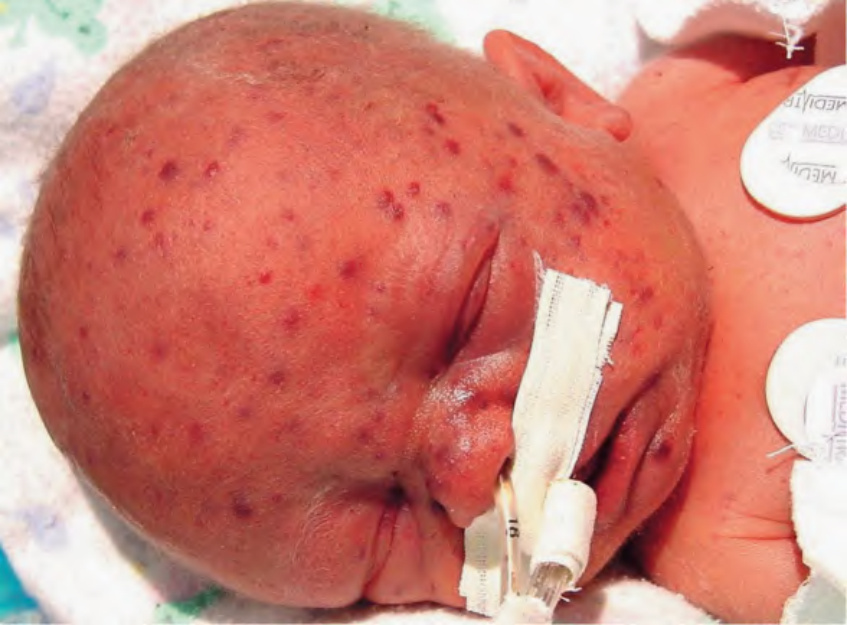
Fig. 80.22 TORCH syndrome due to cytomegalovirus. Multiple purpuric papules of dermal hematopoiesis. Courtesy Mary S. Stone, MD.
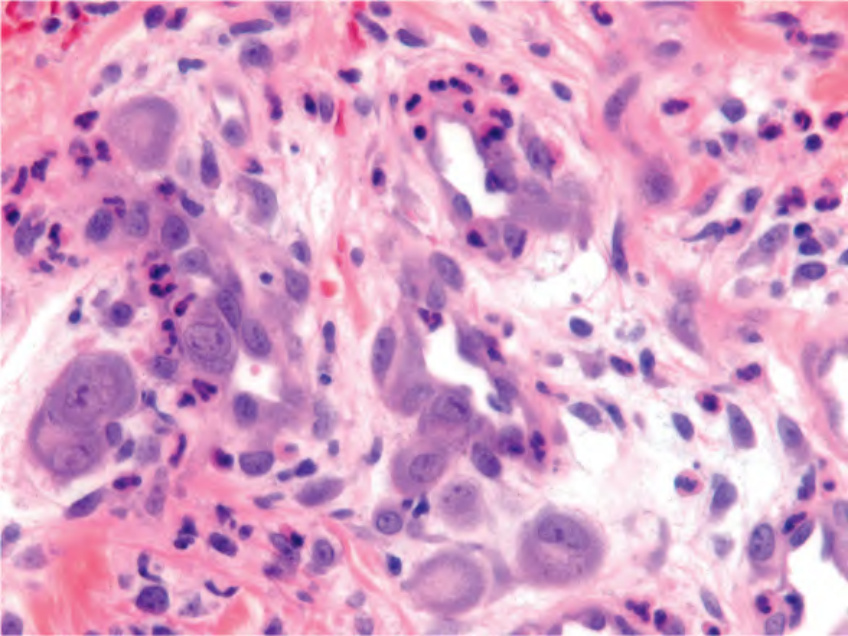
Fig. 80.23 Histology of cytomegalovirus (CMV) infection. Enlarged endothelial cells with a prominent intranuclear inclusion are typical of CMV infection. Courtesy Angela Galan, MD.
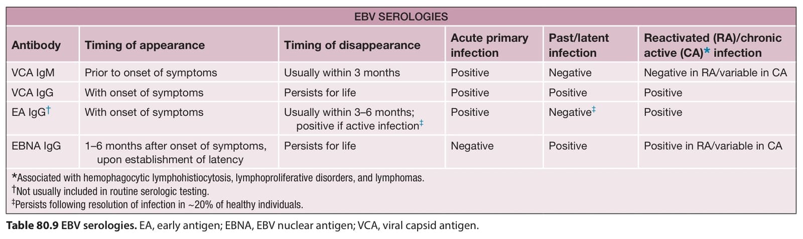
Table 80.9 EBV serologies. EA, early antigen; EBNA, EBV nuclear antigen; VCA, viral capsid antigen.
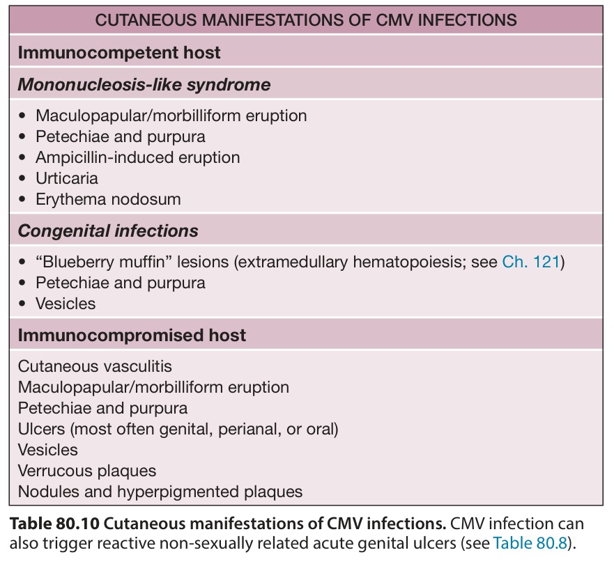
Table 80.10 Cutaneous manifestations of CMV infections. CMV infection can also trigger reactive non-sexually related acute genital ulcers (see Table 80.8).
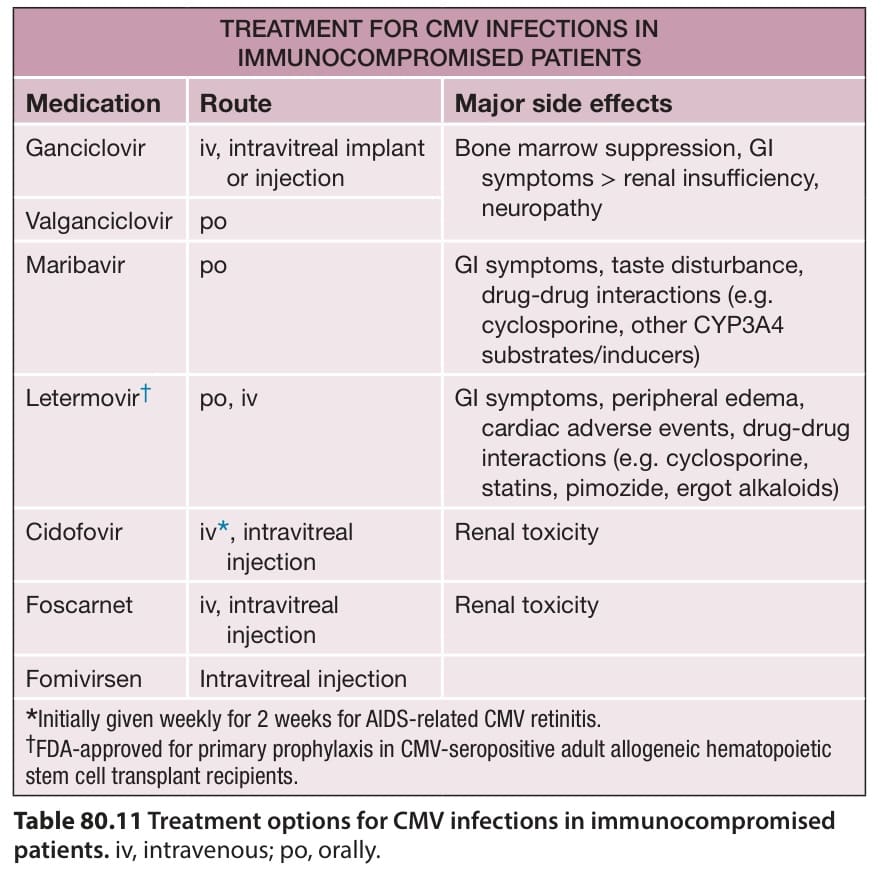
Table 80.11 Treatment options for CMV infections in immunocompromised patients. iv, intravenous; po, orally.