ENTEROVIRUS INFECTIONS
Key features
Most common in summer and fall
Exanthems include erythematous, vesicular, and petechial types
Meningitis and encephalitis possible
Introduction
The enteroviruses, a subgroup of the picornavirus family, cause a wide array of illnesses associated with exanthems. The non-polio enteroviruses include echoviruses and coxsackievirus types A and B, with >100 genetically distinct types that infect humans. These viruses consist of a single-stranded RNA genome with an unenveloped capsid.
Epidemiology
Enteroviral infections occur worldwide and are usually transmitted human-to-human via fecal–oral or respiratory routes, usually with a 3- to 6-day incubation period. They can also be spread from mother to infant in the peripartum period. Exposure to virus in fecally contaminated water in swimming pools or via ingestion of oysters may be responsible for some infections. Clinical attack rates are highest in young children, and infections occur more frequently in lower socioeconomic groups. In temperate climates, infections are most common in the summer and fall. However, coxsackievirus A6 outbreaks frequently affect adults and can occur in the winter.
Pathogenesis
Enteroviruses infect epithelial surfaces of the pharynx and lower alimentary tract. They then replicate in lymphoid tissues and disseminate via an initial minor viremia. Subsequently, replication may occur at multiple anatomic sites, including the CNS, heart, liver, adrenals, respiratory tract, skin, and mucous membranes. This replication correlates with the appearance of clinical symptoms. A major viremia occurs during the period of viral multiplication in the secondary infection sites.
Clinical Features and Differential Diagnosis
Fig. 81.3 depicts the potential clinical manifestations of an enteroviral infection.
The most distinctive exanthematous illness due to enteroviruses is hand-foot-and-mouth disease (HFMD). This disorder is characterized by a vesicular eruption on the palms and soles (Fig. 81.4A) in conjunction with an erosive stomatitis. The dorsal aspect of the hands and feet may be involved as well as the buttocks and perineum; oral
lesions are most common on the tongue, buccal mucosa, palate, uvula, and anterior tonsillar pillars. Onychomadesis occasionally occurs 1–2 months after HFMD as a consequence of temporary nail matrix arrest related to the viral infection.
Patients with HFMD often present with fever and malaise, and they may have a mild prodrome prior to the onset of the exanthem. Numerous coxsackievirus serotypes have been implicated, most commonly type A16; in addition, type A10 represents a major cause of outbreaks in Europe and Asia. Although HFMD generally runs a benign and self-limited course, since 1998 there have been multiple epidemics of severe enterovirus 71 infection, primarily in the Asia-Pacific region, which have resulted in cardiopulmonary and neurologic complications and even deaths in affected young children.
Globally beginning in 2008 and since 2011 in the US, coxsackievirus A6 (CVA6) has emerged as a major cause of both outbreaks and sporadic cases of severe and atypical HFMD. This variant is characterized by more widespread vesicular, papulovesicular, and sometimes bullous or petechial eruptions that frequently involve the perioral area, arms, legs, and trunk as well as the palms, soles, and buttocks (Fig. 81.4B–G). Oral lesions develop in approximately half of patients, and delayed acral desquamation and onychomadesis are common. The eruption may be accentuated in areas of pre-existing skin injury or eczematous dermatitis, with the latter referred to as “eczema coxsackium” (see Fig. 81.4F). The differential diagnosis may include eczema herpeticum, varicella, disseminated zoster, Gianotti–Crosti syndrome, erythema multiforme, and autoimmune bullous diseases. In addition, patients with HFMD due to CVA6 are often febrile but do not usually have neurologic or other complications.
Herpangina is a febrile illness, primarily in children 3–10 years of age, characterized by painful vesicles and erosions on the soft palate, uvula, tonsils, pharynx, and buccal mucosa. An exanthem is usually absent. Coxsackievirus groups A and B, as well as echoviruses, are the usual pathogens. Coxsackievirus B5 infection has also been associated with conjunctival hyperemia.
In addition to HFMD and herpangina, enteroviruses cause a diverse spectrum of exanthems that may be morbilliform, rubelliform, roseolalike, urticarial, scarlatiniform, vesicular, pustular, and/or petechial. The majority of enteroviral exanthems feature erythematous macules and papules in a widespread distribution. Petechiae are fairly common, especially with echovirus 9 infection, and may lead to consideration of a more serious infection such as meningococcemia. Non-polio enteroviruses often infect newborns, producing a nonspecific erythematous exanthem and additional features such as fever, gastroenteritis, hepatitis, pneumonia, and meningoencephalitis.
In 1969, Cherry et al. described four children with echovirus 25 or 32 infection who had fever and transient vascular papules; histologically, dilated blood vessels were seen. The term eruptive pseudoangiomatosis was subsequently proposed, and additional cases have been described in adults as well as children, with spontaneous resolution within a few weeks. Associated infections have included other enteroviruses, cytomegalovirus (CMV), Epstein–Barr virus (EBV), adenovirus, parvovirus B19, and SARS-CoV-2 (vaccination as well as infection) .
While classic HFMD and herpangina are fairly straightforward clinical diagnoses, other less specific exanthematous illnesses due to enteroviruses may have a broad differential diagnosis. Important factors that may point to the etiology include the season, exposure history, geographic location, and clinical signs and symptoms.
Confirmation of an enteroviral infection may be accomplished via viral culture, which has low sensitivity (especially for CVA6), or polymerase chain reaction (PCR)-based assays; determination of the specific type of enterovirus via gene sequencing following RT-PCR is currently done only at research and public health laboratories (e.g. the US Centers for Disease Control [CDC]). Viral testing can be performed from vesicular fluid (preferred for HFMD), throat swabs, stool samples, and cerebrospinal fluid (CSF). Serologic diagnosis is difficult due to the large number of different serotypes and the time delay, which make it clinically less useful. Table 81.1 lists types of serologic assays.
Pathology
Skin biopsy is generally not a useful diagnostic procedure in enteroviral infections. Histopathologic features of the vesicular lesions of HFMD include epidermal necrosis with intraepidermal vesicles, a lack of inclusion bodies or multinucleation, and a nonspecific dermal inflammatory infiltrate.
Treatment
For most enteroviral exanthems, the course is self-limited, the prognosis is excellent, and supportive care suffices. However, in certain patient populations, e.g. immunosuppressed individuals or neonates, an enteroviral infection may be associated with potentially life-threatening complications. Pleconaril, a drug that interferes with enterovirus attachment and uncoating by binding to the protein capsid, has been demonstrated to be effective both in vitro and in clinical studies, including a randomized, placebo-controlled trial in neonates with enteroviral sepsis; it therefore has potential as a specific antiviral therapy for serious enteroviral infections. Three inactivated enterovirus-71 vaccines are licensed for clinical use in China, and other enteroviral vaccines are under development.
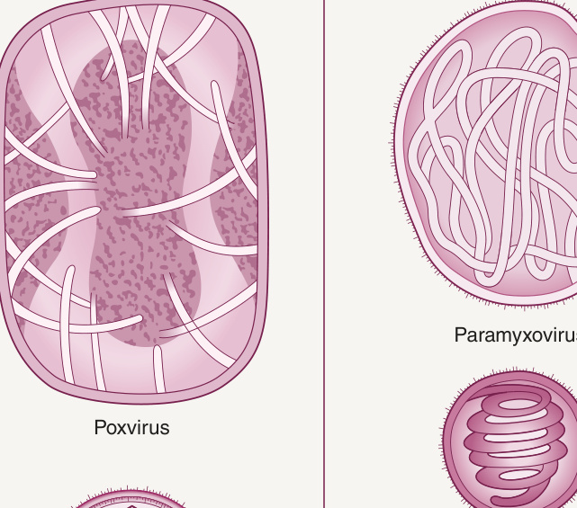
Fig. 81.1 Relative size and shape of selected viruses infecting humans.Adapted from Hsiung’s Diagnostic Virology, Yale University Press, 1994.
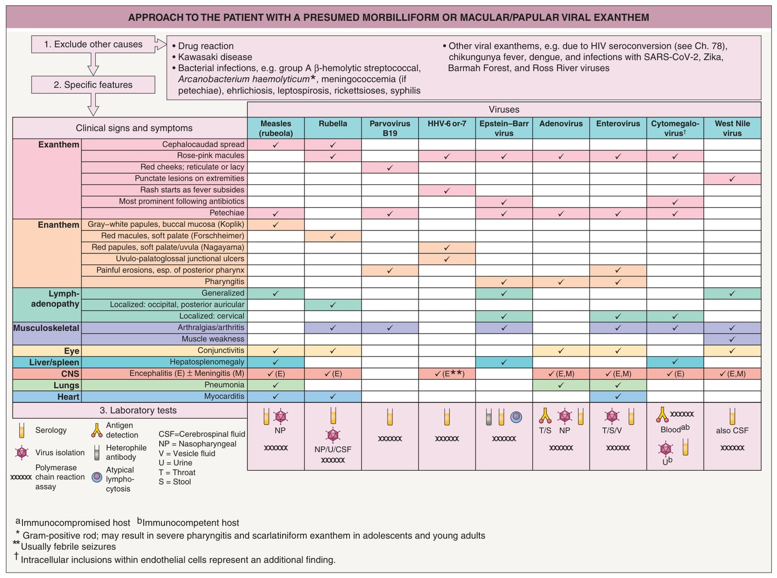
Fig. 81.2 Approach to the patient with a presumed morbilliform or macular/papular viral exanthem.
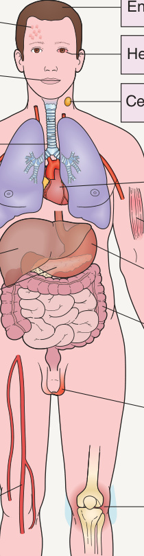
Fig. 81.3 Clinical manifestations of enterovirus infection.
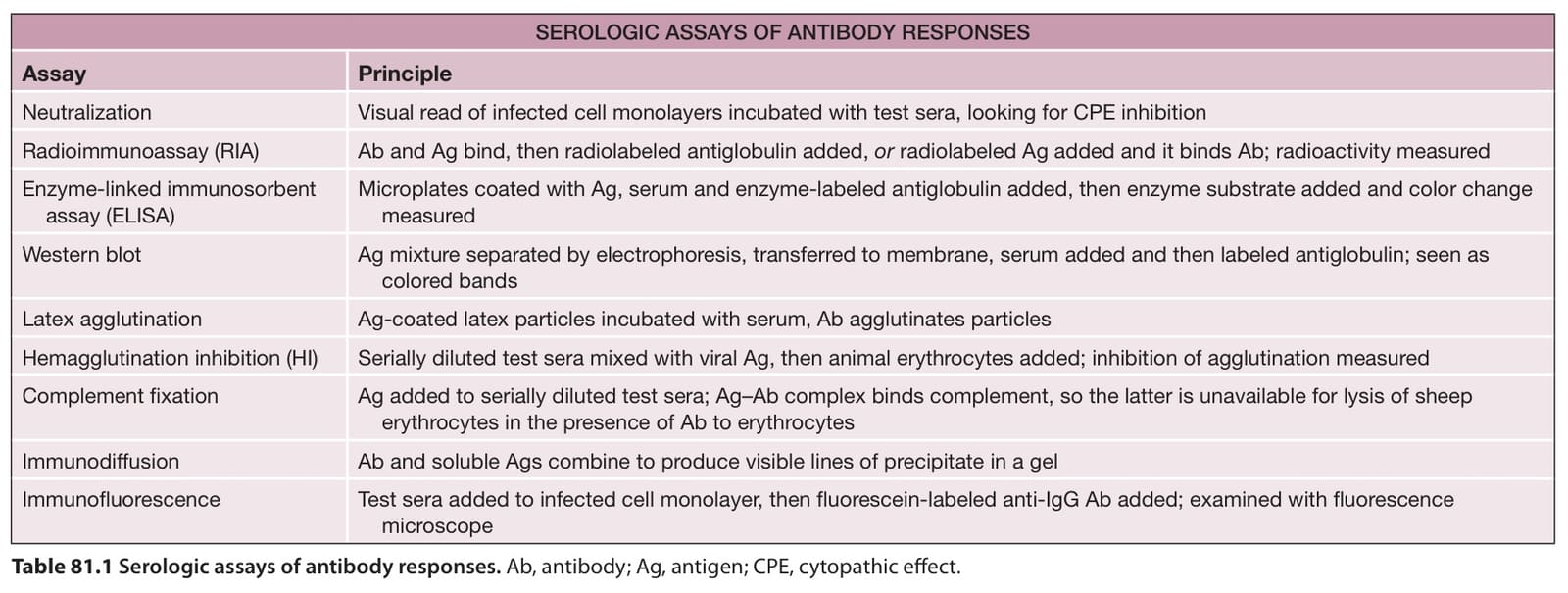
Table 81.1 Serologic assays of antibody responses. Ab, antibody; Ag, antigen; CPE, cytopathic effect.