RUBELLA
Synonym: German measles
Key features
Exanthem of rose-pink macules with cephalocaudad spread
Tender lymphadenopathy, especially occipital and posterior auricular
Joint involvement common
Fetal infection is associated with congenital anomalies
Introduction and History
Rubella is caused by an RNA virus in the Togaviridae family. Although usually a mild, self-limited disease in children and adults, it can cause major complications when transmitted to a fetus in utero, including miscarriage, stillbirth, and severe congenital malformations.
Epidemiology
The disease is spread via respiratory droplets, with an incubation period of 16–18 days. The incidence of rubella has markedly declined since the licensure of the MMR (measles, mumps, and rubella) vaccine in 1969. Rubella was declared as eliminated from the US in 2005 and from the Americas in 2014; currently all US cases reported since 2012 (<10 per year) were acquired in another country. Rubella has a worldwide distribution and occurs most often in the spring.
Pathogenesis
The primary site of infection is the nasopharynx, followed by spread to regional lymph nodes and a subsequent viremia.
Clinical Features and Differential Diagnosis
Rubella typically presents with a mild prodrome that includes fever, headache, and upper respiratory symptoms. In children, many cases are subclinical. One to 5 days following the prodrome, an eruption of erythematous macules and papules appears on the face, spreading in a cephalocaudad direction. Erythematous petechial macules may also be present on the soft palate (Forchheimer spots). The eruption is often accompanied by tender lymphadenopathy, especially of the occipital, posterior auricular, and cervical regions. The cutaneous eruption tends to fade in 2–3 days in the same order as it appeared.
Rubella may result in arthralgias and arthritis, especially in postpubertal girls and women; less frequent complications include hepatitis, myocarditis, pericarditis, hemolytic anemia, and thrombocytopenia. Encephalitis occurs in ~1 in 6000 infected individuals.
Rubella can be diagnosed by detection of anti-rubella IgM antibodies or a fourfold increase in specific IgG antibodies; virus isolation or detection via RT-PCR from a nasal wash, throat swab, or urine sample is also possible. The exanthem of rubella is nonspecific and the differential diagnosis includes other viral exanthems (e.g. adenovirus, enterovirus,
measles, EBV) and toxin-mediated eruptions such as scarlet fever. The occipital lymphadenopathy and the typical progression of the eruption can be helpful in diagnosing the disease.
Congenital rubella occurs when a nonimmune pregnant woman transfers the virus to the fetus. Birth defects are most commonly observed when infection is during the first 16 weeks of pregnancy. Congenital rubella syndrome is characterized by cataracts, deafness, congenital heart defects (patent ductus arteriosus, ventricular septal defects), and CNS abnormalities (microcephaly, developmental delay). A “blueberry muffin baby” presentation due to dermal hematopoiesis is occasionally observed (see Ch. 121).
Both wild type and vaccine-derived rubella virus have been identified and found to persist for years in macrophages within cutaneous granulomas in individuals with immunodeficiencies characterized by
Pink macules and minimally elevated papules. Courtesy Louis A. Fragola, Jr, MD.
T cell lymphopenia and/or defective lymphocyte cytotoxicity. The latter include ataxia telangiectasia, Griscelli syndrome type 2, and a variety of combined immunodeficiency syndromes (see Ch. 60). Clinically, these patients present with pink–violet to red–brown papules, nodules, and plaques, often with crusting or ulceration, that favor the extremities and face but may be widespread. Rubella virus has also been identified within cutaneous granulomas (palisaded and necrotizing type) involving the left upper arm (a typical vaccination site) in clinically immunocompetent adults; localized lesions associated with vaccine-derived rubella developed 2 to >10 years after the last MMR vaccine administration.
Pathology
Reported histopathologic findings of rubella include a superficial perivascular infiltrate with mild spongiosis. Atypical lymphocytes may be evident in the cutaneous infiltrate as well as in peripheral blood.
Treatment
Treatment of rubella is supportive. The current recommended immunization schedule for rubella vaccine, which is given in conjunction with the measles and mumps vaccines (MMR vaccine), is an initial dose at 12–15 months and a second dose at 4–6 years. In a pregnant woman who has been exposed to rubella, serologic testing (IgG and IgM) should be performed. Prophylaxis via administration of IM/IVIg to rubellasusceptible women exposed to confirmed rubella early in pregnancy may be considered, but it does not reliably prevent fetal infection. If rubella-specific IgM antibodies or a diagnostic rise in IgG anti-rubella antibodies (the latter not useful following IM/IVIg administration) are detected, the patient should be offered prenatal counseling.
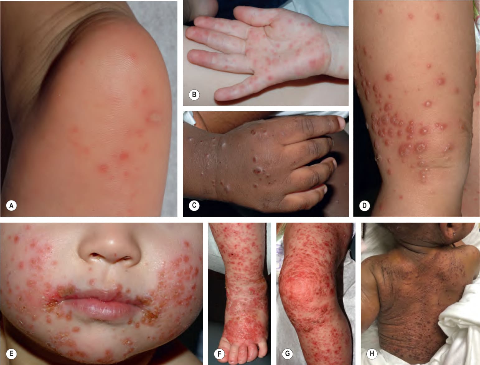
Fig. 81.4 Hand-foot-and-mouth disease.A Vesicle and erythematous macules on the sole in classic hand-foot-and-mouth disease. B–G Coxsackievirus A6 infection presenting as: multiple erythematous macules and papules with some targetoid lesions resembling early erythema multiforme on the palm (B); vesicles and papulovesicles on the dorsal hand (C) and thigh (D); perioral monomorphic crusted lesions resembling eczema herpeticum (E); more extensive “eczema coxsackium” in toddlers (F & G) and an infant (H) with atopic dermatitis. A–G, Courtesy Julie V. Schaffer, MD. H, Courtesy Henry Heaton, MD.
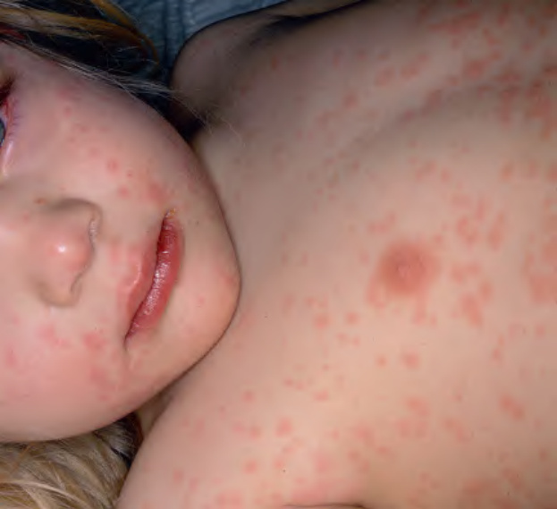
Fig. 81.5 Measles.
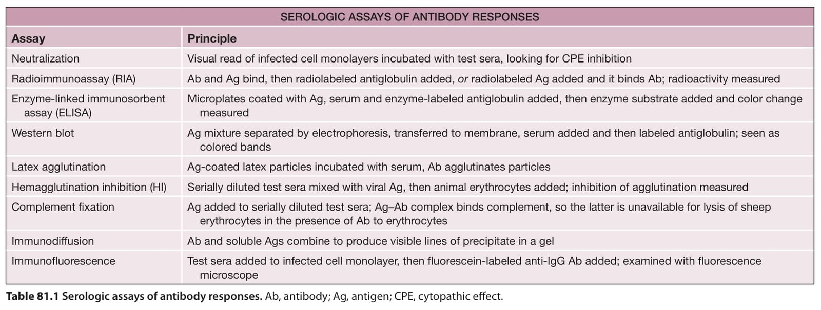
Table 81.1 Serologic assays of antibody responses. Ab, antibody; Ag, antigen; CPE, cytopathic effect.