ERYTHEMA INFECTIOSUM
Synonyms: Fifth disease “Slapped cheek” disease Parvovirus B19 infection
Key features
“Slapped cheeks” followed by lacy extremity-predominant eruption, especially in children
Eruptions in adolescents and adults may be petechial and/or have an acral or periflexural distribution
Marked affinity of parvovirus B19 for erythrocyte precursors
Fetal infection may result in anemia, fetal hydrops, or death
Introduction and History
Human parvovirus B19 (B19), the only parvovirus known to infect humans, was accidentally discovered in 1975 by Cossart and colleagues,
who were screening blood samples for hepatitis B virus. The small, single-stranded DNA virus, which was named after the panel of sera in which it was found (number 19, row B), was identified as the etiology of erythema infectiosum in 1983. Subsequent elucidation of the pathogenesis of human B19 infections has led to its association with a wide variety of clinical findings (Table 81.2).
Epidemiology
Infection with B19 occurs in a seasonal pattern, with a peak incidence of erythema infectiosum during the winter and spring. Transmission is via respiratory secretions, blood products, and vertically from mother to fetus; the incubation period is between 4 and 14 days. B19 infection occurs worldwide and is most common in school-aged children. A cyclical pattern of epidemics seems to have peaks about every 6 years, and individual community epidemics last 3–6 months. The seroprevalence of B19 antibodies increases in direct proportion with age, with immunity in 2%–15% of children 1–5 years of age, 15%–60% of those 5–19 years of age, and 30%–80% of adults.
Pathogenesis
B19 has a strong tropism for erythroid progenitor cells, and the erythrocyte “P antigen” (globoside) is the cellular receptor to which the virus binds. In fact, individuals who lack the P antigen (blood group Pk or p phenotypes) seem to be naturally resistant to B19 infection.
After initial infection of the respiratory tract, a B19 viremia occurs, ending when the anti-B19 IgM antibody appears ~8–10 days post inoculation (Fig. 81.6). During the viremic period, reticulocytopenia occurs for 7–10 days. One week after the appearance of IgM antibody, anti-B19 IgG appears and coincides with the appearance of the rash and arthralgia. An immune-mediated process is suggested by the temporal disconnect between the viremia and the clinical symptoms.
The anemia and reticulocytopenia tend to be inconsequential in the normal host but may result in transient aplastic crises in at-risk individuals, such as those with disorders resulting in enhanced red blood cell destruction or decreased production; thrombocytopenia, neutropenia, and pancytopenia may also occur (see Table 81.2). Fetal infection from intrauterine virus transmission causes fetal anemia of varying severity (see below).
Clinical Features and Differential Diagnosis
The most common illness caused by B19 infection is erythema infectiosum, which most often occurs in children 4–10 years of age. Mild prodromal symptoms such as a low-grade fever, myalgias, and headache may develop 7–10 days before the characteristic exanthem appears. The initial stage of the exanthem consists of bright red macular erythema of the cheeks, with sparing of the nasal bridge and circumoral regions (Fig. 81.7A). One to 4 days later, the second stage appears as erythematous macules and papules on the extremities and to a lesser extent the trunk, progressing to form a lacy, reticulated pattern (Fig. 81.7B,C). This
Volunteers were infected with parvovirus B19 and the virologic, hematologic, and clinical events that followed were recorded. Reprinted from Cohen J, Powderly WG. Infectious Diseases, 2nd edn. St Louis: Mosby, 2004.
exanthem typically lasts 1–3 weeks or occasionally longer; it fluctuates in intensity during this period, often with exacerbations upon exposure to sunlight or overheating. The differential diagnosis of erythema infectiosum may include scarlet fever, enteroviral infection, and rubella, but the diagnosis is usually straightforward when the characteristic eruption is present. Although the evanescent exanthem of systemic juvenile idiopathic arthritis (Still disease) is also accentuated by heat, it is accompanied by episodic high fevers, has a more chronic course, and often exhibits the Koebner phenomenon.
In adolescents and adults with B19 infection, the exanthems tend to be petechial and often have an acral or periflexural distribution (see Table 81.2). A distinctive papular-purpuric gloves and socks syndrome (PPGSS) was first described in 1990 and subsequently found to be associated with acute B19 infection, although other infectious etiologies have also been reported, e.g. coxsackievirus B6, HHV-6, and EBV. PPGSS most often develops in the spring, and although it has a predilection for young adults, PPGSS may occur at any age, including in children. The hallmark findings are edema and erythema of the hands and feet, especially the palms and soles, in association with petechiae and purpura (Fig. 81.8). Occasionally, there is extension onto the dorsal surfaces of the hands and feet, and patients may complain of burning and pruritus. Additional manifestations can include an enanthem of erosions and petechiae involving the palate, pharynx, and tongue, as well as fever and mild prodromal symptoms.
Therapy of PPGSS is symptomatic, and spontaneous resolution usually occurs over 1–2 weeks. Although few studies have addressed the immune response in PPGSS, based on limited data it appears that patients develop the mucocutaneous lesions while still viremic, which implies contagiousness and therefore has different epidemiologic implications than the classic eruption of erythema infectiosum.
Arthralgia or arthritis occurs in up to 10% of patients with erythema infectiosum and is usually self-limited, typically resolving in 1–3 weeks but sometimes lasting for months. It tends to affect the small joints of the hands as well as the wrists, knees, and ankles. Joint involvement related to B19 infection is more common in adults, especially women,
occurring in up to 30%–60% of those with acute infection. Arthropathy may be seen without an accompanying exanthem.
The manifestations of fetal B19 infection range from self-limited anemia to hydrops, spontaneous miscarriage, or stillbirth. The greatest susceptibility is associated with infections acquired before 20 weeks’ gestation, and most fetal losses occur between 20 and 28 weeks’ gestation. Third-trimester fetal demise may also occur, but it is less often associated with fetal hydrops. The overall risk of fetal loss in the setting of acute B19 infection during pregnancy is only 2%–6%, and the majority of infants born to B19-infected mothers are delivered asymptomatic at term. There is no evidence for long-term neurodevelopmental sequelae for surviving infected infants, and congenital malformations have not been conclusively demonstrated.
When diagnostic confirmation of B19 infection is needed, detection of serum anti-B19 IgM antibody is the preferred method, and its presence indicates infection within the previous 2–4 months. PCR-based assays may be helpful in diagnosing infection in immunocompromised patients.
Pathology
Skin biopsy is generally not useful in the diagnosis of B19 infection. In patients with PPGSS, histologic evaluation demonstrates a lymphocytic dermal infiltrate with frequent extravasation of erythrocytes. Anti-B19 antibodies can highlight infected endothelial cells in superficial dermal blood vessels.
Treatment
No specific antiviral therapy is available for B19 infections. Children with erythema infectiosum usually feel well and often require no treatment. NSAIDs are useful if symptomatic arthropathy is present. Patients who develop an aplastic crisis may require red blood cell transfusions. Pregnant women with confirmed primary B19 infection during the first two trimesters should undergo serial fetal ultrasonography, and management of severely affected fetuses is possible with in utero transfusions and other interventions.
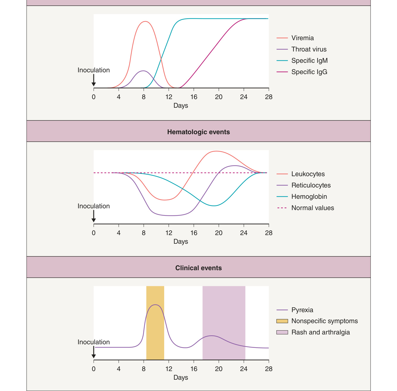
Fig. 81.6 Course of parvovirus B19 infection.
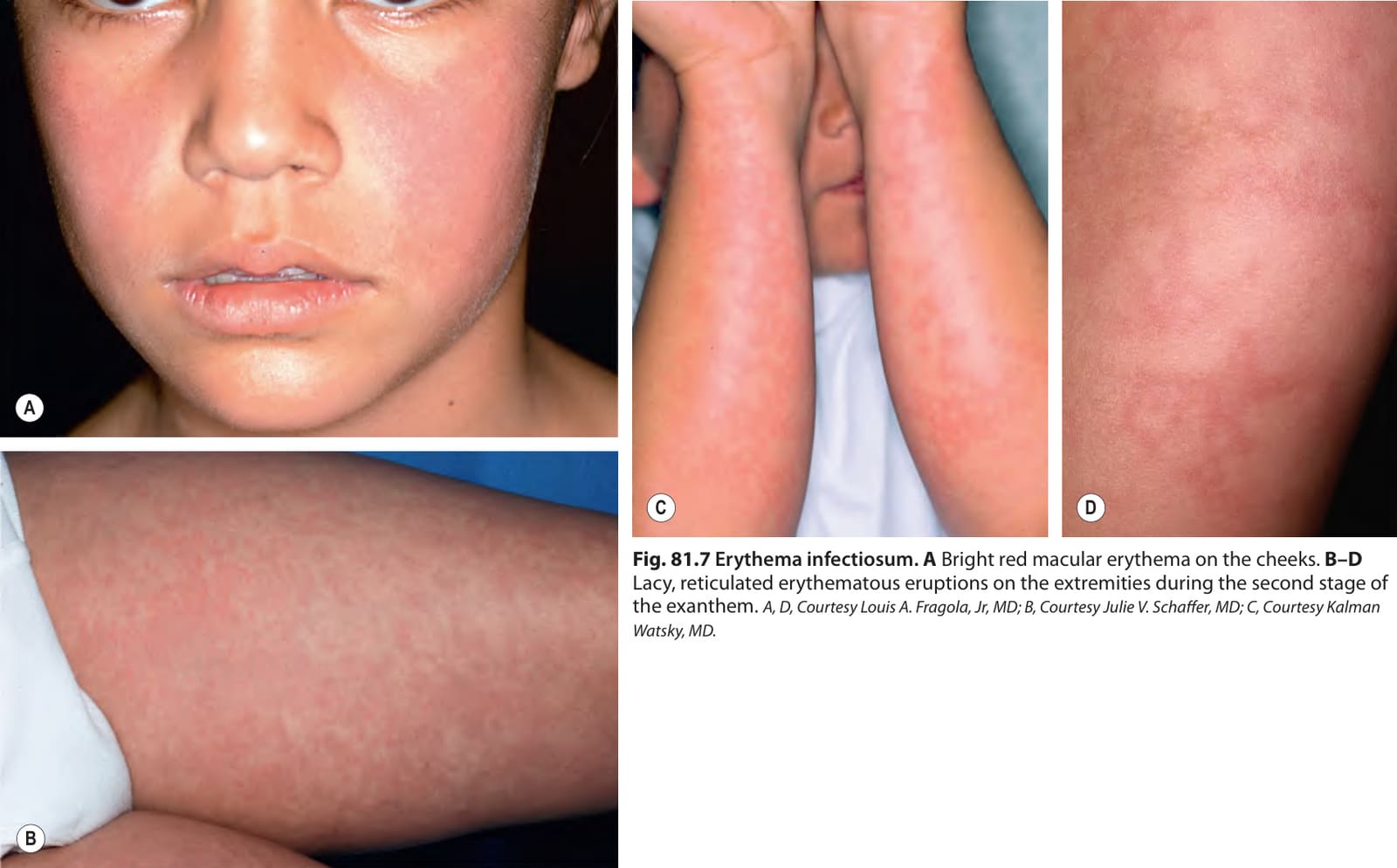
Fig. 81.7 Erythema infectiosum.A Bright red macular erythema on the cheeks. B–D Lacy, reticulated erythematous eruptions on the extremities during the second stage of the exanthem. A, D, Courtesy Louis A. Fragola, Jr, MD; B, Courtesy Julie V. Schaffer, MD; C, Courtesy Kalman Watsky, MD.
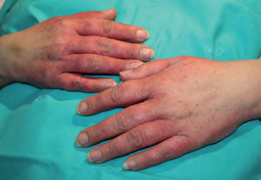
Fig. 81.8 Papular-purpuric gloves and socks syndrome. Erythema and edema with associated petechiae and small purpuric papules on the dorsal aspect of the fingers and hands. Courtesy Luis Requena, MD.
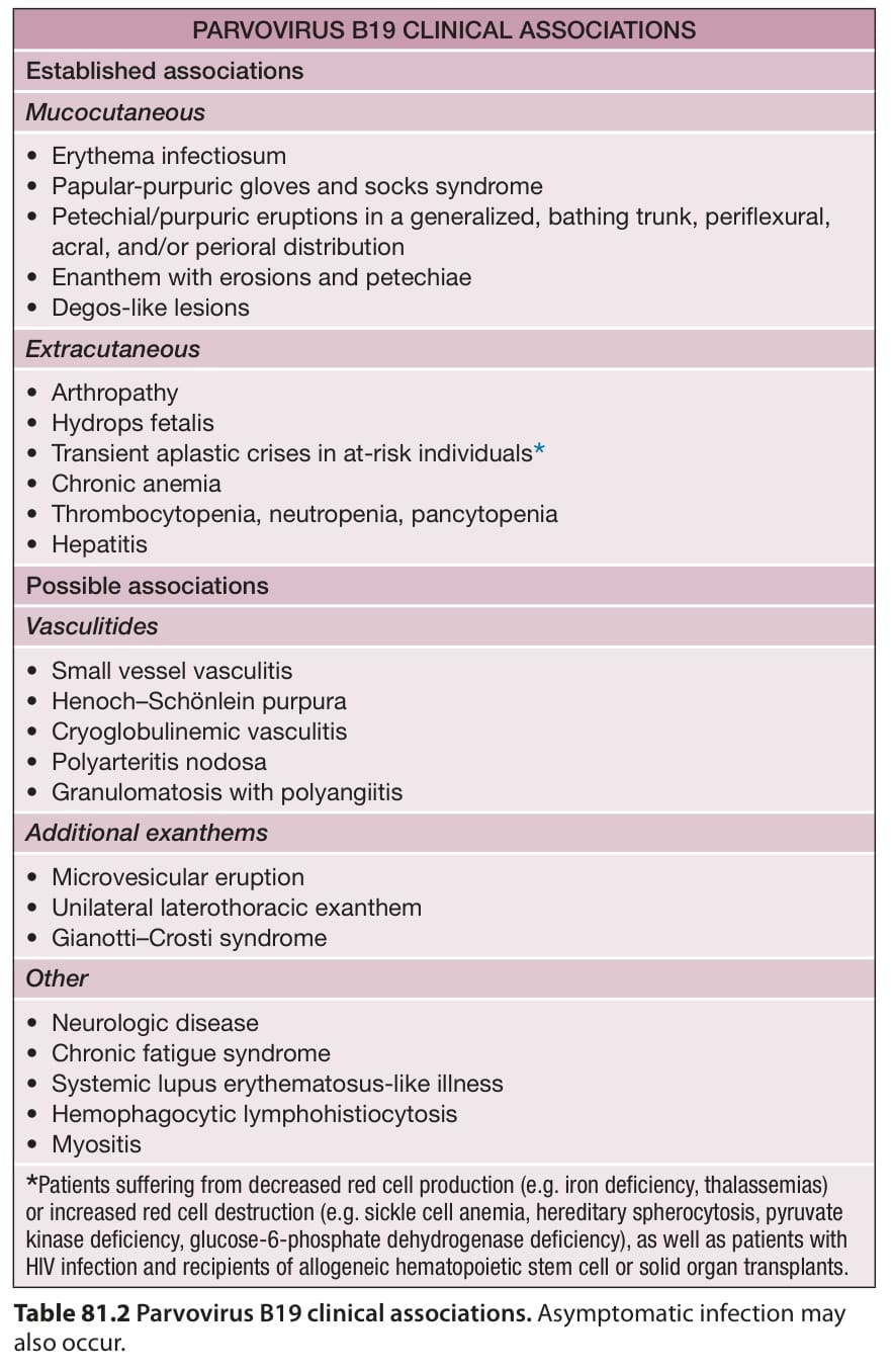
Table 81.2 Parvovirus B19 clinical associations. Asymptomatic infection may also occur.