UNILATERAL LATEROTHORACIC EXANTHEM
Synonym: Asymmetric periflexural exanthem of childhood
Key features
Initial unilateral distribution, most often affecting the axilla and lateral trunk
Subsequent generalization, but maintains unilateral predominance
Spontaneous resolution over 3–6 weeks
Presumed to be viral in etiology, although exact cause unknown
Introduction and History
This disorder was first described in 1962, and the term “unilateral laterothoracic exanthem” (ULTE) was introduced in 1992. ULTE is initially characterized by a unilateral periflexural (often axillary) location, with spread in a centrifugal pattern to the contralateral side over time.
Epidemiology
ULTE occurs most commonly in toddlers and preschool-aged children, with an age range of 6 months to 10 years and a male : female ratio of 1 : 2. It has most often been reported in Europe and North America, and the majority of cases occur in the spring.
Pathogenesis
The etiology of ULTE remains unknown despite multiple attempts to identify a causative infectious agent. The seasonal pattern, associated prodrome, reports of familial cases, and lack of response to systemic antibiotics suggest a viral etiology. In studies in which screening for multiple viruses (e.g. EBV, CMV, HHV-6, HHV-7) was performed, no etiologic agent has been consistently demonstrated. A relationship to infection with Spiroplasma, parvovirus B19, EBV, and SARS-CoV-2 has been noted in individual cases.
Clinical Features and Differential Diagnosis
The eruption typically begins with a unilateral distribution (Fig. 81.9), most commonly in the axillary region, followed in frequency by the trunk, arm, and thigh. It is usually morbilliform or eczematous in nature and tends to spread to contralateral areas while maintaining a unilateral predominance.
The exanthem may be preceded by a low-grade fever, diarrhea, and/or rhinitis in up to 60% of patients, and pruritus is common. Lymphadenopathy may also be present. The eruption typically lasts 3–6 weeks and then resolves spontaneously, usually without recurrence.
Contact dermatitis is often initially suspected, and the differential diagnosis also includes a nonspecific viral exanthem, a drug eruption, atypical pityriasis rosea, scabies, miliaria, and tinea corporis. If there is prominent involvement of the extremities, Gianotti–Crosti syndrome may also be considered. An eczematous eruption with a distribution pattern similar to ULTE can occur in association with mollusca contagiosa located in the same areas. Laboratory evaluation is generally unnecessary.
Pathology
Histologic features are nonspecific and include mild spongiosis, exocytosis of lymphocytes into the epidermis, and a superficial perivascular infiltrate that is predominantly lymphocytic. The dermal infiltrate may be accentuated around eccrine ducts.
Treatment
Supportive treatment is indicated in symptomatic patients, although topical corticosteroids tend to be of little help. Parents should be reassured that the exanthem will resolve spontaneously in about 3–6 weeks.
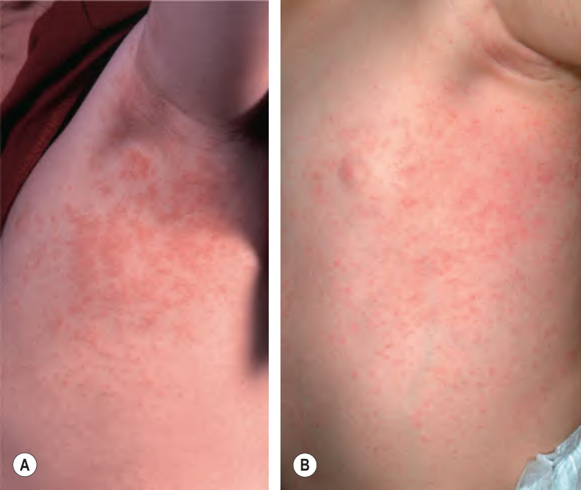
Fig. 81.9 Unilateral laterothoracic exanthem. Erythematous macules and papules involving the left axilla and upper flank (A) and a slightly more extensive distribution on the left lateral trunk (B).