GIANOTTI–CROSTI SYNDROME
Synonyms: Papular acrodermatitis of childhood Papulovesicular acrolocated syndrome
Key features
Symmetric papular eruption on the extremities, face, and buttocks
EBV and globally hepatitis B virus represent the most common triggers
Introduction and History
Gianotti–Crosti syndrome (GCS) was first described by Gianotti and Crosti in the 1950s. This childhood condition is characterized by the acute onset of a symmetric papular eruption on the face, extremities, and buttocks. In 1964, Gianotti and Crosti reported coexistent anicteric hepatitis, and in the 1970s an association with hepatitis B virus was described. Subsequent reports demonstrated that GCS can result from a variety of viral triggers (Table 81.3).
The term “papulovesicular acrolocated syndrome” was used by Gianotti in 1985 to distinguish a more vesicular variant that is characterized by a lack of hepatitis, more pruritus, and a prolonged course. However, differences in clinical manifestations are not generally related to the etiologic agent, and this variant is now included within the spectrum of GCS.
Epidemiology
GCS has a worldwide distribution and occurs most often in young children during the spring and early summer. In one large series, the age range was 6 months to 14 years, with a mean of 2 years.
Pathogenesis
GCS is regarded as a self-limited cutaneous response to various infections. Hepatitis B virus and EBV are the most frequently reported agents, with EBV representing the most commonly documented cause in the US. Other potential etiologic associations are summarized in Table 81.3. Although the pathogenic mechanisms in GCS are not clear,
there is some suggestion that immunizations or immune imbalance may enhance the risk of developing the exanthem following certain infections.
Clinical Features and Differential Diagnosis
GCS is often preceded by an upper respiratory syndrome with mild constitutional symptoms. The exanthem begins abruptly, with monomorphic, skin-colored to pink–red, edematous papules that are
symmetrically distributed on the face, buttocks, and extensor surfaces of the extremities (Fig. 81.10). In some patients, lesions may be limited to the face. The trunk is usually spared, although some extension onto the trunk does not exclude the diagnosis. Lesions tend to be asymptomatic and may occasionally be vesicular or purpuric. Systemic manifestations include a low-grade fever and lymphadenopathy (mainly inguinal and axillary) that can persist for a few months; hepatomegaly and splenomegaly occur less often.
The differential diagnosis of GCS may include a drug eruption, papular urticaria, other viral exanthems, erythema multiforme, and molluscum contagiosum. An inflammatory response to molluscum contagiosum, which often heralds clearance of the lesions, can present with an eruption of monomorphic erythematous papules that favor the elbows and knees; this “id” reaction-like phenomenon can resemble GCS but tends to be extremely pruritic and to respond to treatment with topical corticosteroids.
Pathology
The histopathologic findings are nonspecific and can include spongiotic and lichenoid changes as well as a lymphocytic vasculitis. Exocytosis of lymphocytes within the lower epidermis and increased numbers of cytotoxic T lymphocytes in the infiltrate may be observed.
Treatment
The treatment for GCS is supportive, and topical corticosteroids are often of little benefit. A thorough history and physical examination should be performed, and laboratory tests for hepatitis or specific viral agents (e.g. hepatitis B virus, EBV) should only be obtained if clinically indicated. Spontaneous resolution usually occurs within 3–6 weeks, although the eruption may occasionally persist for 8 weeks or longer.
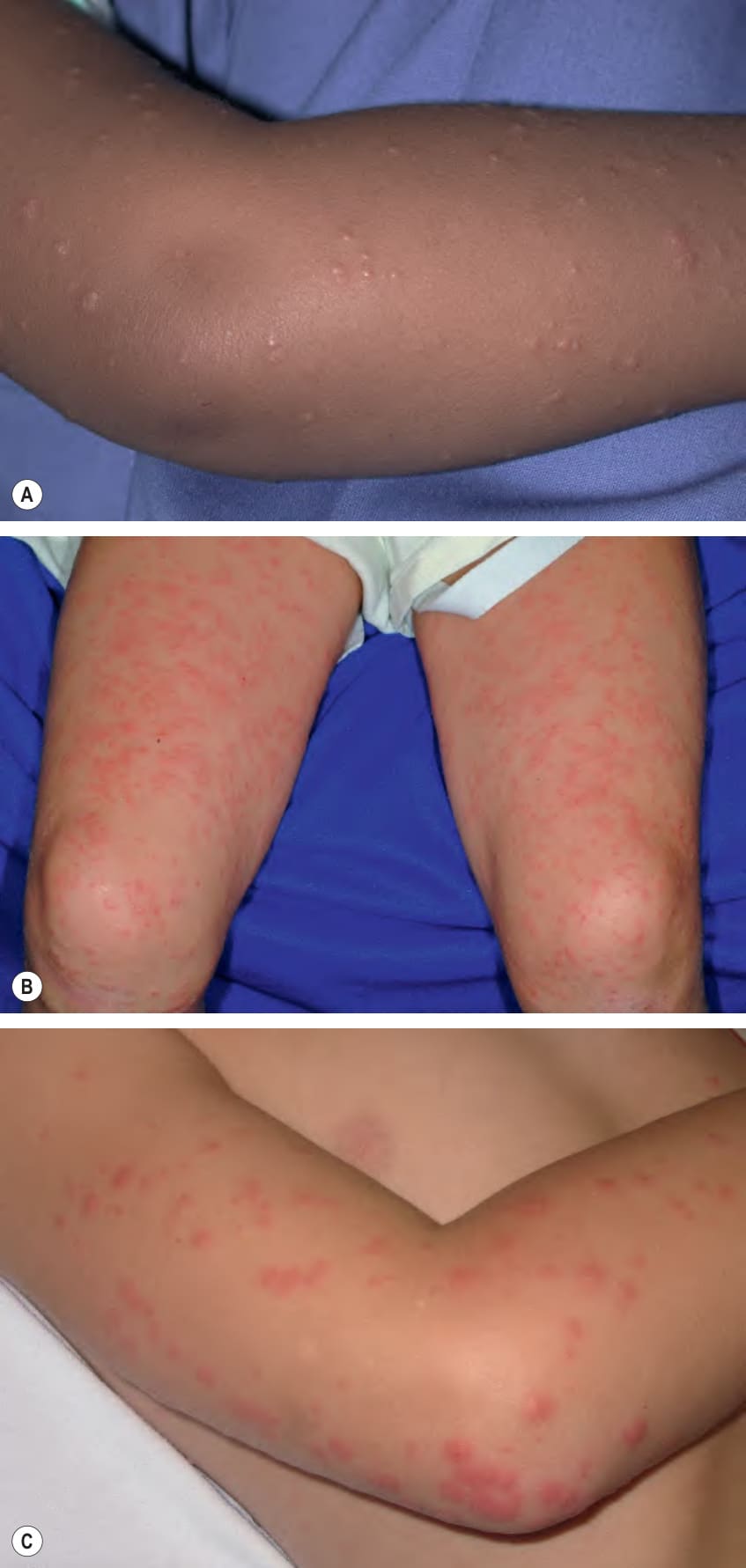
Fig. 81.10 Gianotti–Crosti syndrome.A Monomorphic, small erythematous papules on the elbow. B A more exuberant eruption of coalescing edematous, erythematous papules on the thighs and knees. C Larger edematous, erythematous papules on the elbow.
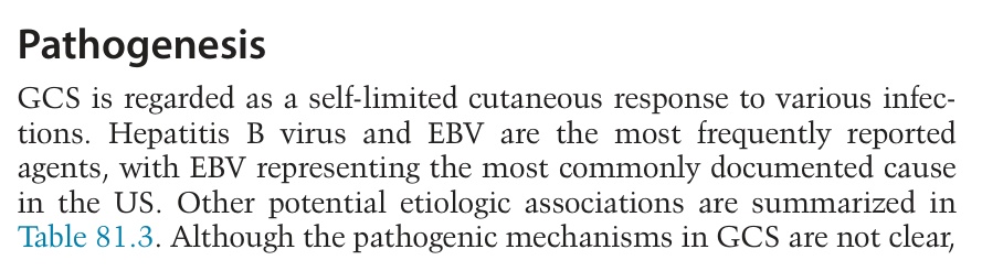
Table 81.3 Potential etiologies reported in association with Gianotti–Crosti syndrome. A pruritic Gianotti–Crosti syndrome-like eruption can also occur in patients with molluscum contagiosum (see text). Most common viral etiologies are in bold.