POXVIRUS INFECTIONS
Key features
Although the last reported case of smallpox was in 1978, it is still considered a bioterrorism threat
Molluscum contagiosum represents the most common poxvirus infection in humans
The family Poxviridae are large, brick- or ovoid-shaped, double-stranded DNA viruses that are characterized by cytoplasmic replication. Diseases caused by poxviruses include smallpox, vaccinia, cowpox, mpox (previously referred to as monkeypox), tanapox, orf, milker’s nodules, and molluscum contagiosum (Table 81.4). The most historically significant of these was smallpox, which at one time was endemic throughout the world and responsible for millions of human deaths. The last epidemic of smallpox in the US was in 1949, and in 1980, the World Health Assembly declared the world free from smallpox. However, two reference collections remain in the US and Russia. The infection that has clinical features most similar to smallpox is varicella; a comparison of these two conditions is provided in Table 81.5.
Because of concerns regarding smallpox being used as a bioterrorism agent, extensive recommendations have been adopted for handling of a suspected outbreak (emergency.cdc.gov/agent/smallpox/). The current strategy of ring vaccination involves isolation of patients with suspected infection and immunization of all identified contacts as well as their contacts. If given within 3–4 days following exposure, smallpox vaccination may prevent or reduce the severity of disease. If possible, first responders and healthcare workers who come into contact with a suspected patient should have previously been vaccinated. Tecovirimat and brincidofovir are both FDA-approved for the treatment of smallpox in adults and children (weighing ≥13 kg and including neonates, respectively). Recently, tecovirimat access was expanded to include patients with other orthopoxvirus infections, including mpox. Both of these agents as well as the JYNNEOS™ (Modified Vaccinia Ankara) vaccine were utilized in the 2022 international mpox outbreak (see Table 81.4).
With the eradication of smallpox, molluscum contagiosum (MC) became the only remaining poxvirus infection to specifically afflict humans. The MC virus (MCV) is a member of the Molluscipox genus of Poxviridae, and the MCV I and MCV II subtypes result in indistinguishable skin lesions. MC is a common, self-limited condition in children. It also occurs in adults, usually as a sexually transmitted disease, and in immunocompromised hosts, most notably HIV-infected individuals. Transmission is via skin-to-skin contact and, less commonly, fomites.
MC lesions are firm, umbilicated, pearly papules with a waxy surface. They can occur anywhere on the skin surface but are most common in skin folds (e.g. axillae, neck); on the lateral trunk, thighs, and buttocks; and in the genital region (Fig. 81.13A). Facial lesions can also occur and are often especially distressing to the patient (Fig. 81.13B). Widespread, large, and occasionally deforming lesions may be seen in the setting of immunosuppression, particularly AIDS (see Ch. 78).
An associated molluscum dermatitis is common (Fig. 81.13C), especially in children with atopic dermatitis. Inflammation of MC lesions can occur, often with a pustular or furuncle-like component (Fig. 81.13D); this signals the development of a host immune response. As noted above, an id reaction-like eruption of pruritic erythematous papules that favor the elbows and knees occasionally develops in conjunction with an inflammatory response to MC.
The differential diagnosis of MC may include adnexal tumors, verrucae, condylomata acuminata, basal cell carcinoma, juvenile xanthogranuloma, melanocytic nevi (especially Spitz nevi), papular granuloma annulare, and pyogenic granuloma. In immunocompromised hosts, infectious processes such as cryptococcosis or histoplasmosis can mimic MC. Inflammatory reactions to mollusca may resemble nummular dermatitis, staphylococcal furunculosis, Gianotti–Crosti syndrome, or unilateral laterothoracic exanthem (ULTE). Histologic evaluation of an MC lesion reveals large, intra-cytoplasmic inclusion bodies (molluscum bodies, Henderson– Patterson bodies) within epidermal keratinocytes, which increase in size as they move toward the skin surface (Fig. 81.13E). A variably dense dermal infiltrate may occasionally have pseudolymphomatous features.
MC resolves spontaneously in immunocompetent children, with the time interval between onset and clearance of the lesions ranging from several months to several years. However, treatment may be requested when there are numerous or cosmetically significant lesions. Therapeutic options include curettage, manual expression, cryotherapy, topical chemovesicants, keratolytics, and cidofovir (Table 81.6). Although imiquimod has been used anecdotally for MC, efficacy was not demonstrated in large controlled studies. In-office use of cantharidin was recently FDA-approved for the treatment of MC in individuals ≥2 years of age, with complete MC clearance in ~50% of patients after 4 applications at 3-week intervals (see Ch. 129). Although cantharidin has the benefit of non-traumatic application, it may require more visits than curettage. Berdazimer 10.3% gel is a nitric oxide-releasing agent shown in randomized controlled studies to result in complete MC clearance in ~30% of patients treated daily for 12 weeks. Treatment of molluscum dermatitis with a topical corticosteroid may help to reduce associated pruritus and prevent autoinoculation from scratching.
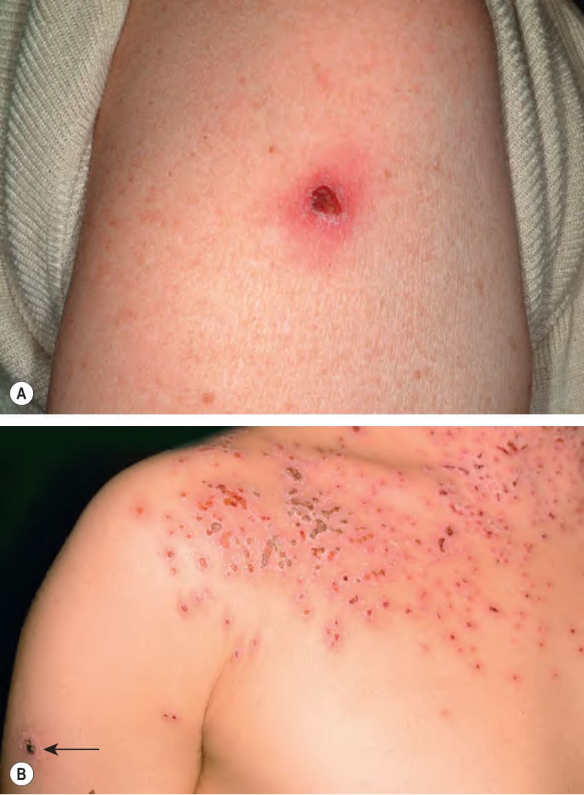
Fig. 81.11 Smallpox vaccine.A Crusted papule at the site of vaccination, 14 days following administration. B Eczema vaccinatum in a patient with atopic dermatitis. Spread from the vaccination site (arrow) to areas of eczema occurred due to the impaired skin barrier. C Erythema multiforme-like reaction associated with marked erythema and edema at the vaccination site. B, C, Courtesy Louis A. Fragola, Jr, MD.
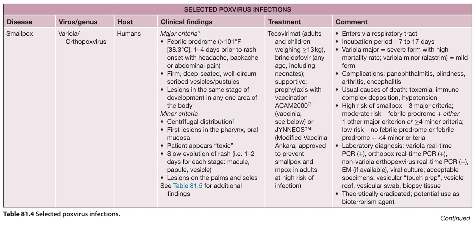
Table 81.4 Selected poxvirus infections. Continued
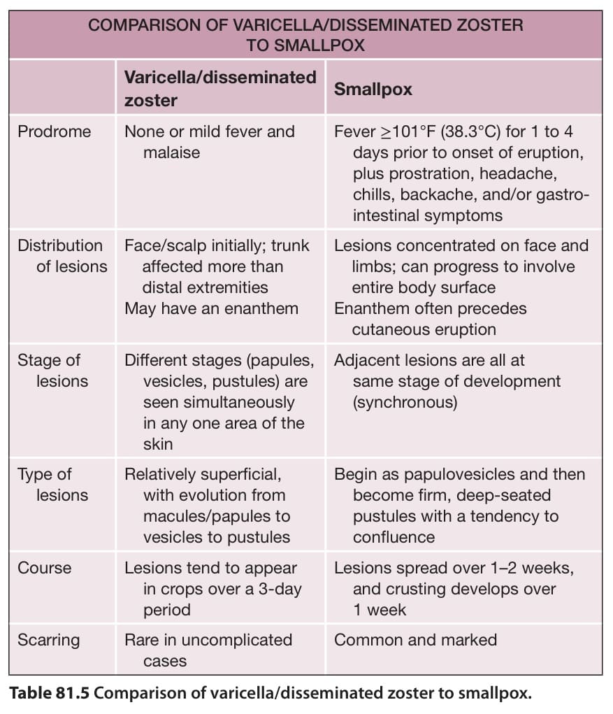
Table 81.5 Comparison of varicella/disseminated zoster to smallpox.
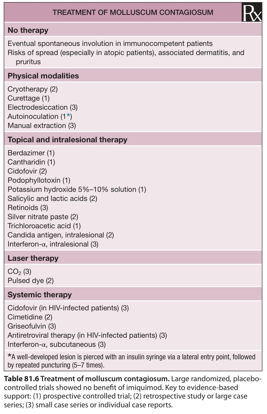
Table 81.6 Treatment of molluscum contagiosum. Large randomized, placebocontrolled trials showed no benefit of imiquimod. Key to evidence-based support: (1) prospective controlled trial; (2) retrospective study or large case series; (3) small case series or individual case reports.