POLYOMAVIRUS INFECTIONS
Synonyms: Trichodysplasia spinulosa: Viral-associated trichodysplasia (of immunosuppression) “Cyclosporine-induced” folliculodystrophy Follicular dystrophy of immunosuppression Pilomatrix dysplasia Trichodysplasia of immunosuppression
Human polyomaviruses (HPyVs) were initially described in 1971, when JC and BK viruses were identified in immunosuppressed patients with progressive neurodegenerative disease. HPyVs were subsequently linked to several skin disorders, beginning in 2008 with the discovery that most Merkel cell carcinomas are driven by the Merkel cell polyomavirus (see Ch. 115). Trichodysplasia spinulosa is an increasingly recognized entity that was first reported in 1999 and found in 2010 to be caused by the trichodysplasia spinulosa-associated polyomavirus (TSPyV). This condition occurs primarily in solid organ transplant recipients receiving immunosuppressive medications and patients undergoing chemotherapy for a leukemia or lymphoma. Pruritic and dyskeratotic dermatoses have also been linked to infections with HPyV6 and 7 in immunocompromised individuals (e.g. with solid organ transplantation or HIV infection). Most recently, HPyV9 infection was found to underlie widespread keratotic skin lesions and fatal pulmonary disease in solid organ transplant recipients.
Trichodysplasia spinulosa presents with numerous erythematous to skin-colored papules with a central spiny projection on the face (especially the mid portion), ears, and less often extremities and trunk (Fig. 81.15A). Associated alopecia of the eyebrows and eyelashes, or less often other facial and body hair, as well as thickening of the skin resulting in a leonine appearance may develop. Histologic evaluation is characterized by dilated anagen follicles with abnormal maturation and eosinophilic keratinocytes containing large trichohyaline granules. Large eosinophilic inclusions within affected keratinocytes show positive immunohistochemical staining for the polyomavirus middle T antigen, and electron microscopy demonstrates intranuclear icosahedral viral particles. The diagnosis can be confirmed by PCR-based viral detection in skin scrapings or a biopsy specimen. Trichodysplasia spinulosa may improve following reduction or discontinuation of immunosuppressive medications, and successful treatment with topical cidofovir, oral valganciclovir, and leflunomide has been described. Trichodysplasia spinulosa-like hyperkeratosis has also been described in a patient with basal cell nevus syndrome treated with vismodegib.
Pruritic and dyskeratotic dermatoses (PDDs) are characterized by scaly pink to gray–brown papules, patches, and/or plaques coalescing on the trunk and extremities, with associated pruritus and/or burning. Additional reported findings have included diffuse involvement with islands of sparing, alopecia, spiny perifollicular papules, and palmar pits or spicules (Fig. 81.15C). HPyV9 infection-associated eruptions presented with keratotic, pink–violet to brown papules and plaques that began acrally and spread to the proximal extremities and trunk;
keratotic spines and lesions resembling keratoacanthomas were noted in one patient. Histopathologically, both of these disorders are characterized by a “peacock plumage” pattern of dyskeratotic cells and irregular columns of parakeratosis. Successful treatment of PDDs with acitretin ± topical cidofovir has been described.
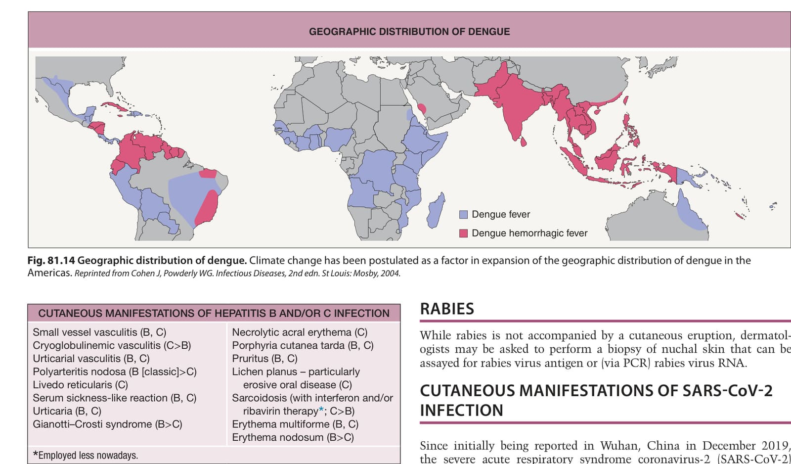
Fig. 81.14 Geographic distribution of dengue. Climate change has been postulated as a factor in expansion of the geographic distribution of dengue in the Americas. Reprinted from Cohen J, Powderly WG. Infectious Diseases, 2nd edn. St Louis: Mosby, 2004.
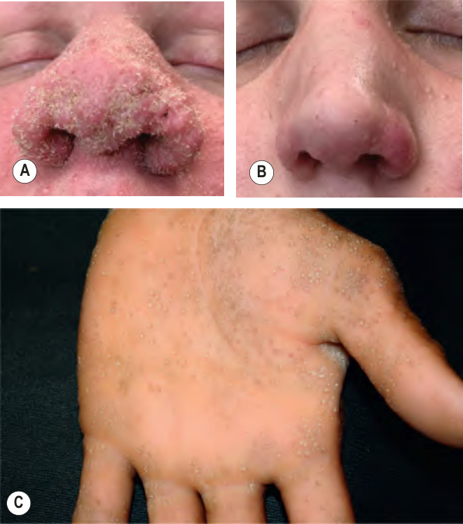
Fig. 81.15 Cutaneous polyomavirus infections.A Trichodysplasia spinulosa presenting as coalescing pink folliculocentric papules with central spiny projections on the nose. B Same patient, post-treatment with topical cidofovir. C Human polyomavirus 6 (HPyV6) associated spicules on the palm of a renal transplant recipient. A, B, Courtesy Stephen K. Tyring, MD, PhD; C, Courtesy Jeffrey Callen, MD.
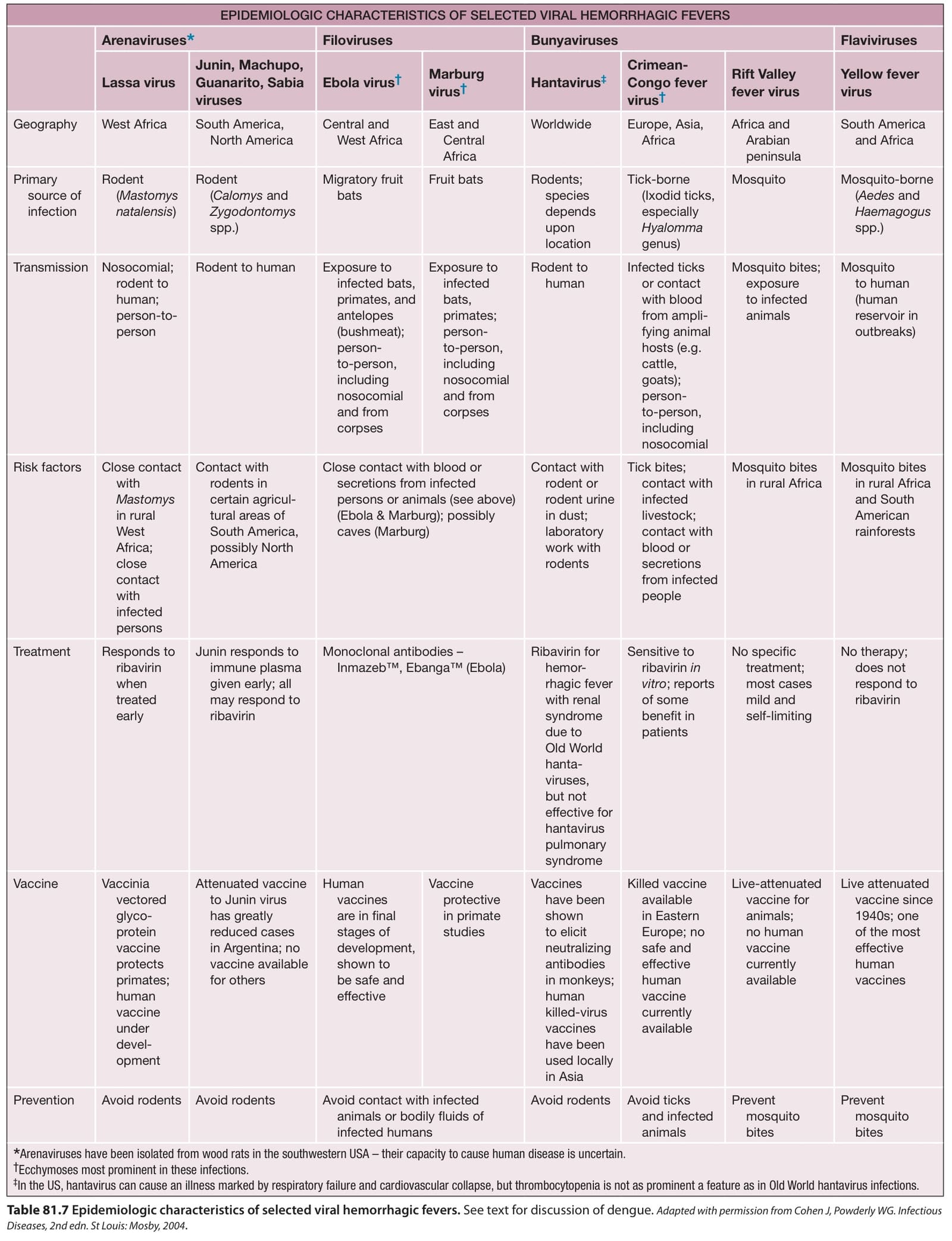
Table 81.7 Epidemiologic characteristics of selected viral hemorrhagic fevers. See text for discussion of dengue. Adapted with permission from Cohen J, Powderly WG. Infectious Diseases, 2nd edn. St Louis: Mosby, 2004
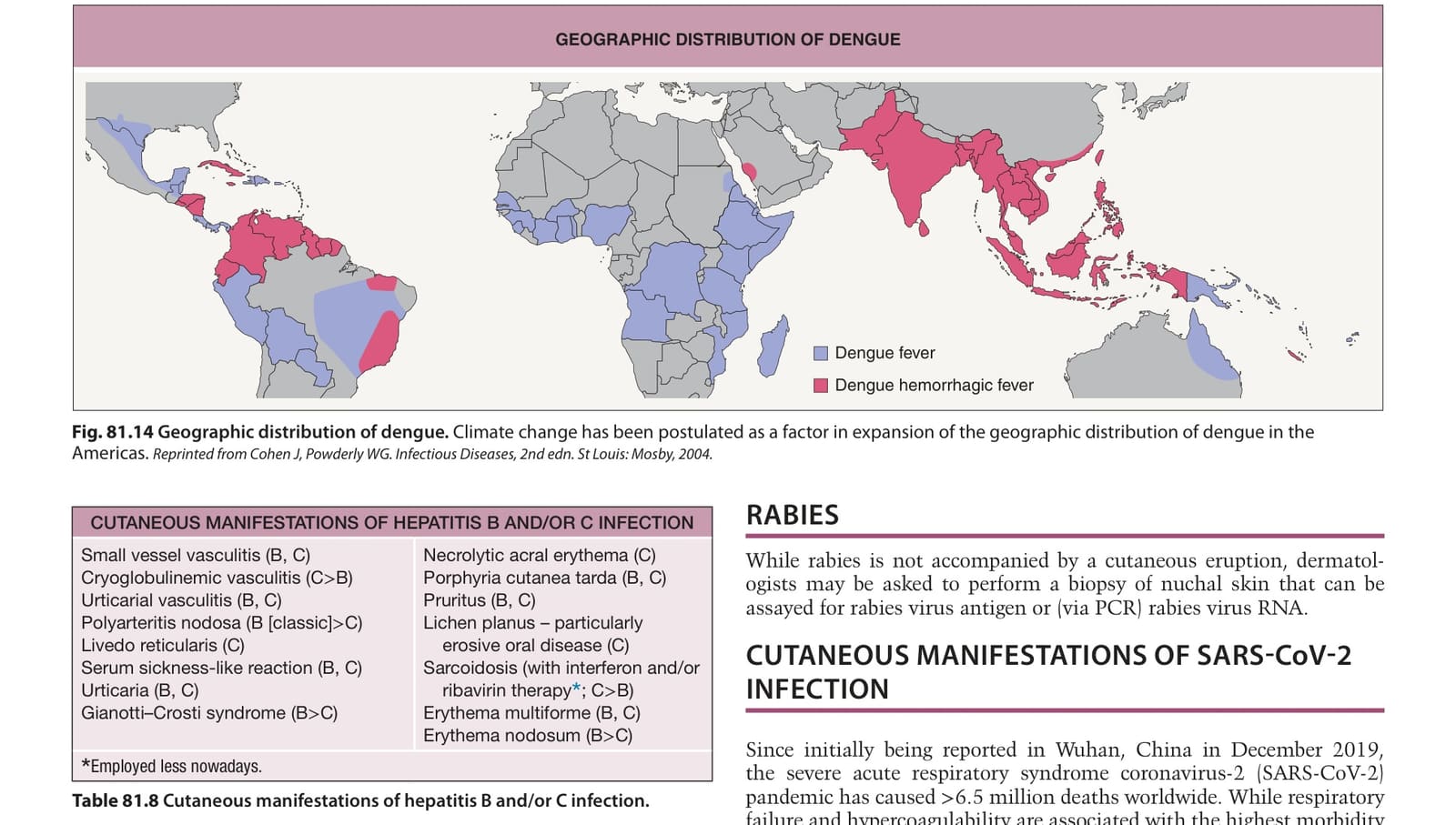
Table 81.8 Cutaneous manifestations of hepatitis B and/or C infection.