KAWASAKI DISEASE
Synonym: Mucocutaneous lymph node syndrome
Key features
Fever, conjunctival injection, oral mucosal changes, acral swelling, and cervical lymphadenopathy
Polymorphous exanthem, often with early perineal involvement
Represents the most common cause of acquired heart disease in children in the US
Introduction and History
Kawasaki disease (KD) is an acute febrile multisystem disease that primarily affects children ≤5 years of age. Initially described by Kawasaki in Japanese children, KD has subsequently been reported worldwide, and in the US it represents the leading cause of acquired heart disease in the pediatric population. Coronary artery aneurysms or ectasias develop in 15%–25% of untreated KD patients, resulting in substantial morbidity and mortality. The clinical manifestations of KD can mimic a variety of other diseases, in particular viral exanthems, and the need to promptly treat KD in order to prevent potentially lifethreatening complications makes differentiation among these entities essential.
2023 case definition and comparison with Kawasaki disease. CDC, Centers for Disease Control and Prevention; IL, interleukin; IFN, interferon; MAS, macrophage activation syndrome. Adapted from references 88 & 89
Epidemiology
The peak incidence of KD is in children ≤2 years of age, and 85% of patients with KD are ≤5 years of age; however, adults with KD have also been reported. The condition is 1.5 times more common in boys than in girls. A peak incidence in late winter to early spring has been observed in North America. Epidemics have occurred in both Japan and the US and are characterized by a wave-like spread over a 2- to 4-year period. KD is more common in children of Asian descent. Male sex and an age of <1 year or >6 years appear to be risk factors for the development of cardiovascular sequelae, which may be related to delayed diagnosis in the latter group.
Pathogenesis
The etiology of KD remains unknown, but several observations suggest an infectious cause, including the clinical features, seasonal variation in incidence, community outbreaks, rarity in infants under 6 months of age with potentially protective maternal antibodies, and infrequent recurrences. However, conventional microbial cultures and serologic studies have failed to identify a causative agent. A pathogenic role for a bacterial toxin that acts as a superantigen has been hypothesized; although this remains unproven, it is supported by the increased prevalence of superantigen-encoded genes in the stool of KD patients compared to other febrile or healthy children.
In fatal cases of KD, IgA-producing plasma cells have been found in the coronary arteries, pancreas, kidney, and upper respiratory tract; the latter finding is similar to that seen in children with fatal respiratory viral infections. Oligoclonal KD antibodies synthesized in vitro have been shown to bind to cytoplasmic antigen in the proximal bronchial epithelium, coronary arteries, and macrophages of KD patients. Furthermore, electron microscopic studies have provided evidence of virus-like particles within intracellular inclusion bodies in the ciliated bronchial epithelium of patients with KD. These findings are suggestive of a respiratory infectious agent with tropism for vascular tissue.
KD is characterized by marked activation of the immune system, with increased numbers of activated lymphocytes (both T and B) and mononuclear cells as well as increased levels of proinflammatory cytokines. A functional polymorphism in the inositol 1,4,5-trisphos phate 3-kinase C gene (ITPKC) has been shown to be significantly associated with KD susceptibility and an increased risk of coronary artery lesions in children from the US and Japan. ITPKC acts as a negative regulator of T cell activation through the nuclear factor of activated T cells (NFAT) signaling pathway; the polymorphism, which reduces the efficiency of ITPKC splicing, may contribute to immune hyperreactivity in Kawasaki disease.
Clinical Features and Differential Diagnosis
The diagnostic criteria for KD include fever lasting at least 5 days plus the presence of four of the five following criteria:
●bilateral non-purulent conjunctival injection
●oropharyngeal changes including diffuse hyperemia, strawberry tongue, and lip fissures
●cervical lymphadenopathy (usually unilateral)
●peripheral extremity changes including erythema, edema, and eventual desquamation of the hands and feet
●a polymorphous exanthem. The diagnosis of KD should be strongly considered in any child presenting with prolonged, unexplained fever in association with skin findings. The fever in KD is usually high-spiking (>39°C; 102°F), and it may not respond to antipyretics.
A cutaneous eruption is seen in >80% of patients with KD. It is usually composed of macular and papular erythematous lesions, often in a morbilliform pattern, but may also be erythema multi-forme-like, urticarial, scarlatiniform, or even pustular (Fig. 81.16A,E). Petechiae and crusting are unusual findings and vesiculobullous lesions are rarely if ever seen. In some patients, ulceration at the site of a bacillus Calmette–Guérin (BCG) vaccination has been observed during the disease course. A characteristic early cutaneous finding is erythema of the perineum, which often desquamates within 48 hours (Fig. 81.16A–C). Edema and brawny induration of the hands and feet are also common early in the disease course (Fig. 81.16D), with eventual desquamation that is prominent in the periungual regions. Gangrene of the peripheral extremities occasionally occurs. Subacute cutaneous manifestations of KD can include a psoriasiform eruption and transverse orange–brown (pseudo)chromonychia thought to result from nail bed splinter hemorrhages.
The conjunctival injection of KD is typically bulbar with sparing of the limbus (an avascular zone around the iris); it is not associated with increased tearing or an exudate. Keratitis and photophobia are uncommon and should suggest an alternative diagnosis. Oropharyngeal changes consist of dry, fissured lips, a “strawberry tongue” (Fig. 81.17), and in some patients diffuse hyperemia of the oral mucous membranes.
Hemorrhagic crusting and erosions of the vermilion portion of the lips can also be seen in primary gingivostomatitis due to herpes simplex virus, pemphigus vulgaris, and paraneoplastic pemphigus. Histologic examination of the flaccid sterile bullae of staphylococcal scalded skin syndrome shows cleavage at or below the stratum granulosum (see inset).
Cardiac involvement can include myocarditis, pericardial effusions, congestive heart failure, and coronary aneurysms; the latter represent the source of greatest morbidity in KD. Valvular disease, usually mitral or aortic, may also occur. Tachycardia, murmurs, gallops or distant heart sounds may be noted on cardiac examination. Other possible sites of involvement include the CNS (extreme irritability, aseptic meningitis, cranial nerve palsies, sensorineural hearing loss), gastrointestinal tract (abdominal pain, diarrhea, hepatic dysfunction, obstructive jaundice, gallbladder hydrops), musculoskeletal system (arthralgias, arthritis) and the genitourinary tract (urethritis, meatitis, sterile pyuria).
“Incomplete” (“atypical”) KD may be diagnosed in patients with prolonged fever plus coronary artery disease detected by echocardiography or coronary angiography, but fewer than four additional criteria (see above). This presentation tends to be more common in infants, and the diagnosis may be difficult to establish. An algorithm for evaluation of suspected incomplete KD has been proposed and was updated in
2017. For children with fever of unknown etiology for ≥5 days who meet 2–3 KD clinical criteria and have an elevated ESR (≥40 mm/hour) and/or CRP level (≥3.0 mg/dl), echocardiography should be performed and treatment initiated if there are signs of cardiac involvement. Both echocardiography and (regardless of the findings) treatment are recommended for those individuals with persistent fever, 2–3 KD clinical criteria, and elevated acute phase reactants who meet ≥3 supplemental laboratory criteria. The latter include hypoalbuminemia, anemia, elevated alanine aminotransferase, white blood cell count ≥15 000/mm (often with neutrophilia), thrombocytosis after 7 days of illness, and sterile pyuria; thrombocytopenia is occasionally observed, although not included as a criterion. Because infants <6 months of age meet KD diagnostic criteria less often and have a greater risk of coronary artery disease, echocardiography is recommended for all patients in this age group with fever of unknown etiology for ≥7 days and an elevated ESR and/or CRP level.
Electrocardiographic findings associated with cardiac involvement in KD include decreased R-wave voltage, ST-segment depression, and T-wave flattening or inversion. Higher serum levels of N-terminal pro-brain natriuretic peptide (NT-proBNP) are associated with increased risk of coronary artery disease and IVIg resistance, and NT-proBNP may also have utility as a diagnostic marker in young infants. Macrophage activation syndrome is a rare complication of recalcitrant KD that presents with hepatosplenomegaly, cytopenias, hypofibrinogenemia, and marked hyperferritinemia (see Table 91.1).
Although ~25%–50% of patients diagnosed with multisystem inflammatory syndrome in children (MIS-C) also meet criteria for KD, the two entities have distinct epidemiological, immunological, and clinical features (see Table 81.10). The differential diagnosis of KD also includes viral exanthems (e.g. adenoviral, enteroviral, EBV, measles), scarlet fever, toxic shock syndrome, staphylococcal scalded skin syndrome, erythema multiforme, drug eruptions, serum sickness-like reaction, recurrent toxin-mediated perineal erythema, systemic juvenile idiopathic arthritis, annular erythema of infancy, infantile polyarteritis nodosa (cutaneous arteritis), and periodic fever syndromes. Fig. 81.17 shows the facial findings that differentiate KD from staphylococcal scalded skin syndrome and erythema multi-forme major.
Pathology
Histologic findings are nonspecific and include dermal edema and a perivascular mononuclear infiltrate.
Treatment
During its acute phase, the treatment for KD is aimed at decreasing inflammation, and the current first-line therapy is IVIg administered in a single infusion of 2 g/kg over 8–12 hours. This regimen was demonstrated to be more efficacious in preventing coronary aneurysms than the prior recommendation of multiple daily infusions. However, the mechanism of action of IVIg in the treatment of KD is unknown. With the utilization of IVIg, the incidence of coronary aneurysms decreases from 25% to ≤5%–10%, and mortality from 2% to 0.3%. Aspirin is also recommended during the acute phase, with initial administration of 30–100 mg/kg daily (maximum 4 g/day) divided into four doses for 14
days or until afebrile for 48–72 hours, followed by a maintenance dosage of 3–5 mg/kg daily, usually for 6 to 8 weeks.
Approximately 10%–20% of children with KD have persistent or recrudescent fevers after receiving a single dose of IVIg, and these individuals have a greater risk of coronary artery disease. A second dose of IVIg may be given to patients who fail to become afebrile within 24–48 hours after completion of the first dose. Management options for high-risk or refractory KD include corticosteroids and infliximab administered along with or following failure of IVIg therapy, respectively. One large randomized, double-blind trial found that initial treatment with pulsed corticosteroids plus conventional therapy (IVIg and aspirin) showed no benefit over conventional therapy alone in terms of hospital days, days of fever, rates of retreatment with IVIg, or adverse events. However, another randomized controlled study found that the addition of pulsed corticosteroids to initial IVIg therapy in patients at high risk of having refractory KD was associated with fewer days of fever and a lower likelihood of coronary artery dilation. Randomized controlled studies have shown that infliximab is well tolerated and equally or more effective than a second dose of IVIg in pediatric patients with IVIg-resistant KD, although it did not decrease the likelihood of resistant disease when given as initial therapy together with IVIg and aspirin.
Long-term cardiac follow-up of KD patients is recommended, as adults with a history of KD may have vascular endothelial dysfunction and an increased risk for early-onset atherosclerosis.
Additional figures available in our eBook (see inside front cover for access code).
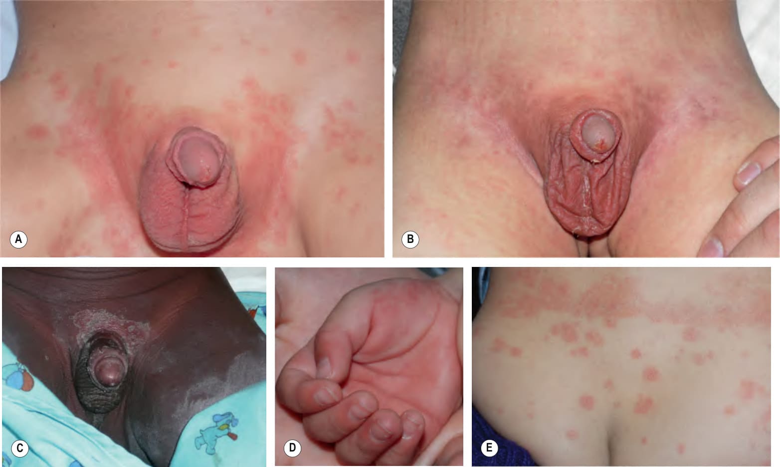
Fig. 81.16 Kawasaki disease. Erythema multiforme-like lesions becoming confluent in the perineal region on the second day of fever (A), followed by desquamation 2 days later (B). C Erythema (less evident due to the patient’s darkly pigmented skin) and desquamation in the genital area. Involvement of the skin in this location is a characteristic early finding in Kawasaki disease. D Erythema and edema of the palm early in the disease course. E Pink papules coalescing along the waistline. A, B, D, E, Courtesy Julie V. Schaffer, MD.
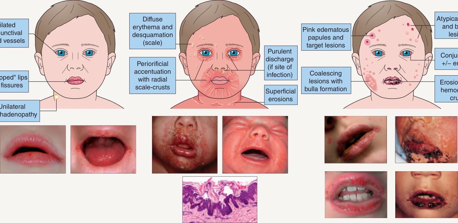
Fig. 81.17 Facial findings in Kawasaki disease, staphylococcal scalded skin syndrome, and erythema multiforme major/Stevens–Johnson syndrome.
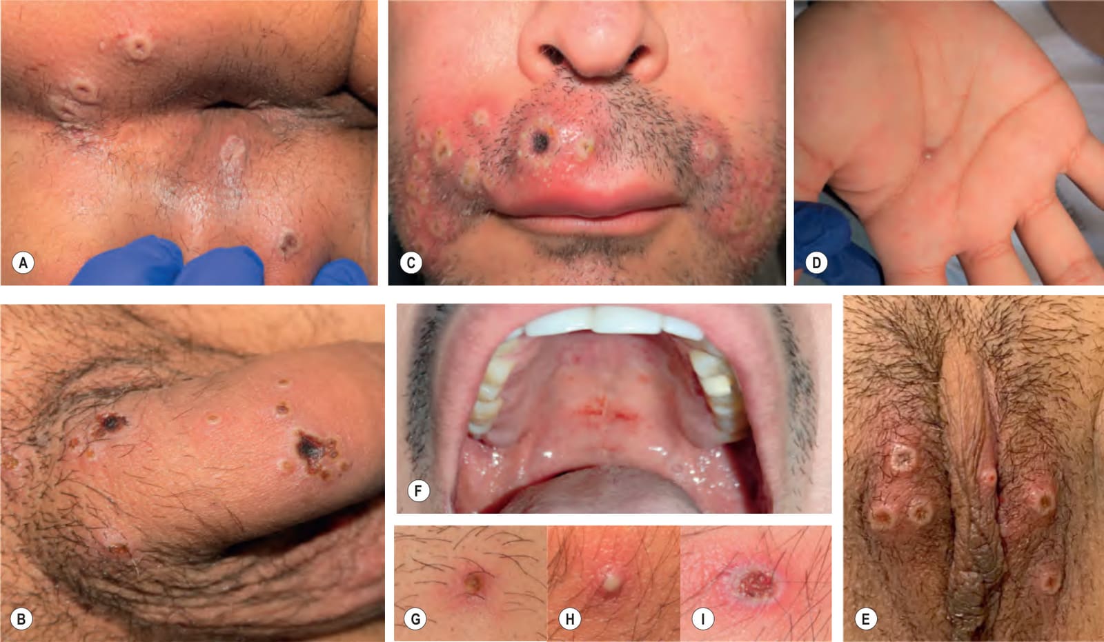
Fig. 81.18 Clinical spectrum of mpox.A-E Umbilicated vesicles, some of which have become necrotic with central hemorrhagic crusts, clustered in the oral and anogenital regions; lesions can also be randomly scattered. A scalloped border is occasionally seen as in herpes simplex viral infection (B). F Intraoral erosions. G-I Primary lesions vary from edematous erythematous papules to pustules to vesicles with a central hemorrhagic crust. Courtesy, Luis Requena, MD.
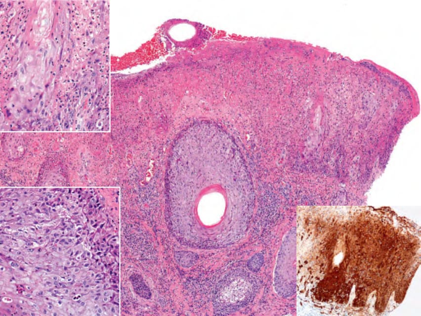
Fig. 81.19 Histopathologic findings in mpox. In addition to epidermal necrosis, hemorrhage, and erosion, there is a brisk lymphohistiocytic infiltrate in the dermis admixed with neutrophils. Note the viral changes (enlarged cells with pale cytoplasm and scattered apoptosis) in the keratinocytes of the epidermis (left upper inset) and hair follicle (left lower inset). Immunohistochemical staining for orthopox antigens can assist in diagnosis (right lower inset). Courtesy, Luis Requena, MD.
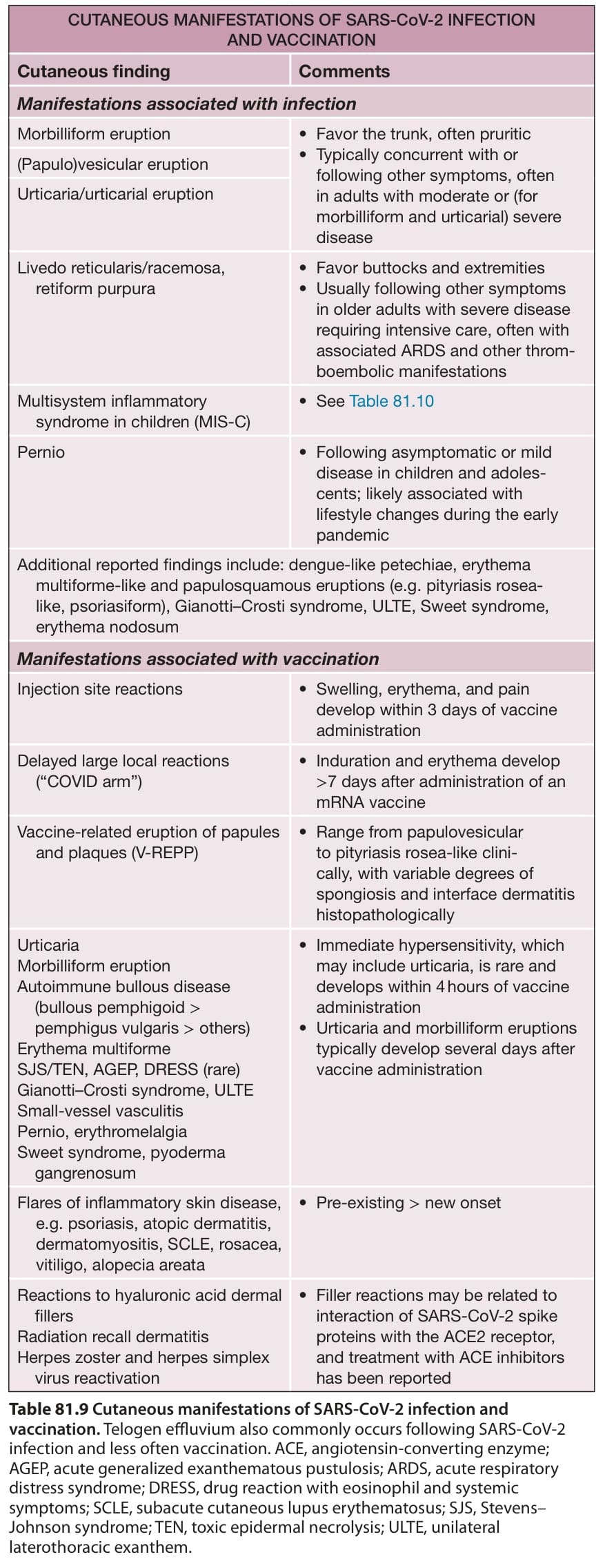
Table 81.9 Cutaneous manifestations of SARS-CoV-2 infection and vaccination. Telogen effluvium also commonly occurs following SARS-CoV-2 infection and less often vaccination. ACE, angiotensin-converting enzyme; AGEP, acute generalized exanthematous pustulosis; ARDS, acute respiratory distress syndrome; DRESS, drug reaction with eosinophil and systemic symptoms; SCLE, subacute cutaneous lupus erythematosus; SJS, Stevens– Johnson syndrome; TEN, toxic epidermal necrolysis; ULTE, unilateral laterothoracic exanthem.
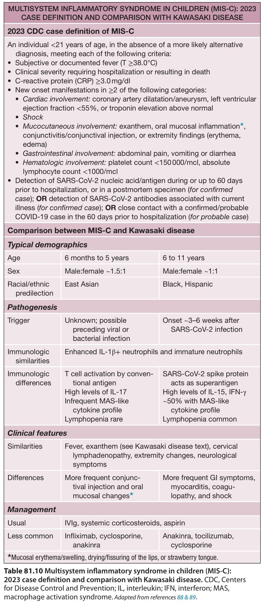
Table 81.10 Multisystem inflammatory syndrome in children (MIS-C):