SYPHILIS
Synonym: Lues
Key features
Sexually acquired, chronic infection caused by Treponema pallidum that is characterized by a variety of clinical manifestations and involvement of multiple organ systems
Can also be transmitted before birth (congenital)
Intermittently active disease with primary, secondary, and tertiary stages as well as a latent period of variable length that occurs before the onset of tertiary syphilis
Mucocutaneous manifestations vary from genital ulcers to widespread papulosquamous eruptions to granulomatous nodules
Syphilis facilitates the transmission of HIV, especially in countries with a high rate of both infections
Overall, the incidence of syphilis and other STIs has increased substantially in high-income countries after the introduction of antiretroviral therapy and pre-exposure prophylaxis for HIV
History
Syphilis has been of great historical importance and has played a major role in medicine for centuries. The disease was named after an afflicted shepherd named Syphilus in 1530. Theories on the origin of the disease in the late fifteenth century are controversial. One theory proposes that Christopher Columbus and his crew acquired the disease from Native Americans living in the Caribbean islands and brought it back to a nonimmune population in Europe in 1493, as signs of syphilis have been found in skeletons of pre-Columbian Native Americans. Another theory is that venereal syphilis may have already been endemic but became more widespread and severe as a consequence of the wars
at that time in Europe. Lastly, the environmental theory claims that venereal syphilis is a variant of other diseases caused by Treponema spp. and became modified by environmental factors, especially temperature.
Information on the natural course of untreated syphilis has come primarily from two large studies (Fig. 82.1). The Oslo Study was a prospective investigation of infected, but untreated, individuals conducted between 1890 and 1910, with a follow-up period of 50 years. Approximately one-quarter of infected persons had at least one additional relapse of secondary syphilis, often occurring during the first year (90%). Approximately 15% of infected persons developed benign tertiary syphilis with gummas of the skin (70%), bones (10%), and mucous membranes (10%). Cardiovascular syphilis was reported in ~14% of men and 8% of women, while neurosyphilis was observed in 10% of men and 5% of women. Altogether, the study concluded that 17% of men and 8% of women died as a result of untreated syphilis. The Tuskegee Study was undertaken in 1932 in infected Black men, with regular examinations for short- and long-term consequences of untreated infection. This represented a highly unethical investigation, as penicillin treatment was withheld without written consent. The principal finding was an increased mortality rate in the syphilitic group compared to the controls, with an ~20% loss-of-life expectancy at an interval of 12 years. Specific lesions of late syphilis were found in about 14% of infected men at the 20-year evaluation, and 12% after 30 years, with cardiovascular syphilis and neurosyphilis representing the primary causes of death. The unethical aspects of this study had serious social implications, contributing to mistrust in the healthcare system amongst African-Americans.
Epidemiology
Syphilis is distributed worldwide and is particularly problematic in low-income countries, where it is a leading cause of genital ulcer disease. Worldwide, the rates of primary and secondary syphilis decreased dramatically with the introduction of penicillin treatment after the Second World War. In contrast to Western European countries, an increase in the infection rate was observed during the late 1980s in rural southern and urban regions of the US. Although the number of syphilis cases per year in the US subsequently declined and in 2000 fell to its lowest point since reporting began in 1941, the number of primary and secondary syphilis cases diagnosed per year in men has subsequently increased more than sevenfold (Fig. 82.2). According to the Centers for Disease Control and Prevention (CDC), the incidence rate of primary and secondary syphilis in 2019 in the US was 20.1 cases per 100 000 population in men and 3.9 cases per 100 000 population in women. In the US, the incidence in Hispanic and Black individuals is 2- to 5-fold higher than in other population groups, but the highest risk group is men who have sex with men (MSM) as they accounted for nearly 60% of cases of primary and secondary syphilis in 2019.
With the resurgence of syphilis in Eastern Europe in the 1990s and migration of sex workers from this region to Western Europe, syphilis has also been observed more frequently in Western European countries over the past few decades.
Biology of T. pallidum
T. pallidum is a member of the genus Treponema of the order Spirochaetales, and it was identified in 1905 by Schaudinn and Hoffmann (Table 82.2). The characteristics of this microorganism are outlined in Table 82.3. Fig. 82.3 represents a darkfield photomicrograph of T. pallidum.
Pathogenesis of Untreated Syphilis
Syphilis is a chronic systemic infection that progresses through active and latent stages (Fig. 82.4). Inoculation and penetration occur via mucosal surfaces and abraded skin, followed by attachment to host cells and multiplication of the microorganism. Within a few hours, treponemes disseminate to the regional lymph nodes and internal organs.
Primary stage
The primary lesion develops 10–90 days after infection (median of 3 weeks) as an indolent papule, followed by surface necrosis and the
typical well-circumscribed ulceration that is firm to palpation (chancre). It is accompanied by enlarged regional lymph nodes. Histopathologically, the microorganism is observed among T cells that produce interferon-γ and interleukin-17, which lead to macrophage activation and destruction of a large number of treponemes. Several pathogenic mechanisms have been proposed, including an antigenically inert treponemal cell surface, resistance to phagocytosis, and premature down-regulation of the local host immune response.
Secondary stage
The secondary stage is characterized by dissemination and multiplication of the microorganism in different tissues, either simultaneously with the primary local lesion or up to 6 months after the primary lesion has healed. This stage follows primary syphilis in almost every patient who does not receive appropriate treatment. Circulating immune complexes (which contain treponemal outer membrane proteins), human anti-fibronectin antibodies, and complement are present during this stage of the disease and play a role in the pathogenesis of the different types of lesions.
Secondary syphilis is characterized by a broad spectrum of cutaneous manifestations as well as systemic signs such as malaise, fever, and generalized lymphadenopathy. It lasts several weeks or months, with relapses in ~25% of patients. Pregnant women can infect a fetus via transplacental passage of the microorganism.
Latency
Latency is the period between healing of the clinical lesions and appearance of late manifestations, and it can last for many years. About 70% of untreated individuals will remain in this stage for the rest of their lives and are immune to new primary infection (see Fig. 82.1). Latent syphilis is divided into early (1 year or less) and late (more than 1 year) subsets, and it is characterized by positive serologic tests for specific antibodies (see below) without clinical signs or symptoms. Infectivity may occur intermittently due to the presence of treponemes in the bloodstream, and pregnant women with latent syphilis may infect the fetus in utero.
Tertiary stage
The tertiary stage is also called late syphilis and is characterized by the presence of a small number of organisms and a high cellular immune reactivity against the organism. Signs of late syphilis can be recognized in approximately one-third of untreated individuals several months to years after being infected with treponemes (see Fig. 82.1). The microorganisms may invade the central nervous and cardiovascular systems as well as the skin and other organs (e.g. bones), leading to damage related to host delayed-type hypersensitivity responses, which produce local inflammation and gummas in affected tissues.
Syphilis and HIV
Syphilis and other STIs that produce genital ulcers further increase the risk of acquiring HIV. Reasons for the increased risk of HIV transmission include:
●lack of an epithelial barrier due to ulceration of the skin or mucous membranes
●large numbers of macrophages and T cells with receptors for HIV
●production of cytokines by macrophages stimulated by treponemal lipoproteins. In addition, syphilitic manifestations are altered in HIV-positive patients, with a higher likelihood of neurologic findings and (in those with secondary syphilis) ulcerative lesions.
Clinical Features
Syphilis is usually sexually acquired, but maternal–fetal transmission also occurs and can result in congenital syphilis. Syphilis is an inter-mittent disease with primary, secondary, and tertiary stages as well as a latent period of variable length (divided into early and late subsets) that precedes the onset of tertiary syphilis (see Fig. 82.4). The definitions of early and late syphilis by the CDC and World Health Organization (WHO) differ slightly. Early syphilis includes the primary and secondary stages as well as early latency (CDC: acquired <1 year previously; WHO: acquired <2 years previously). Late syphilis extends from late latency (CDC: acquired >1 year previously; WHO: acquired >2 years previously) through the tertiary stage.
Primary syphilis
The chancre usually presents as a single, indolent, round or oval, indurated ulcer (Fig. 82.5) that is associated with regional adenopathy. Some patients report a preceding painless papule that enlarged and ulcerated a few days later. The time of onset ranges from 10 to 90 days (average 3 weeks) after T. pallidum exposure. Untreated, chancres heal within a few weeks (Fig. 82.6).
Asymptomatic infections are commonly due to unrecognized chancres, especially when located in the cervical region in women; in these cases, syphilis is more frequently diagnosed during the secondary stage. The same applies to chancres located in the anal, perianal, or rectal areas (see Fig. 82.5D), seen more commonly in MSM. The mechanism of spontaneous healing without treatment is not well understood and seems to depend on local immunity.
Laboratory diagnosis of primary syphilis includes the following:
●Detection of T. pallidum DNA via PCR from a swab of a chancre is the most sensitive and specific method.
●Darkfield microscopic examination of fluid from the surface of the chancre is also a sensitive, and for the genital region a specific, method for confirming the presence of T. pallidum (see Fig. 82.3).
●Immunohistochemical staining of biopsy specimens (or Warthin- Starry staining if immunohistology not available).
●Antibodies to cardiolipin are present in ~80% of individuals at the onset of clinical symptoms. Cardiolipin, a component of mammalian cells, seems to be modified by treponemes such that antibodies are produced by the infected person and can be measured by the rapid plasma reagin (RPR) or Venereal Disease Research Laboratory (VDRL) assay.
●Antibodies to surface proteins of T. pallidum as detected by hemagglutination assays (T. pallidum hemagglutination test [TPHA], microhemagglutination assay for antibodies to T. pallidum [MHA-TP]), or fluorescent treponemal antibody absorption (FTA-ABS) assay are present in 90% of patients with clinical symptoms of primary syphilis. Since antibodies usually remain positive for life, a differentiation between primary syphilis and a prior infection may not be possible and PCR or darkfield examination should be undertaken.
Secondary syphilis
The secondary stage of the disease results from the hematogenous and lymphatic dissemination of treponemes after a few weeks or months (3–10 weeks; Fig. 82.7). It is characterized by recurrent disease activity,
with mucocutaneous as well as systemic manifestations. Prodromal symptoms include low-grade fever, malaise, sore throat, adenopathy, myalgias, weight loss, and sometimes a headache from meningeal irritation (Table 82.4).
During the second stage of syphilis, the most commonly observed clinical presentation (80%) is a generalized, non-pruritic papulosquamous eruption (Fig. 82.8). Lesions can range from 1–2 mm to 15–20 mm in diameter, and they vary in color from pink to violaceous to red–brown. Mucosal lesions range from small, superficial ulcers that resemble painless aphthae to large gray plaques (Fig. 82.9A,B). Condylomata lata are often observed in the moist regions of the anogenital area due to local spreading of the microorganisms (Fig. 82.9C,D). Lymph node enlargement is present in the majority of patients. Focal neurologic findings occasionally occur as does bone pain.
Additional cutaneous presentations of secondary syphilis include annular or figurate plaques with central hyperpigmentation on the face (Fig. 82.10A), non-scarring “moth-eaten” alopecia, split papules at the oral commissures (Fig. 82.10B), granulomatous nodules and plaques (Fig. 82.10C,D), and crusted necrotic lesions (Fig. 82.10E). Malignant syphilis (lues maligna) is extremely rare; the disseminated lesions resemble primary chancres (Fig. 82.10F).
Without treatment, the lesions resolve over several weeks to months. Relapse occurs in ~20% of patients within 1 year, often with mucosal or mucocutaneous manifestations in the anogenital area.
Laboratory diagnosis of secondary syphilis includes the following:
●Presence of treponemes by darkfield examination of serous exudates from localized lesions of the skin and mucous membranes (the exception being the oral cavity; see below).
●Serologic tests are more useful in secondary, as compared to primary, syphilis. Cardiolipin antibodies (e.g. RPR or VDRL tests) as well as specific antibodies are always positive in patients with secondary syphilis; a mistakenly or temporarily negative non-treponemal test rarely occurs due to the prozone phenomenon or HIV infection, respectively (see Table 82.8).
●Immunohistochemical staining of biopsy specimens (or Warthin- Starry staining if immunohistology not available).
Widespread exanthem of pink papules (A), subtle minimally inflamed lesions localized to the arms in an HIV-positive man (B), and generalized papulosquamous lesions (C). Genital lesions can resemble psoriasis (D) while lesions on the palms and soles (E, F) often have a collarette of scale. B,D, Courtesy Kalman Watsky, MD.
●Spirochetes can be detected by immunohistochemistry in the majority of biopsy specimens (see Fig. 0.33). Management considerations for patients diagnosed with secondary syphilis include the following:
●All patients should be tested for HIV infection, and testing should be repeated in those who fail to respond to treatment.
●Patients with ocular symptoms (e.g. photophobia, visual changes) should undergo ophthalmologic evaluation, including slit-lamp examination, to assess for uveitis/iritis, neuroretinitis, and optic neuritis.
●A lumbar puncture and cerebrospinal fluid (CSF) analysis should be performed in patients with ocular or neurologic (e.g. headache, hearing loss, cranial neuropathies) signs/symptoms and when treatment failure is suspected (e.g. persistent/recurrent signs/symptoms or failure of non-treponemal test titers to decline fourfold within 6–12 months).
●In patients with symptoms of otosyphilis (e.g. hearing loss, vertigo), an otologic examination is needed. However, CSF evaluation in patients with otosyphilis does not assist in clinical management, so lumbar puncture is not recommended.
●Patients with HIV disease are at increased risk for neurosyphilis, especially if they have a CD4 count <350 cells/ml and/or an RPR titer of ≥1 : 32; however, unless neurologic symptoms are present, CSF examination in HIV-positive patients has not been associated with improved clinical outcomes.
Latent syphilis
After a period of 3–12 weeks, untreated secondary syphilis typically resolves spontaneously, leaving the patient in an asymptomatic state. Diagnosis is based on a positive syphilis serology test result in the setting of no clinical evidence of treponemal infection. The two stages are:
●Early latent stage: within 1 year of the onset of latency (CDC definition), with the possibility of recurrence of the disease. About 90% of secondary relapses occur within the first year.
●Late latent stage: after 1 year from the onset of the disease (CDC definition); relapses after 1 year are very infrequent. This disease state may last for many months or years. Experience regarding the outcome of late latent syphilis in the absence of treatment is based upon several studies. About one-third of infected individuals have a non-reactive RPR test, no sign of reactivation during the remainder of their lives, and only the specific antibody assays (e.g. MHA-TP, FTA-ABS) remain positive (see Fig. 82.1). Another one-third of patients remain clinically asymptomatic for the rest of their lives, but antibodies against cardiolipin (e.g. RPR, VDRL) persist together with a positive MHA-TP or FTA-ABS assay. In the remaining one-third, clinical symptoms of tertiary syphilis occur.
Diagnosis of latent syphilis is established by reactivity in an RPR or VDRL assay, a positive MHA-TP or FTA-ABS test, and the absence of any clinical signs of the disease. The diagnosis is easy to establish, if there is a history of a chancre or skin manifestations that fit the clinical picture of syphilis. In many cases, however, the medical history provides no useful information regarding the presence of clinical symptoms in the previous weeks to months or whether the patient was previously treated. In such instances, differentiation between early and late latency is not possible and patients are regarded as having late latent syphilis from a management standpoint (see below). Response of latent syphilis to treatment is indicated by a decline in the RPR or VDRL titer. Persistently high (e.g. lack of a fourfold decline within 12–24 months) or rising titers are a sign of treatment failure, and examination of the CSF should be performed.
Tertiary (late) syphilis
Classic tertiary syphilis, as a consequence of untreated syphilis, is nowadays rare. It has a variable range of manifestations that appear months to years after initial infection (Fig. 82.11). In particular, these include involvement of the skin, bones, CNS, heart, and great vessels. Approximately one-half of patients with tertiary syphilis have “benign” late syphilis with the development of gummas, about one-quarter develop cardiovascular manifestations, and one-quarter develop neurologic symptoms (see Fig. 82.1).
In tertiary syphilis, non-treponemal serologic tests are usually positive with high titers. Patients with tertiary syphilis (or any symptoms related to late syphilis) should undergo lumbar puncture and CSF examination to determine if neurosyphilis is present before therapy is initiated.
The most common feature of late syphilis is gummas, which are locally destructive lesions in the skin, bones, liver, and other organs. The gummas in the skin are nodular or noduloulcerative lesions, often with an arciform pattern (Fig. 82.12). They remain for weeks to months
Adapted from Fritsch P, Zangerle R, Stary A. Venerologie. In: Fritsch P (ed.). Dermatologie und Venerologie. Berlin: Springer, 2004:865–86.
and then involute with scarring, but eventually new nodules reappear. With treatment, resolution of the lesions is prompt and complete. A solitary gumma that is located subcutaneously may become necrotic, similar to a “cold” abscess, resulting in ulceration of the skin or mucous membranes as well as destruction of underlying bones.
Bones are affected as frequently as the skin. Within the bone, a gumma is destructive and is typically accompanied by periostitis and osteitis. Clinical manifestations include pain, swelling, and limited range of motion. Other sites that can be affected by gummas include the tongue and oral cavity, upper respiratory tract, myocardium, and digestive as well as nervous systems.
Cardiovascular syphilis has a late onset, with a latent period of 15–30 years, and it occurs in about 8%–10% of individuals with untreated infection. During the early stage of the disease, T. pallidum demonstrates a predilection for the vasa vasorum of the proximal aorta and produces transmural inflammatory lesions, which lead to endarteritis of the vessels. The organisms then remain there in a latent state for many years.
Neurosyphilis
Although classically a manifestation of tertiary syphilis, neurosyphilis can occur at any stage of the disease. Neurosyphilis is the reflection of a chronic (or sometimes acute) form of meningitis, with vascular and parenchymatous sequelae in the cerebrum and spinal cord (Table 82.5). Asymptomatic neurosyphilis is defined by the presence of abnormalities in the CSF (see below), but in the absence of any neurologic symptoms or clinical findings. While in some individuals the CSF abnormalities may resolve spontaneously or persist into advanced age, asymptomatic neurosyphilis can progress to late symptomatic neurosyphilis.
The basic pathomechanism for cerebrovascular syphilis is an infarction secondary to syphilitic endarteritis; this can result in hemiparesis or hemiplegia. Parenchymatous neurosyphilis, including general paresis, dementia paralytica, and paretic neurosyphilis, is due to direct invasion of the cerebrum by treponemes. It is a rare and late form of neurosyphilis, with dementia and neurologic symptoms such as paralysis progressing, if untreated, until death occurs.
Tabes dorsalis, which affects up to a third of patients with neurosyphilis after a long latent period, has a number of typical clinical signs and symptoms, including diplopia, lightning pains (painful paresthesias, especially of the extremities), loss of vibratory and position sense, reduced reflexes in the legs, ataxia, sphincter dysfunction, visceral crisis (abdominal, rectal, and laryngeal pain), and Argyll Robertson pupils (abnormal pupillary light reflexes: loss of direct and consensual; retention of accommodation). Tabes dorsalis may burn out even without treatment.
In the mid 1970s, the first cases of so-called “atypical neurosyphilis” were described. With the subsequent recognition of co-infections with syphilis and HIV, it became apparent that neurosyphilis is common in HIV-infected individuals, and patients present with manifestations of early-onset neurosyphilis.
In addition to the clinical findings, the diagnosis of neurosyphilis (including acute syphilitic meningitis, meningovascular syphilis, and parenchymatous neurosyphilis) is based upon reactive blood and CSF antibody testing, which are almost always positive. CSF abnormalities include elevation of pressure, protein concentration, and immunoglobulin level as well as a mononuclear pleocytosis. The presence of specific antitreponemal antibodies in the CSF is mandatory, but it is not proof of the diagnosis of neurosyphilis, since IgG antibodies can diffuse into the CSF or result from contamination of the CSF by blood. Establishment of local antibody synthesis is possible by using the CSF-IgG index, obtained by dividing the CSF to serum IgG ratio by the CSF to serum albumin ratio. A result >0.7 is indicative of IgG synthesis in the brain due to local inflammation. The intrathecal T. pallidum
antibody index is calculated by dividing the TPHA titer in the CSF by the CSF to serum albumin ratio multiplied by a factor of 10, and an index >100 is indicative of the synthesis of treponemal-specific antibodies within the CNS. The presence of nonspecific antibodies, e.g. a positive VDRL test or RPR assay in CSF, is observed in most, but not all, cases, and a negative test does not exclude the presence of neurosyphilis.
Congenital syphilis
Congenital syphilis was initially described in 1497 and is therefore the oldest recognized congenital infection. In 1906, it was demonstrated via the Wassermann test that transmission of syphilis to the fetus required an infection in the pregnant woman.
There was an upsurge in congenital syphilis in Eastern Europe in the late 1990s, which paralleled the increased incidence of syphilis in adults and adolescents in this region. In the early 1990s, an increased frequency of congenital syphilis was also observed in some inner-city minority populations in the US. From 2015–2019, the number of reported cases of congenital syphilis in the US nearly quadrupled. The risk of mother-to-child transmission of untreated syphilis and its potential consequences are outlined in Table 82.6. Therefore, all women should be screened serologically for syphilis at the first prenatal care visit.
Infants generally present with symptoms during the neonatal period or within the first 3 months of life (Fig. 82.13). At the latest, they develop symptoms within the first 2 years of life. Typical manifestations are marasmic syphilis (i.e. cachexia) and skin lesions similar to those of acquired secondary syphilis (Fig. 82.14), except that they may be bullous (pemphigus syphiliticus) and tend to be more erosive. Additional clinical findings are “snuffles” (bloody or purulent mucinous nasal discharge), perioral and perianal fissures, lymphadenopathy, and hepatosplenomegaly. Skeletal involvement (i.e. osteochondritis) may result in pseudoparalysis of Parrot because of reduced movement of the extremities due to pain; other manifestations include anemia, thrombocytopenia, syphilitic pneumonitis (pneumonia alba), hepatitis, nephropathy, and congenital neurosyphilis.
Late congenital syphilis in a child or adolescent corresponds to tertiary syphilis in an adult and is not infectious. The stigmata represent the delayed consequences of localized inflammation at the sites of treponemal infection (Table 82.7). In about one-third of children, an interstitial keratitis is seen; this finding together with typical dental abnormalities (Hutchinson teeth) and neural deafness forms the Hutchinson triad.
A confirmed diagnosis of congenital syphilis requires laboratory demonstration of treponemes or child-specific antibodies. Serodiagnosis of congenital syphilis poses a difficult problem because IgG antibodies that are present may have been acquired transplacentally from the mother. A serum titer for a non-treponemal test that is fourfold higher than the mother’s titer is suggestive of infection, but infected neonates
may have lower titers. The detection of chromatographically separated 19S-antibodies in the FTA-ABS-19S-IgM test has a sensitivity of about 90%, similar to the IgM-capture ELISA. More recently, it has been shown that the detection of spirochetemia by PCR can improve the sensitivity of the diagnosis of congenital syphilis in neonates.
In late congenital syphilis, diagnosis is based on clinical findings in association with reactive serologic tests.
Laboratory Diagnosis of Syphilis
The diagnosis of syphilis is based on the direct detection of treponemes or treponemal DNA by microscopy or molecular biologic techniques as well as various serologic tests that assess antibody responses to either cardiolipin (non-treponemal tests) or treponemal antigens (treponemal tests). Diagnosis of the different stages of syphilis depends on the inter-pretation of laboratory test results, the presence of clinical signs and symptoms, and their history. All patients who have syphilis should also be tested for HIV infection.
The detection of the microorganism depends on microscopic examination and molecular assays, since T. pallidum cannot be routinely cultured in vitro. For propagation and experimental studies, growth in rabbit testicles is possible.
Microscopic examination
Darkfield microscopy permits the definite diagnosis of syphilis by visual detection of motile spirochetes from lesions of the skin (see Fig. 82.3). It requires careful specimen collection, ideally consisting of serous fluid free of red blood cells. Identification is easy for an experienced microbiologist because of the typical episodic movements of the microorganism. Due to the presence of saprophytic spirochetes in the oral cavity, there is limited utility for darkfield examination of serous exudate from lesions in this site. Immunologic detection of the microorganism by the indirect fluorescent antibody test, which utilizes fluorescein-labeled anti-T. pallidum antibodies, is especially useful for oral lesions.
In the case of a negative result from microscopic examination, repeat testing is recommended. Serologic tests are also indicated.
Polymerase chain reaction-based assays
PCR-based assays for detection of T. pallidum DNA can be useful in special circumstances, e.g. neurosyphilis, congenital syphilis, extragenital primary syphilis, and are increasingly being employed for more classic presentations.
Serologic tests for syphilis
The VDRL and RPR tests and related assays such as the unheated serum reagin (USR), reagin screen test (RST), and toluidine red unheated serum test (TRUST) detect antibodies to cardiolipin. The latter is a component of mammalian cells that is incorporated and modified by treponemes, which results in the development of host antibodies against it (comparable with autoantibodies directed against phospholipids; see Ch. 23). All these tests measure IgG and IgM antibodies against this lipoprotein-like material released from damaged host cells and from treponemes.
Titers of these antibodies correlate with disease activity and are useful in screening and monitoring treatment (Fig. 82.15). Qualitative non-treponemal tests are suitable for screening purposes, and reactive results have to be confirmed by antibody titer. The performance of a quantitative non-treponemal test is generally requested even with a positive darkfield examination or PCR assay, in order to provide a baseline for longitudinal evaluation after antibiotic therapy. A fourfold decrease in the antibody titer indicates successful treatment, while a fourfold increase indicates relapse or reinfection. In the case of early and efficacious treatment, non-treponemal assays usually become negative. Limitations of the tests are listed in Table 82.8.
The major indication for treponemal tests for syphilis is confirmation of reactive non-treponemal tests. IgM and IgG antibodies are usually detected in these assays by the end of the fourth week after infection. Accurate quantitative evaluation is not useful. The specific treponemal tests generally remain positive indefinitely, except in the case of treatment of very early syphilis where they do revert to negative. The specificity is very high and biologic false-positive reactions seldom occur. The sensitivity varies with the stage of syphilis: between 70% and 100% in primary syphilis, 100% in secondary and latent syphilis, and about 95% in late syphilis. There is no differentiation between antibodies to T. pallidum and other treponemes and spirochetes.
Specific assays include the following:
●TPHA, MHA-TP, T. pallidum particle agglutination test (TPPA): these assays measure antibodies directed against surface proteins of T. pallidum (sonicated fragments of treponemes) attached to rabbit erythrocytes as antigen carriers. Automatization is useful for largescale testing and screening purposes. A positive result means that the patient had or still has active syphilis, but no assessment about the activity of the disease can be concluded from the qualitative result. Ninety percent of patients are positive at the time they seek medical care for a chancre.
●FTA-ABS assay: the serum reacts with the whole treponeme and forms antigen–antibody complexes visualized by fluoresceinisothiocyanate. To avoid nonspecific reactions with antibodies
directed against saprophytic treponemes, a further step with Reiter treponemes is included to absorb nonspecific antibodies. The differentiation into IgM and IgG is possible by including selective anti-Ig antibodies.
●FTA-ABS-19S-IgM test: the isolated IgM antibody fraction is separately tested and provides a higher specificity than other tests. It is restricted to special situations, e.g. congenital syphilis or the differentiation of relapsing syphilis from reinfection.
●Solid phase hemadsorption test (SPHA) or IgM ELISA: the SPHA test is used for the detection of specific IgM antibodies that attach to the solid phase of microtiter plates by reacting with the treponemal antigen on rabbit erythrocytes as antigen carriers. IgM antibodies can also be measured by ELISA technology. These tests are useful for the diagnosis of congenital syphilis, neurosyphilis (positive, but low-titer), and reinfection.
Pathology
In primary syphilis, there is ulceration and a diffuse dermal infiltrate of plasma cells, lymphocytes, and histiocytes. Endothelial swelling is also present. Spirochetes may be detected with Warthin–Starry or immunohistochemical staining.
In secondary syphilis, there is great variability in the histopathological pattern, reflecting the variable clinical appearance of the disease. The epidermis may be normal, psoriasiform, necrotic, or ulcerated. Dermal infiltrates of plasma cells, lymphocytes, and histiocytes can be perivascular, lichenoid, nodular, or diffuse (Fig. 82.16A). Older lesions of secondary syphilis may be granulomatous and can resemble sarcoidosis or other granulomatous dermatoses, except for the presence of plasma cells. Endothelial swelling and vascular proliferation can also be seen in secondary syphilis. By immunohistochemistry, spirochetes are identified in the majority of cases (Fig. 82.16B). Lues maligna is characterized by vasculitis.
In tertiary syphilis, tuberculoid granulomas (with or without caseation) are present together with plasma cells. Endothelial swelling is evident, but organisms may be difficult to identify.
Differential Diagnosis
The differential diagnoses for the different stages of syphilis are listed in Table 82.9.
Treatment
Penicillin G is still the treatment of choice for all stages of syphilis, and recommended regimens are listed in Table 82.10. A treponemicidal level of the antimicrobial should be achieved in the serum and (for patients with neurosyphilis) in the CSF. A penicillin level of >0.018 mg/L is considered treponemicidal, but the maximally effective in vitro concentration is considerably higher (0.36 mg/L). No tendency toward penicillin resistance has been detected in T. pallidum. Tetracyclines are used as second-line therapy if penicillin cannot be given; although a single 2 g dose of azithromycin can be effective, treatment failures due to macrolide-resistant T. pallidum have been reported.
Recommended treatment regimens in the settings of pregnancy and congenital syphilis are summarized in Table 82.11. Although HIV-infected patients are at increased risk for neurologic complications and may have higher rates of treatment failure, prevention of these outcomes by altered therapeutic regimens has not been demonstrated. The CDC and International Union Against Sexually Transmitted Infections (IUSTI) currently recommend that HIV-infected individuals receive the same syphilis regimens as HIV-negative patients. Treatment guidelines and additional information are provided by the CDC (www. cdc.gov/std/treatment-guidelines/STI-guidelines-2021.pdf) and IUSTI (www.iusti.org). Of note, a Jarisch–Herxheimer reaction characterized by the acute onset of fever, headache, and myalgias can occur upon treatment of early syphilis.
The IUSTI recommends that non-treponemal tests (e.g. VDRL, RPR) be performed 1, 2, 3, and 6 months following antibiotic treatment of early syphilis, then every 6 months for up to 2 years post treatment; the CDC suggests clinical and serologic evaluation at 6 and 12 months for uncomplicated cases, with more frequent and extended evaluation (e.g. at 3, 6, 9, 12, 24 months) for HIV-infected patients. Evaluation of late syphilis at 6-month intervals for up to 3 years is recommended. In the setting of abnormal CSF findings, a CSF examination is recommended at 6-month intervals until cell counts are normal and the CSF-VDRL is negative. Evaluation of sexual partners and reporting are mandatory in many countries.
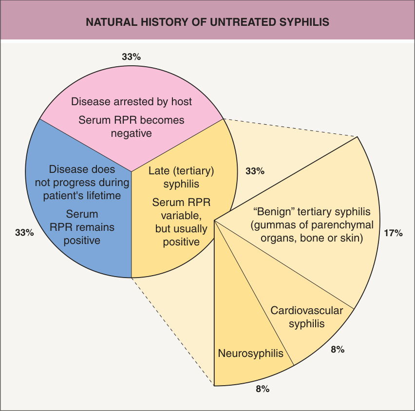
Fig. 82.1 Natural history of untreated syphilis. The data represent a composite of several studies. The specific antibody assays (e. g. MHA-TP, FTA-ABS) remain positive in at least 95% of all patients. RPR, rapid plasma reagin.
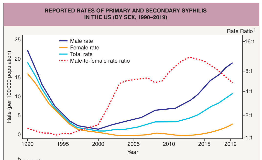
Fig. 82.2 Reported rates of primary and secondary syphilis in the US (by sex, 1990–2019). Note the elevated male : female ratio since 2000. Unfortunately, the use of pre-emptive antiretroviral therapy has been associated with a reduction in the use of condoms, including in men who have sex with men. From the Centers for Disease Control and Prevention: Sexually Transmitted Disease Surveillance 2019. https://www.cdc.gov/std/statistics/2019/std-surveillance- 2019.pdf.
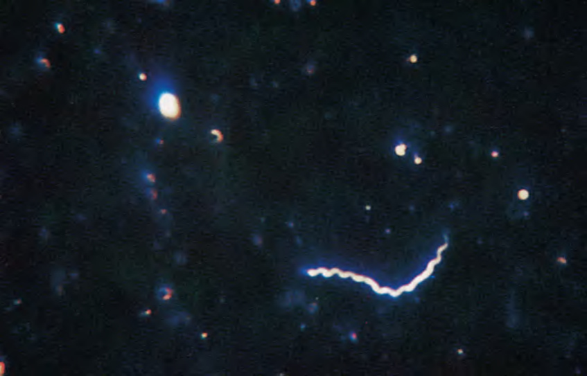
Fig. 82.3 Positive darkfield examination. Treponemes are recognized by their characteristic corkscrew shape, and deliberate forward and backward movement with rotation about the longitudinal axis. From Morse SA, et al. Atlas of Sexually Transmitted Diseases and AIDS, 3rd edn. London: Mosby, 2003.
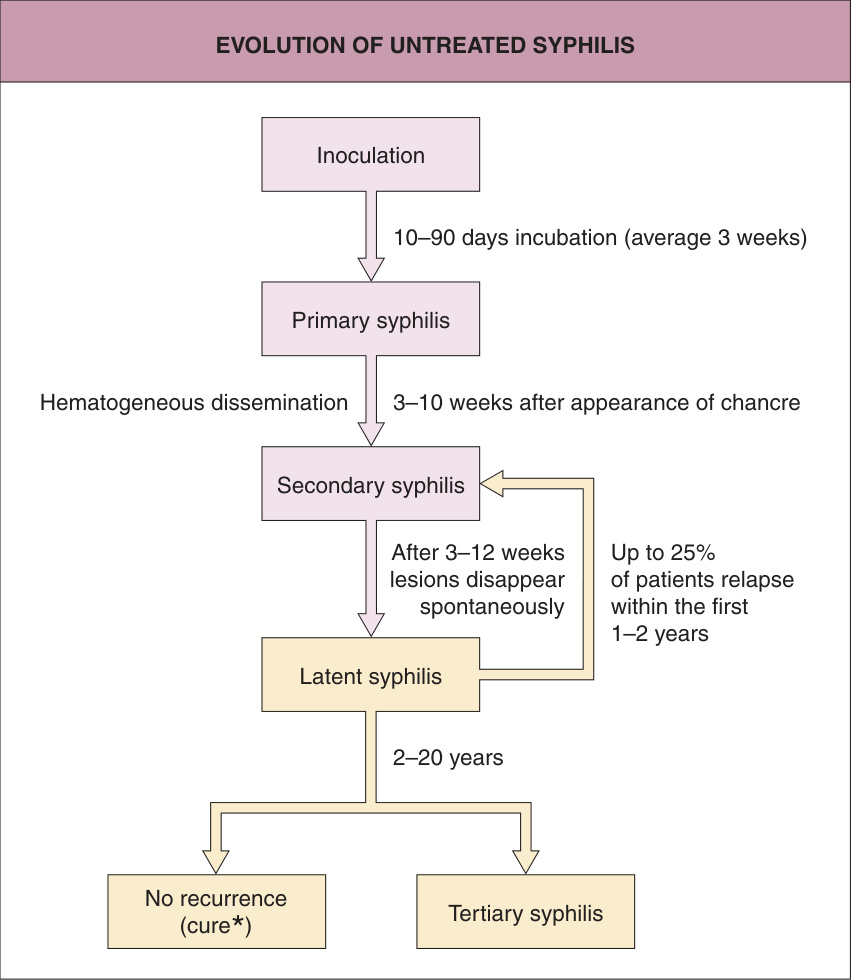
*Fig. 82.4 Evolution of untreated syphilis. Or clinically asymptomatic. Adapted from Rein MF, Musher DM. Late syphilis. In: Rein MF (ed). Atlas of Infectious Diseases, Vol V: Sexually Transmitted Diseases. New York: Current Medicine, 1995:10.1–10.13.
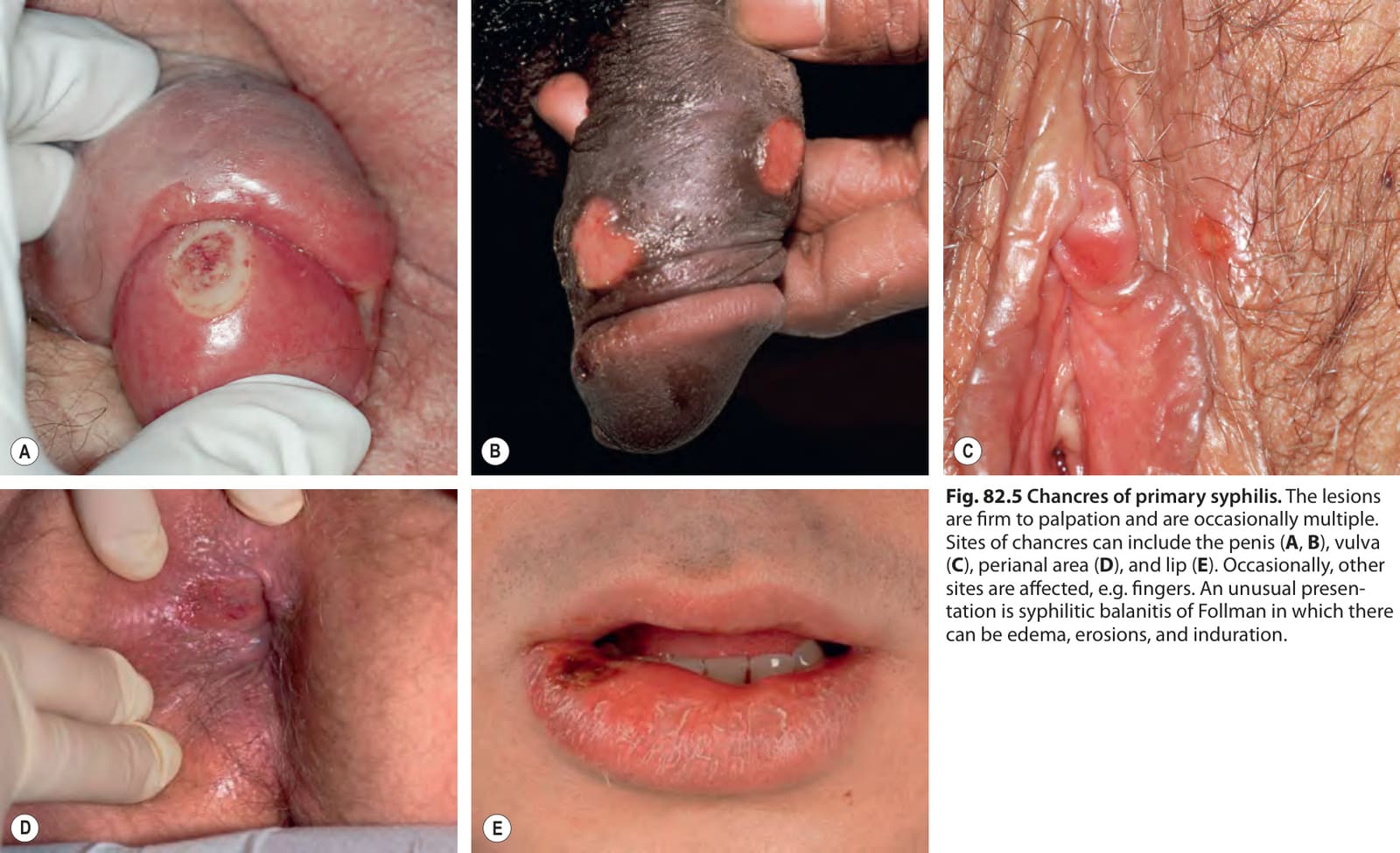
Fig. 82.5 Chancres of primary syphilis. The lesions are firm to palpation and are occasionally multiple. Sites of chancres can include the penis (A, B), vulva (C), perianal area (D), and lip (E). Occasionally, other sites are affected, e. g. fingers. An unusual presentation is syphilitic balanitis of Follman in which there can be edema, erosions, and induration.
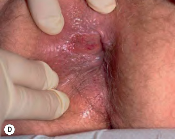
Fig. 82.6 Clinical manifestations of syphilis.Adapted from Fritsch P, Zangerle R, Stary A. Venerologie. In: Fritsch P (ed). Dermatologie und Venerologie. Berlin: Springer, 2004:865–86.
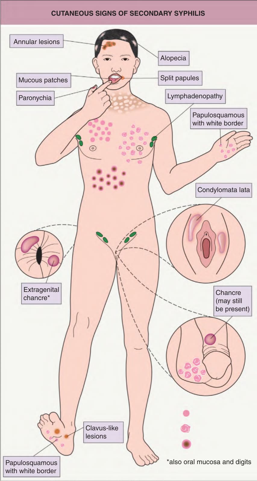
Fig. 82.7 Cutaneous features of secondary syphilis. Additional symptoms include low-grade fever, malaise, sore throat, myalgias, bone pain, and headaches.
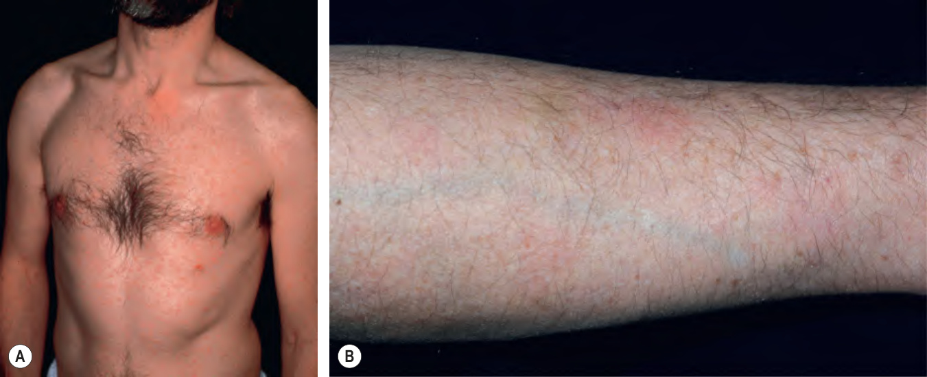
Fig. 82.8 Secondary syphilis.
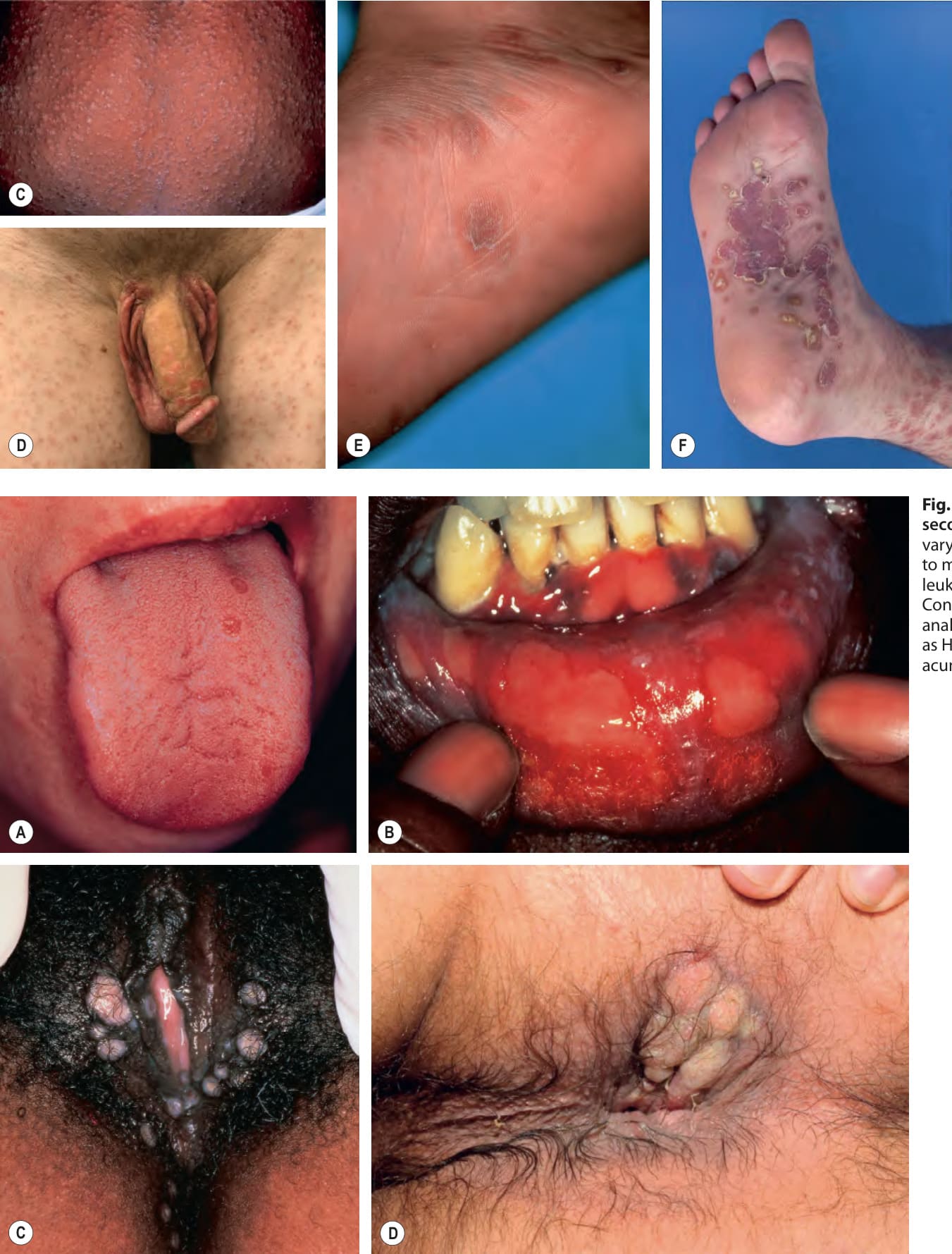
Fig. 82.9 Orogenital lesions of secondary syphilis. Oral lesions can vary from small superficial ulcers (A) to mucous patches (B). Occasionally, leukokeratosis or nodules are seen. Condylomata lata in the vulvar (C) and anal (D) areas may be misdiagnosed as HPV infection (i. e. condylomata acuminata).
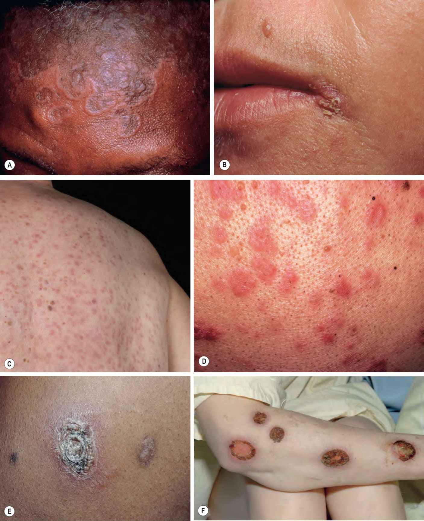
Fig. 82.10 Less common manifestations of secondary syphilis.A Annular and polycyclic plaques with central hyperpigmentation on the forehead. B Split papule at the oral commissure. C, D Two elderly patients with granulomatous lesions, varying from numerous, monomorphic, dull pink papules to papulonodules and plaques of varying sizes. E Necrotic lesion with scale-crust in a patient with AIDS. F “Malignant” syphilis with multiple necrotic, ulcerated and crusted lesions associated with severe constitutional symptoms. C, Courtesy Department of Dermatology, Medical University of Graz; E, Courtesy Judit Stenn, MD.
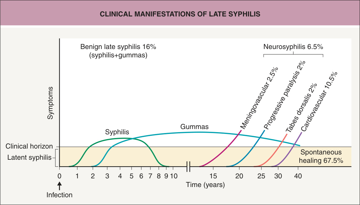
Fig. 82.11 Clinical manifestations of late syphilis.
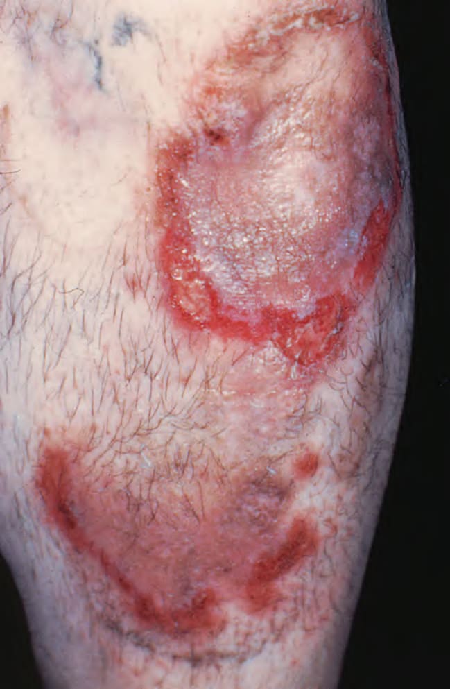
Fig. 82.12 Cutaneous gummas of tertiary syphilis. Arciform, erythematous eroded plaques with central scarring.
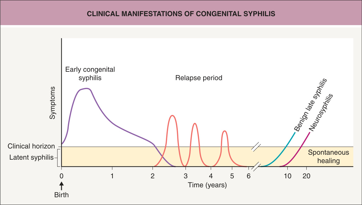
Fig. 82.13 Time course for clinical manifestations of congenital syphilis.Adapted from Fritsch P, Zangerle R, Stary A. Venerologie. In: Fritsch P (ed). Dermatologie und Venerologie. Berlin: Springer, 2004:865–86.
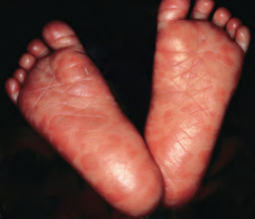
Fig. 82.14 Congenital syphilis. Red–brown plaques on the plantar surface.
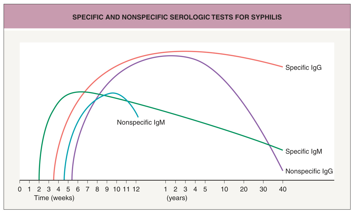
Fig. 82.15 Specific and nonspecific serologic tests for syphilis. These curves represent untreated patients. Non-treponemal tests are nonspecific and treponemal tests are specific. Adapted from Fritsch P, Zangerle R, Stary A. Venerologie. In: Fritsch P (ed). Dermatologie und Venerologie. Berlin: Springer, 2004:865–86.
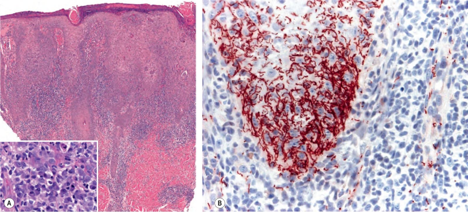
Fig. 82.16 Histology of secondary syphilis.A Psoriasiform epidermal hyperplasia and a dense dermal infiltrate of plasma cells (see inset), lymphocytes, and histiocytes are observed. B Positive immunohistochemical staining with an anti-Treponema pallidum antibody; note the characteristic elongated, twisted shape of the spirochetes. Courtesy Lorenzo Cerroni, MD.
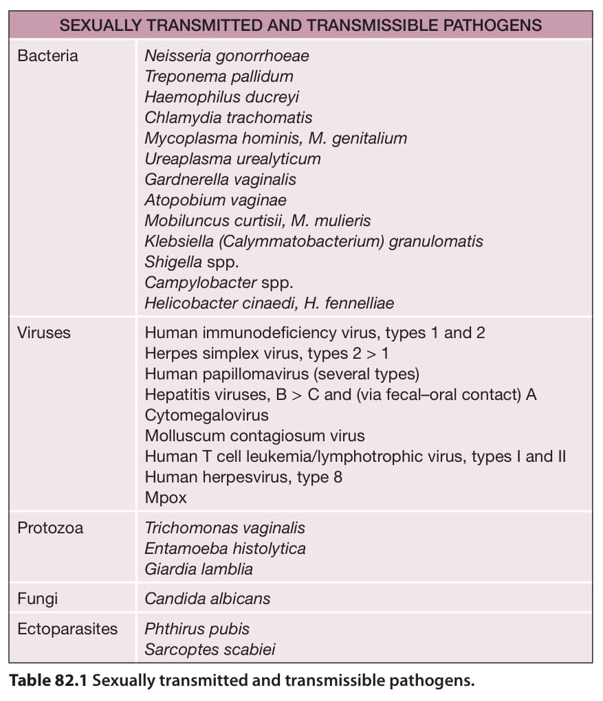
Table 82.1 Sexually transmitted and transmissible pathogens.
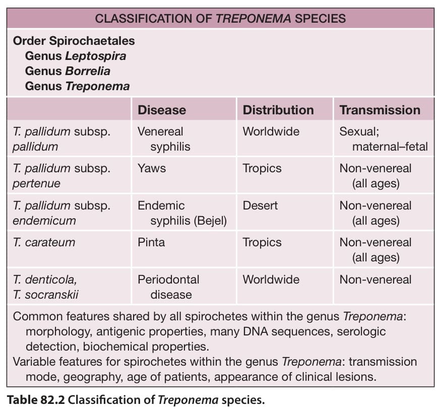
Table 82.2 Classification of Treponema species.
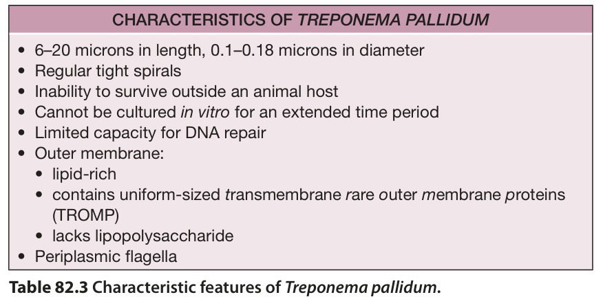
Table 82.3 Characteristic features of Treponema pallidum.
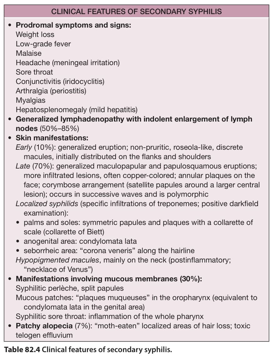
Table 82.4 Clinical features of secondary syphilis.
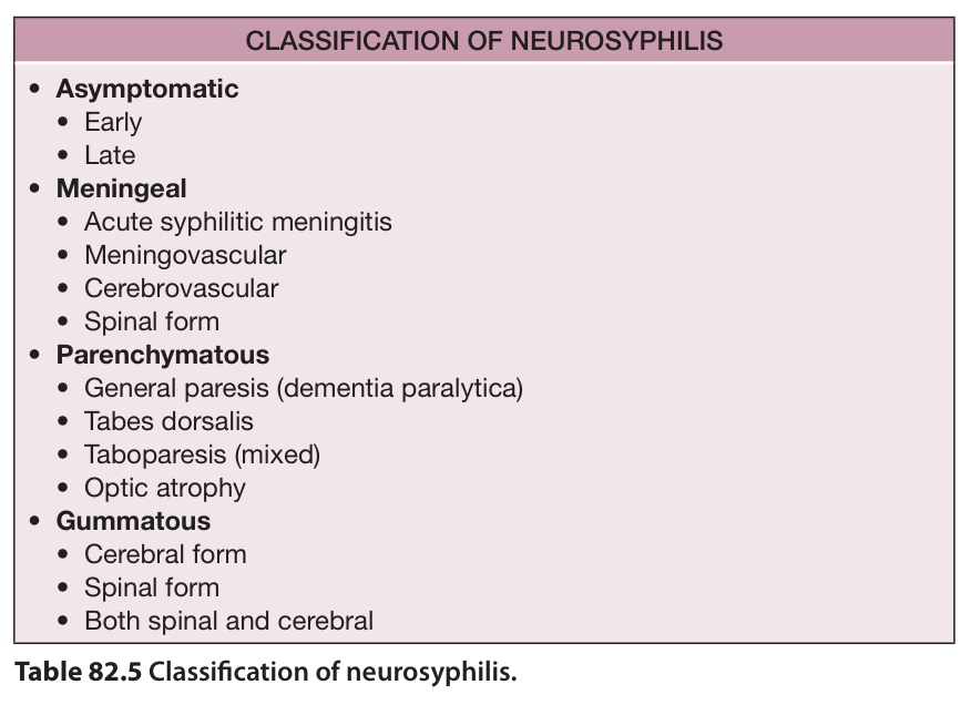
Table 82.5 Classification of neurosyphilis.
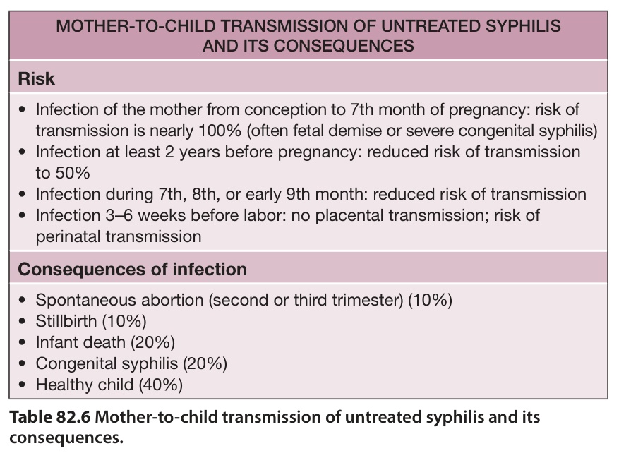
Table 82.6 Mother-to-child transmission of untreated syphilis and its consequences.
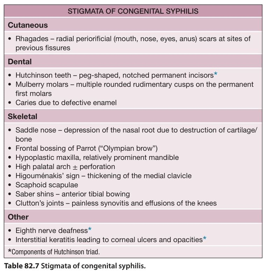
Table 82.7 Stigmata of congenital syphilis.
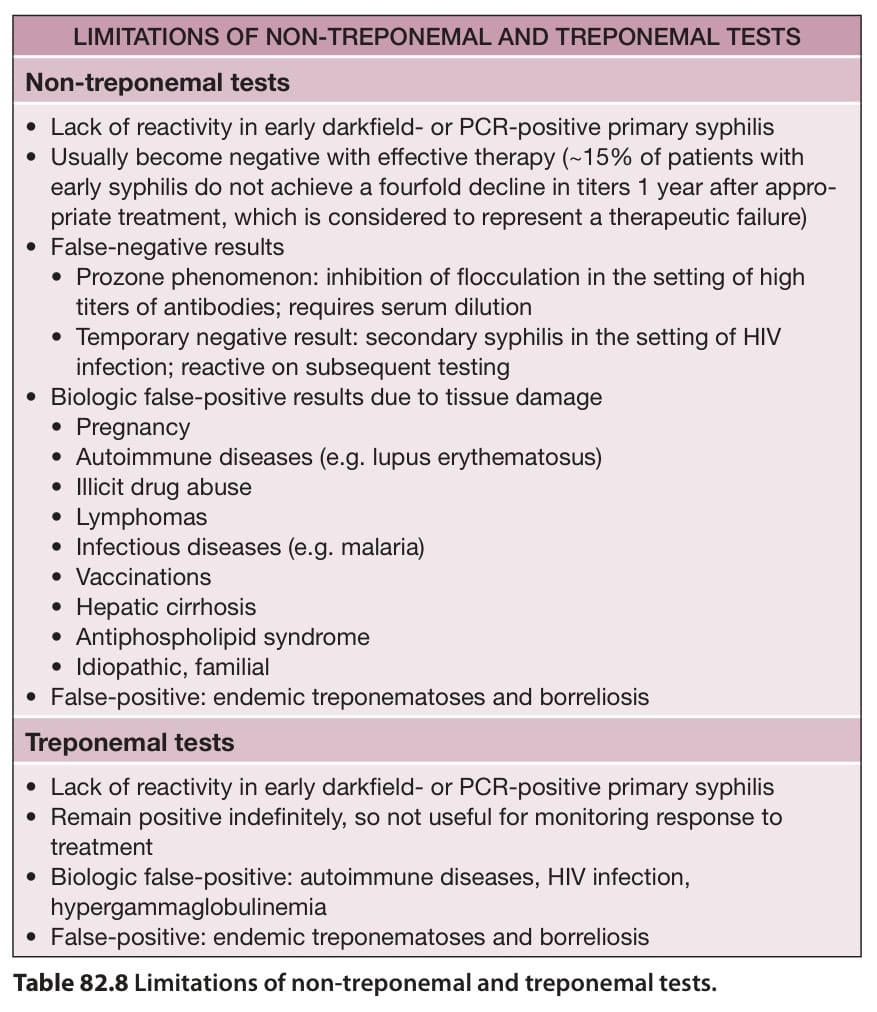
Table 82.8 Limitations of non-treponemal and treponemal tests.
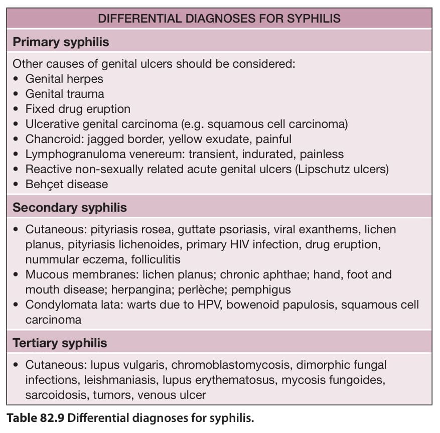
Table 82.9 Differential diagnoses for syphilis.
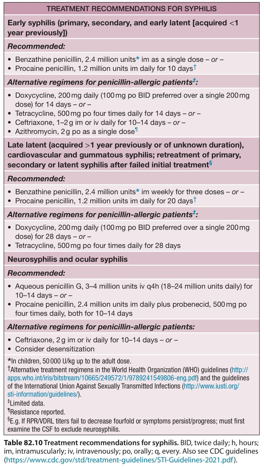
Table 82.10 Treatment recommendations for syphilis. BID, twice daily; h, hours; im, intramuscularly; iv, intravenously; po, orally; q, every. Also see CDC guidelines (https://www. cdc. gov/std/treatment-guidelines/STI-Guidelines-2021. pdf).
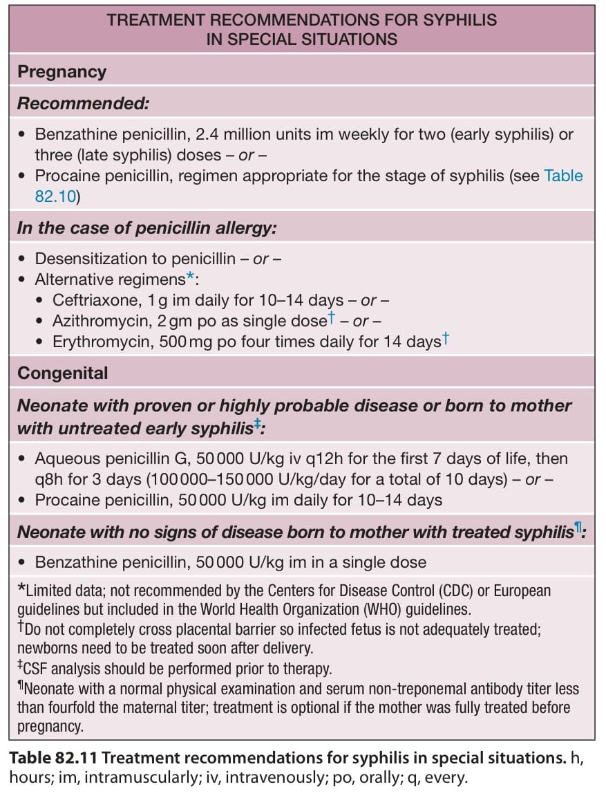
Table 82.11 Treatment recommendations for syphilis in special situations. h, hours; im, intramuscularly; iv, intravenously; po, orally; q, every.