GONORRHEA
Synonyms: Gonorrhea: “clap” Disseminated gonococcal infection: arthritis–dermatosis syndrome
Key features
Gonorrhea is the most common reportable STI in high-income countries
It is caused by Neisseria gonorrhoeae, which infects the mucosal surfaces of the human genital tract (as well as the anus, rectum, and oropharynx) after direct – usually sexual – contact with an infected person
Cutaneous pustules as well as systemic symptoms such as arthritis and fever can result from hematogenous dissemination, which occurs in only a small percentage of infected individuals
Overall, the incidence of gonorrhea declined during the latter portion of the 20th century as a result of safer sexual practices to avoid transmission of HIV, but afterwards the incidence has increased substantially in high-income countries following the introduction of antiretroviral therapy and pre-exposure prophylaxis for HIV
Over the past several decades, resistance of N. gonorrhoeae to various antimicrobials has increased in frequency
History
Gonorrhea was mentioned in ancient literature from China, Egypt, the Roman Empire and Greece, as well as in the Old Testament. Over the centuries, several names have been used to denote infection with N. gonorrhoeae, including: “strangury”, as used by Hippocrates; “clap”, derived from the district of prostitution called “Les Clapiers” in Paris; “gonorrhea”, chosen by Galen (130 CE) to describe the urethral exudate as a “flow of seed”; and “M. Neisser”, recognizing Albert Neisser, who discovered the microorganism in 1879 in stained smears from vaginal, urethral, and conjunctival exudates.
Culture of N. gonorrhoeae was first described by Leistikow and Lüffler in 1882, and it was improved in 1964 by Thayer and Martin, who devised selective growth conditions on special agar plates. Understanding the differences in the virulence of gonococci and studies of its molecular biology have led to greater insight into the pathology of the organism. Treatment of gonococcal infections was problematic until sulfonamides were introduced in 1936 and penicillin in 1943. For decades, gonorrhea was easily treated with just a single dose of one antibiotic. However, over the past several decades, an increase in antimicrobial resistance to penicillin and other antibiotics, such as tetracyclines and quinolones, has been observed and has become problematic. Declining in vitro susceptibility to cefixime led to a change in treatment guidelines, such that dual therapy with ceftriaxone and azithromycin became the recommended therapeutic regimen for uncomplicated gonorrhea. However, a more recent and worrisome development is elevated MICs (minimum inhibitory concentrations) to azithromycin and scattered reports of ceftriaxone-resistant, multi-drug-resistant N. gonorrhoeae30a.
Epidemiology
During the past century, the incidence of gonorrhea was influenced by politics, with peaks at the ends of the First and Second World Wars. A sudden decrease in reported cases occurred after the introduction of penicillin and other effective antimicrobials. Subsequently, the sexual freedom of the 1970s led again to an increase in the incidence of gonorrhea. However, with the advent of the AIDS epidemic, sexual practices again changed due to concerns regarding transmission of HIV, and this had a remarkable impact on the incidence of gonorrhea. Total gonorrhea rates in the US peaked in the mid 1970s and remained relatively low and stable from the 1990s to the mid 2010s.
Political changes in Eastern Europe led to an increased rate of gonorrhea (as well as syphilis) in the 1990s, especially in sex workers and those with sexual exposure to them. Migration of sex workers with a high infection rate also increased the number of reported cases of gonorrhea in other regions of Europe, especially in urban areas and high-risk groups. This probably reflects an increase in unsafe sexual behavior, particularly among young people and in high-risk populations. At the same time, the incidence of gonorrhea decreased in many countries (see above) due to safer sexual practices to avoid transmission of HIV. However, following the introduction of antiretroviral therapy and especially pre-exposure prophylaxis (PrEP) for HIV, there has been a reemergence of infections with N. gonorrhoeae as well as other STIs, especially amongst MSM. Of note, the national lockdowns imposed to prevent spread of SARS-CoV2 were associated with a sudden reduction in reported cases. Nonetheless, the trend of an increasing number of reported cases of gonorrhea, driven primarily by MSM, is expected to continue; for example, in many European countries the number of cases among women has also been increasing continuously since 2009 (ECDC Surveillance report: Annual epidemiological report for 2018).
Similar to other venereal diseases, the incidence of gonorrhea varies with age and is highest in the 15- to 24-year-old age group, in which there is a higher rate in women than in men. In the US, African- Americans have a 10-fold higher rate of gonococcal infections than White individuals. This may reflect different sexual behavior patterns and exposures as well as a better reporting system in public health institutions, which are more often utilized by African-Americans.
Transmission
N. gonorrhoeae is a pathogen restricted to humans as its only host and transmitted primarily by sexual contact. The most efficient transmission occurs by vaginal or anal intercourse where there is physical contact with the mucosal surface of a sexual partner with an asymptomatic or mildly symptomatic infection. An exception is indirect contact among prepubescent girls sharing contaminated objects. There is no evidence that gonococci can be spread by air droplet transmission as occurs with meningococci. Vertical transmission can occur from an infected mother to the newborn during parturition, and this may lead to gonococcal conjunctivitis, pneumonia, or even vulvovaginal infection.
Biology of N. gonorrhoeae
Gonococci are Gram-negative diplococci that typically grow in pairs. They exhibit multiple colony types when grown on hemoglobincontaining media in a 3% CO atmosphere. The outer membrane structure of N. gonorrhoeae is typical for Gram-negative bacteria but, in contrast to N. meningitidis, it lacks the polysaccharide capsule that is responsible for the virulence of meningococci. Surface molecules on the outer membrane are involved in attachment, invasion and host injury, and they also represent important antigenic structures, especially the fibrillar pili (composed of 18 kDa pilin subunits).
Gonococci require iron to grow, with sources including transferrin, lactoferrin, and hemoglobin; acquisition of iron is mediated by binding to host proteins via specific receptors. The microorganism cannot tolerate drying or low temperatures, and its growth is optimal at 35–37°C in a 5% CO atmosphere on a complex growth medium containing inorganic iron, glucose, vitamins, and cofactors as well as antibiotics.
Pathogenesis
Although there is increasing knowledge about the pathogenicity of this microorganism, the exact molecular mechanisms of invasion of gonococci into the host cell are still unknown. Several virulence factors are involved in the processes of adherence, inflammation, and mucosal invasion. Because pili increase adhesion to the host cell and allow movement, they also play an important role in pathogenesis; this may explain why non-piliated gonococci have a reduced ability to cause infections in humans. The adherence of gonococci to the human epithelial host cell and to neutrophils is dependent not just on the pili but also on Opa ligands. Anti-pilus antibodies have been shown to block epithelial adherence and increase phagocytic killing. Expression of the transferrin receptor and full-length lipo-oligosaccharide (LOS) appear to increase infectivity. Gonococci are able to multiply and divide intracellularly, where they are immune to host defense mechanisms. Cellular invasion is favored by expression of certain Opa proteins and non-sialylated LOS.
Gonococci have the ability to cause tissue destruction by production of a variety of enzymes and lipids such as phospholipase, peptidases, and lipid A. This plays a role in their damage of fallopian tubes and the development of postinflammatory arthritis.
Clinical Features
Gonorrhea has a broad spectrum of clinical manifestations in both men and women, including asymptomatic infections, local symptomatic mucosal infections (with or without local complications), and systemic dissemination (Table 82.12). Symptoms vary according to the site of infection and strain of organism.
The incubation period for gonorrhea is relatively short; it only takes 2–5 days until signs and symptoms of an infection with N. gonorrhoeae appear. Up to 10% of infected men and 50% of infected women lack clinical symptoms, which is especially common in rectal and pharyngeal infections.
Gonococcal infection in men
The most common clinical presentation of gonococcal infection in men is an acute anterior urethritis with dysuria and a urethral discharge that is typically purulent and profuse (Fig. 82.17). In about one-quarter of infected men, the urethral symptoms are less pronounced, similar to those of non-gonococcal urethritis, and appear only after urethral manipulation (“stripping”). Without treatment, clinical symptoms disappear in most patients after about 6 months. Local complications can include inflammation of the Cowper and Tyson glands and gonococcal pyoderma; ascension of the infection may lead to epididymitis, prostatitis, and vesiculitis. Patients with gonococcal epididymitis present with unilateral testicular pain and swelling accompanied by urethritis.
Gonococcal infection in women
In about 50% of infected women, gonococcal infection is asymptomatic and therefore remains unnoticed. The primary site of gonococcal infection in women is the endocervical canal, with associated clinical
symptoms consisting of increased vaginal discharge, dysuria, intermenstrual bleeding, and menorrhagia. Clinical inspection shows purulent cervical discharge with erythema and edema; swabbing of the endocervical canal results in a highly characteristic yellow color. Urethral colonization is present in 70%–90% of infected women and is the usual site of infection in women who have had a hysterectomy. Occasionally, inflammation of the Bartholin glands is observed, with acute swelling of the labial folds and appearance of purulent discharge when pressure is applied to the gland.
The most common local complication in women is acute salpingitis or pelvic inflammatory disease (PID) due to ascension of the microorganisms. It occurs in ~10%–20% of infected women (often developing immediately after a menstrual period) and may result in the long-term consequences of infertility, chronic pelvic pain, and ectopic pregnancy. The clinical manifestations of PID vary and include lower abdominal pain, adnexal tenderness, elevation of the ESR, leukocytosis, and fever. Gonorrheic perihepatitis (Fitz-Hugh–Curtis syndrome), with inflammation of the adjacent peritoneal area, is an infrequent complication in which symptoms of PID are accompanied by pain in the right upper quadrant that mimics acute cholecystitis.
Extragenital gonorrhea
This can occur in both men and women after oral sexual exposure. Because it is usually asymptomatic, the infection typically goes undetected and spontaneously resolves within a few weeks. Pharyngeal gonorrhea disproportionately affects MSM and may be an important source of urethral gonorrhea in MSM. It is important to address this site of infection, especially in this high-risk group.
Rectal gonorrhea is mainly seen in MSM and in heterosexual women who practice receptive anal intercourse. Rectal gonorrhea is asymptomatic in at least 50% of patients, but it may result in gonococcal proctitis that is accompanied by inflammation, rectal discharge, anal pruritus, bleeding, tenesmus, and constipation.
Gonococcal ophthalmia is uncommon in adults but still represents a major cause of blindness in some low-income countries. Primarily due to self-inoculation and unusual sexual practices, it initially presents as purulent conjunctivitis and, if left untreated, can rapidly progress to severe keratitis followed by corneal opacification.
Gonococcal infection in newborns is caused by inoculation with N. gonorrhoeae during delivery through an infected birth canal, and most often it presents with purulent conjunctivitis (ophthalmia neonatorum). Due to preventive application of antimicrobial ointment (usually erythromycin) immediately after birth, the rate of this gonococcal infection is currently low.
Disseminated gonococcal infection
The most common clinical manifestation of gonococcal bacteremia is an acute arthritis–dermatosis syndrome, which occurs in ~0.5%–1% of patients with mucosal gonorrhea. Risk factors for disseminated gonococcal infection include: (1) menstruation, with the majority of cases in women developing during or immediately following menses;
(2) deficiencies in the late complement components C5–C9 (see Ch. 60); and (3) inhibitors of the cleavage of C5 to C5a and C5b, e.g. ravulizumab, eculizumab. The classic syndrome consists of fever, joint pain, and a paucilesional eruption of hemorrhagic pustules. Gonococcal tenosynovitis affects primarily larger joints (e.g. knees, elbows, wrists, ankles) and is sometimes visible as erythema overlying the tendons.
The cutaneous lesions consist of scattered pustules, often necrotic, due to an embolic septic vasculitis. They occur primarily on the distal portions of the extremities (Fig. 82.18). With a longer duration of symptoms, positive blood cultures become less common.
Laboratory Diagnosis
Laboratory diagnosis is based on the identification of N. gonorrhoeae in secretions from infected mucous membranes by using stained smears, molecular biologic techniques, and/or bacterial culture (Fig. 82.19). Samples are typically obtained from the endocervical canal in women (after wiping off exudate), the urethra in men and occasionally in women who have had a hysterectomy, and (when indicated) the posterior pharynx. For diagnosis by nucleic acid amplification methods (see below), a vaginal swab or urine sample can also be used. Anorectal specimens should be obtained via direct visualization using anoscopy if possible.
Staining methods
Direct detection of Gram-negative diplococci within neutrophils in Gram- or methylene blue-stained smears provides an immediate diagnosis (Fig. 82.20), which is especially helpful in symptomatic individuals. It also represents a recommended point-of-care test. Gram-stained urethral exudate detects between 95% and 98% of symptomatic infections in men, whereas Gram-stained cervical smears have a sensitivity of about 50%. The specificity of Gram-stained exudates depends on several factors including the experience of the microscopist, proper collection, the presence of acute symptoms, and bacterial load; specificity may approach 100% in an optimal setting. However, because of its lower sensitivity in asymptomatic individuals, a negative Gram stain should not be considered sufficient for excluding infection in such patients. Smears are not helpful for detection of rectal and pharyngeal gonorrhea due to the presence of a large number of other bacteria.
Culture techniques
Isolation of N. gonorrhoeae via culture is still recommended for cases with medicolegal implications. In addition, pustular skin lesions due to gonococcemia should be cultured. Although it does depend on the symptomatic status of the infected person, antibiotic-containing selective media (Thayer–Martin) or selective New York media can have a sensitivity of 80%–95% when genital swabs are directly plated at the time of collection. The specificity of bacterial culture is reported to be ~95% for genital samples. However, sensitivity is much lower for extragenital swabs such as those from the rectal area (<50%). Confirmation of a positive culture by N. gonorrhoeae-specific tests is recommended and is mandatory in cases of extragenital infection.
The advantage of isolating N. gonorrhoeae via culture is the ability to perform antimicrobial resistance (AMR) testing. This is particularly important given the rising number of antibiotic-resistant isolates, and it also allows for regional AMR surveillance. Monitoring antibiotic susceptibility is crucial, especially with the increasingly routine use of non-culture techniques.
Non-culture techniques for gonococcal diagnosis
Molecular techniques for diagnosing gonococcal infections are widely employed and are advantageous over bacterial culture when specimen transport and storage are potential problems. The former are also more sensitive than culture for extragenital sites and when the infection is asymptomatic. In addition, molecular tests can detect gonococci in easy-to-obtain specimens such as urine and are therefore the best test when screening asymptomatic individuals. Another advantage is that multiplex amplification assays can detect Chlamydia trachomatis (CT) and other sexually transmitted microbes such as Mycoplasma genitalium, in addition to N. gonorrhoeae (NG), without the need for an additional specimen (see Fig. 82.19). Their major disadvantage is the lack of antibiotic sensitivity testing (see above).
With respect to overall sensitivity, specificity, and ease of specimen transport, NAATs outperform other tests that are currently available for the diagnosis of gonococcal infections.
NAATs are used to detect gonococcal DNA or RNA, often in addition to chlamydial DNA or RNA. Methods include transcription-mediated amplification to detect rRNA (e.g. Aptima Combo 2® assay for CT/ NG) and strand displacement amplification (e.g. BD ProbeTec™ ET System). The Xpert® CT/NG test kit can detect N. gonorrhoeae within 90 minutes and also co-amplifies CT and NG. NAATs are continuously being enhanced, leading to faster turnaround times, more user-friendly features, and detection of an increasing number of pathogens from a single sample (e.g. Trichomonas vaginalis as well as C. trachomatis and N. gonorrhoeae via the BD MAX™ CT/GC/TV assay). As previously noted, in addition to physician-collected specimens, samples for NAATs can be obtained “non-invasively”, using self-collected vaginal or introital swabs or just urine.
(B) smears of urethral discharge. Neutrophils containing Gram-negative or methylene blue-positive diplococci, consistent with Neisseria gonorrhoeae, are evident. B, Courtesy Harald Moi, MD.
Pathology
Biopsies of skin lesions due to gonococcemia show a septic vasculitis with extravasated erythrocytes and thrombi. Organisms are usually not demonstrated.
Differential Diagnosis
Other causes of urethral and cervical discharge in men and women must be considered, including infections with C. trachomatis, M. genitalium, T. vaginalis, yeasts, and anaerobic bacteria. The skin lesions of disseminated gonococcemia may need to be distinguished from folliculitis (e.g. bacterial), another disseminated infection, a neutrophilic dermatosis, cutaneous vasculitis, or a pustular drug eruption.
Treatment
Although many antibiotics can be used for the treatment of gonorrhea, selection of therapy depends on the susceptibility of the organism that is isolated (or regional patterns), the anatomic site(s) of the infection, clinical symptoms, and the cost (especially in resource-poor settings) as well as potential side effects of the drug. Over the past several decades, strains of gonorrhea resistant to a number of antibiotics, including sulfonamides, penicillin, tetracyclines, erythromycin, spectinomycin, and more recently quinolones, azithromycin, and some cephalosporins, have emerged. Antibiotic resistance of N. gonorrhoeae clearly represents an urgent public health threat.
Over the years, the development of different types of resistance has led to regular revisions of gonococcal treatment recommendations. For example, in 2007, the CDC recommended that fluoroquinolones no longer be prescribed for the treatment of gonococcal infections in the US. Third-generation cephalosporins then became the antibiotics of choice for gonococcal infections (Table 82.13); however, treatment failures (especially with oral cephalosporins in Asian countries) and decreased in vitro sensitivity, e.g. to cefixime, began to appear. In addition to the emergence of a small number of strains with decreased susceptibility to ceftriaxone, the first gonococcal strain with ceftriaxone resistance combined with high-level resistance to azithromycin was reported from England and Australia in 2018. As a result, the WHO declared antimicrobial-resistant N. gonorrhoeae a priority global health issue.
The current CDC treatment guidelines recommend a single dose of 500 mg of intramuscular ceftriaxone for uncomplicated gonococcal infections. Alternative regimens are available when ceftriaxone cannot be used to treat urogenital or rectal gonorrhea. Of note, there is a portable, rapid, on-cartridge magnetofluidic purification and testing (PROMPT) PCR test for determining susceptibility of N. gonorrhoeae to ciprofloxacin that allows for targeted therapy and leads to a reduction
Based on CDC 2021 Guidelines (www. cdc. gov/std/treatment-guidelines/ STI-Guidelines-2021. pdf). BID, twice daily; h, hours; im, intramuscularly; iv, intravenously; po, orally; q, every.
in the use of ceftriaxone. Persons infected with N. gonorrhoeae are frequently co-infected with C. trachomatis and this co-infection is readily diagnosed by combined NAATs (see above). When confirmed or if C. trachomatis infection cannot be excluded, doxycycline is the recommended treatment (see Table 82.13).
For disseminated gonococcal infection, first-line therapy consists of intravenous ceftriaxone (1 g every 24 hours for at least 7 days). Lastly, confirmation of cure, compliance with therapy, and partner notification are legal recommendations for gonococcal treatment and prevention in some countries.
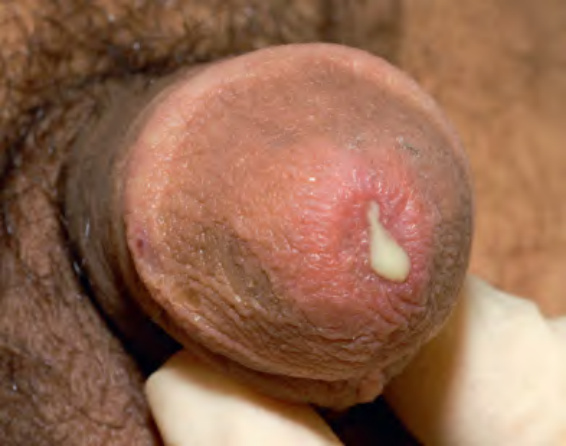
Fig. 82.17 Gonococcal urethritis with a purulent urethral discharge.
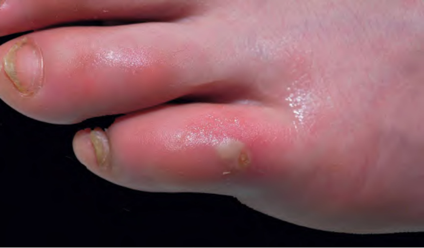
Fig. 82.18 Gonococcemia (arthritis–dermatosis syndrome). Pustule with surrounding erythema on the toe.
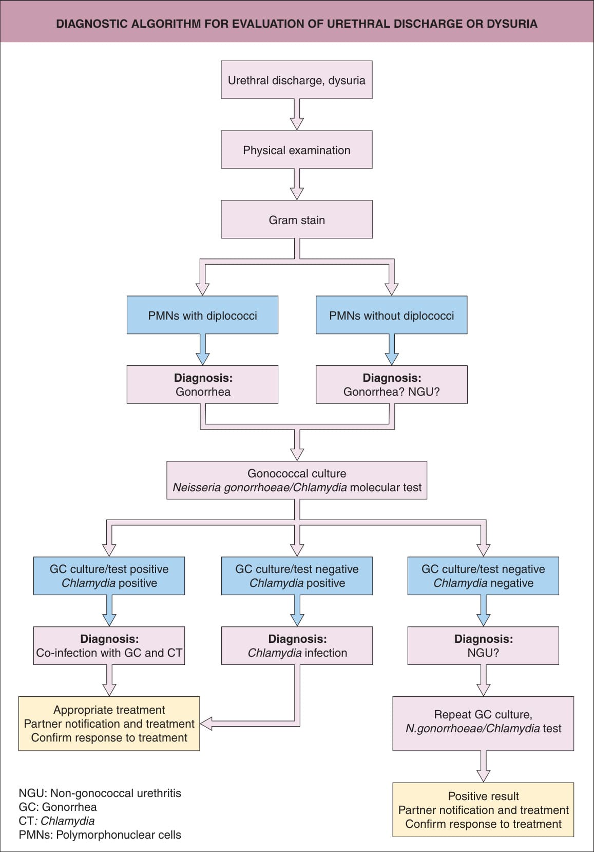
Fig. 82.19 Diagnostic algorithm for evaluation of urethral discharge or dysuria. If cultures are negative, but polymorphonuclear cells are seen on smear, some clinicians will still prescribe doxycycline.
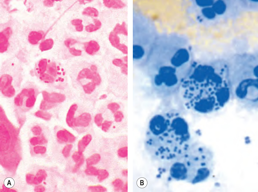
Fig. 82.20 Gonococci in Gram-stained (A) and methylene blue-stained
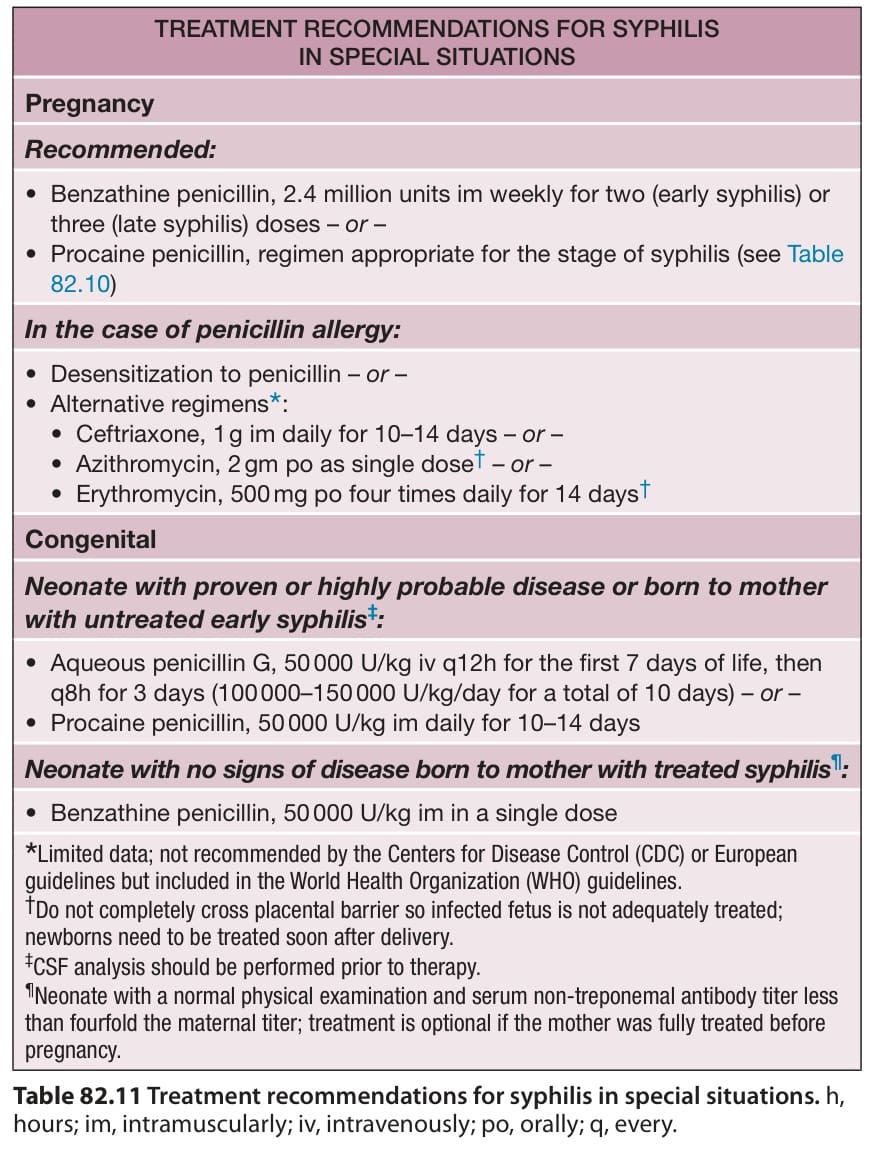
Table 82.11 Treatment recommendations for syphilis in special situations. h, hours; im, intramuscularly; iv, intravenously; po, orally; q, every.
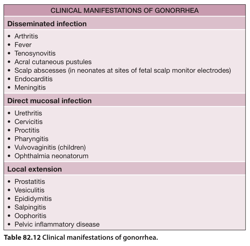
Table 82.12 Clinical manifestations of gonorrhea.
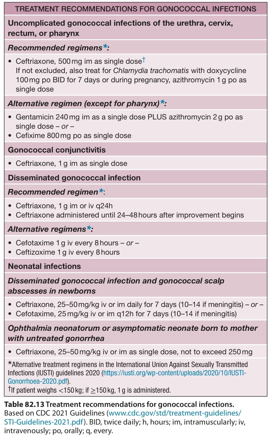
Table 82.13 Treatment recommendations for gonococcal infections.