CHANCROID
Synonyms: Soft chancre Ulcus molle
Key features
Chancroid is characterized by acute genital ulcers and inguinal adenitis
It is caused by the sexually transmitted, Gram-negative, and faculta- tively anaerobic bacterium Haemophilus ducreyi
It is a rare disease in high-income countries, and its occurrence is closely linked with commercial sex workers
History
In 1850, chancroid was first differentiated from syphilis. In 1889, Ducrey described the responsible organism as a streptobacillary rod, which he was able to subcultivate by serial autoinoculations into the forearm of a patient.
Epidemiology
The disease is common in parts of Africa, the Caribbean, and Southeast Asia and is endemic in many low-income countries (Fig. 82.21), with a worldwide annual incidence of about 7 million. Outbreaks are occasionally reported in cities in high-income countries, predominantly within the US. In the US, the number of chancroid cases peaked in the early 1990s and has been very low for the past 25 years. In most of the disease settings, sex workers are the reservoir for the microorganism, and men are much more frequently affected than women (ratio 10 : 1). In some low-income countries, chancroid is the most common cause of infectious genital ulcer disease (Table 82.14), with frequencies even higher than those of genital herpes.
Transmission
The source of infection is almost always sexual contact with an infected person who has genital ulcers. The probability of transmitting chancroid after a single sexual contact is 35%, and untreated infected women are estimated to be infectious for up to 45 days (while the clinical lesions are present). Asymptomatic carriers seem to be rare and there is no evidence that infected individuals harbor the organism for a significantly long period of time without clinical findings.
Chancroid and HIV
Chancroid and other diseases that produce genital ulcers are important risk factors for transmission of HIV. Men infected with HIV more often have had a history of genital ulcers compared with HIV-negative men. The risk of acquiring HIV after having sexual contact with HIV-positive women was highest for uncircumcised men with genital ulcer disease compared with circumcised men with and without genital ulcer disease (29% versus 6% versus 2%). In women, the risk of becoming infected with HIV also increases with the number of episodes of genital ulcer disease.
The presence of increased numbers of CD4+ lymphocytes and macrophages in ulcers due to infection with H. ducreyi provides an ideal opportunity for a latent HIV infection to become productive, with excretion of the virus into ulcer secretions. Genital lesions therefore become both a portal of viral entry for non-infected individuals and viral exit for HIV-infected persons.
Biology of the Organism
H. ducreyi is a Gram-negative, facultatively anaerobic bacillus of small size that shows a typical chaining pattern on Gram stain (Fig. 82.22). It does not share many similarities with other members of the genus Haemophilus.
Pathogenesis
The penetration of the microorganism occurs via microscopic barrier defects in the epidermis. Signs of inflammation develop due to infiltration of lymphocytes, macrophages, and granulocytes in a primarily
Th1 cell-mediated immune response combined with pyogenic inflammation. Chancroid is associated with regional lymphadenitis due to the spread of the infection and resultant pyogenic inflammation.
Clinical Features
The incubation period is 3–10 days; usually, the onset of clinical symptoms occurs between days 4 and 7. In men, the lesion starts with a papule surrounded by erythema and soon progresses to a pustule and then a painful ulcer. The latter typically has a purulent base and soft, undermined edges and is sharply demarcated (Fig. 82.23A,B). In about one-half of infected men, several ulcers develop due to apposition with the initial lesion, and they can coalesce to form giant ulcers. Most lesions are located on the internal or external surface of the prepuce, in the coronal sulcus (particularly in uncircumcised men), or around the frenulum. They are often accompanied by edema of the prepuce.
Occasionally, a so-called “septic sore” can be observed on the shaft of the penis or prepuce, with pus trapped within the skin in the absence of an obvious ulcer. In this case, the pus that can be expressed is full of microorganisms. In contrast to chlamydial infections, chancroid is symptomatic in almost all infected individuals.
Co-infections with syphilis or herpes simplex virus (HSV) are possible. In Kenya, ~4% of men with genital ulcer disease have a co-infection with both T. pallidum and H. ducreyi (ulcus mixtum) and about the same percentage have HSV plus H. ducreyi, which makes etiologic diagnosis based on clinical findings impossible.
In women, the majority of lesions are within the introital area; they may also occur on the cervix or vaginal wall, or in the perianal area, but rarely occur in an extragenital location. Lesions in women are sometimes only mildly symptomatic and are more often multiple.
In both men and women, painful inguinal lymphadenitis may accompany the genital lesions. It is more often observed in men (about 40% of patients) and is usually unilateral (Fig. 82.23C). Inguinal buboes may rupture and lead to inguinal ulceration.
Pathology
Biopsies of chancroid classically have three zones of inflammation beneath the ulcers. The first zone has necrotic debris, fibrin, and neutrophils. The middle zone is an area of granulation tissue, and the deepest zone contains lymphocytes and plasma cells. Gram-negative coccobacilli are only rarely found with tissue Gram or Giemsa stains and are best seen with smears.
Diagnosis
Material is usually obtained with a cotton swab, on which the organism can only survive for a few hours without being refrigerated at 4°C. Gram stain of H. ducreyi in a smear of the exudate shows a “school-of-fish” or “railroad track” pattern of small Gram-negative bacilli (see Fig. 82.22). The finding is not specific because other bacteria may have a similar arrangement, and it is insensitive because the responsible bacteria are only seen in about a third to a half of infected persons.
Accurate diagnosis of chancroid requires isolation of H. ducreyi on special culture media; small, non-mucoid, semiopaque or translucent colonies appear after 24–72 hours if grown at 33–35°C in a 5%–10% CO atmosphere. Further identification methods are needed, such as the porphyrin test, to prove the requirement of hemin (factor X) for growth. There are variations in media (which are not widely available from commercial sources) and the sensitivity of culture is between 60% and 80%, depending on the quality and handling of the specimen, culture conditions, and laboratory experience.
If culture is not possible or is inconclusive, diagnosis can be based on the clinical picture and the exclusion of other microorganisms
that cause genital ulcer disease, such as syphilis or genital herpes, as well as on the epidemiologic data and response to therapy. A genital ulcer disease panel that can detect HSV-1, HSV-2, T. pallidum, and H. ducreyi via real-time PCR is commercially available in some countries.
Other diagnostic procedures, such as antigen detection methods or serologic tests, are still under development and cannot yet be recommended for routine diagnosis.
Differential Diagnosis
Other infections that cause genital ulcers have to be considered (see Table 82.14), as well as trauma, a fixed drug eruption, and, occasionally, carcinoma.
Treatment
Successful treatment can be achieved using several antimicrobial drugs (Table 82.15); ulcers improve within 3 days and typically heal in 14 days or less, depending on the initial lesions. The different treatment regimens cure about 95% of infected individuals, with azithromycin and ceftriaxone requiring only a single dose. Ciprofloxacin is contraindicated in pregnant and lactating women. In patients with HIV infection, the treatment recommendations are the same, but with a longer treatment course; close monitoring may be necessary because of delayed healing and possible treatment failures.
Sexual partners from the 10 days preceding the onset of symptoms should be examined and treatment offered regardless of whether the microorganism can be cultured or clinical symptoms are present.
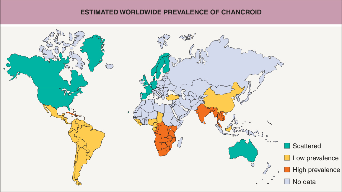
Fig. 82.21 Estimated worldwide prevalence of chancroid.
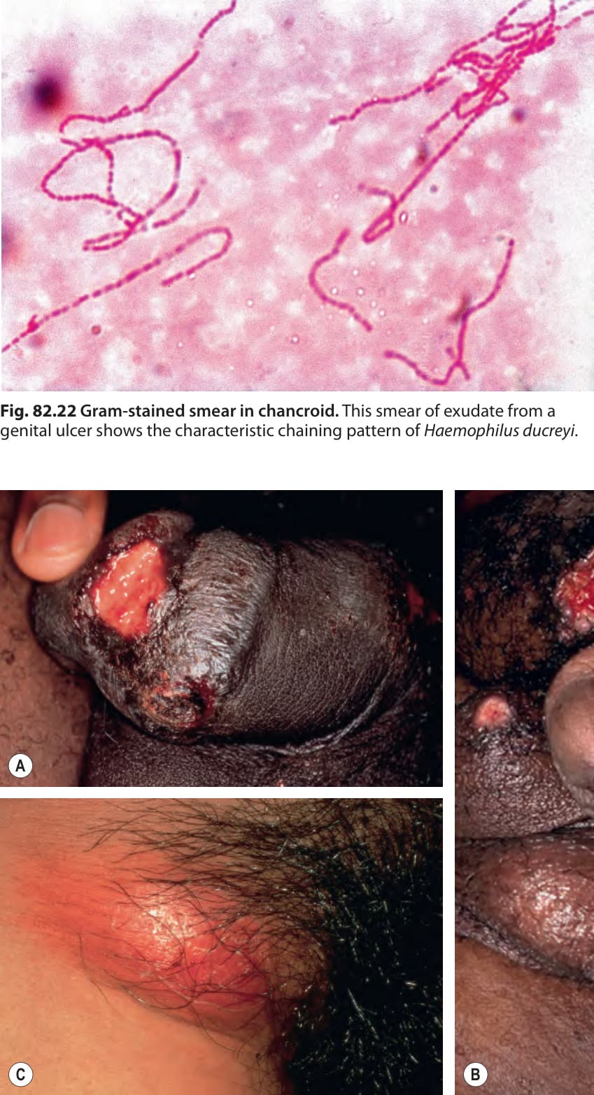
Fig. 82.22 Gram-stained smear in chancroid. This smear of exudate from a genital ulcer shows the characteristic chaining pattern of Haemophilus ducreyi
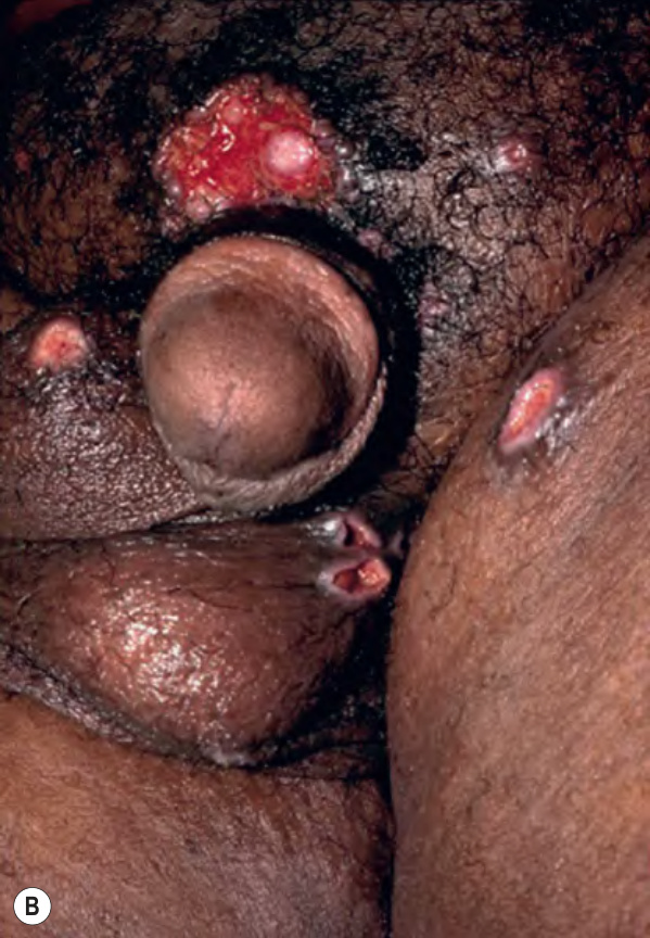
Fig. 82.23 Chancroid.A Well-demarcated painful ulcers on the penis. B Multiple purulent ulcers with undermined borders. C Unilateral lymphadenitis with overlying erythema. B, Courtesy Joyce Rico, MD.
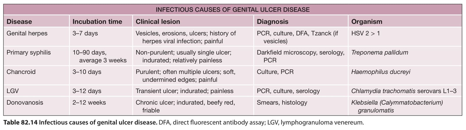
Table 82.14 Infectious causes of genital ulcer disease. DFA, direct fluorescent antibody assay; LGV, lymphogranuloma venereum.
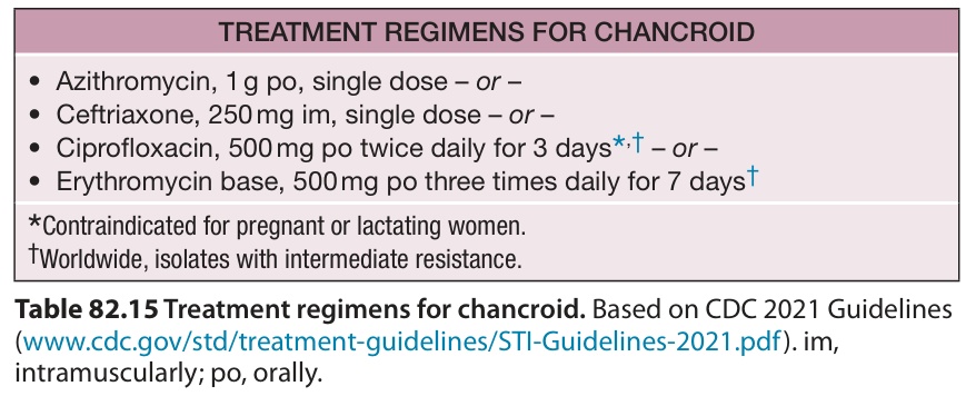
Table 82.15 Treatment regimens for chancroid. Based on CDC 2021 Guidelines (www. cdc. gov/std/treatment-guidelines/STI-Guidelines-2021. pdf). im, intramuscularly; po, orally.