LYMPHOGRANULOMA VENEREUM
Synonyms: Durand–Nicolas–Favre disease Climatic bubo Strumous bubo Poradenitis inguinale Lymphogranuloma inguinale
Key features
Lymphogranuloma venereum (LGV) is an STI caused by Chlamydia trachomatis serovars L1–3 and can be associated with abscesses, fistulas, and involvement of lymphatic tissue
LGV is endemic in some areas of Africa, Asia, and South America
Outbreaks of LGV proctocolitis can occur in high-income countries amongst MSM who also have increased rates of HIV infection
The disease progresses through three stages: (1) initial infection of the genital mucosa; (2) inguinal lymphadenopathy that is usually unilateral; and (3) a firm mass and bubo with spontaneous drainage and involution, often accompanied by proctocolitis and involvement of perirectal or perianal lymphatic tissue
History
Lymphogranuloma venereum (LGV) has been confused with other diseases that cause inguinal lymphadenopathy, especially syphilis, genital herpes, and chancroid. A major diagnostic advantage arose with the introduction of the Frei test, a specific skin test established in
1925. The first isolation of Chlamydia was reported in 1930, enabled
by intracerebral inoculation of monkeys, followed in 1935 by cultivation in fertilized eggs.
Epidemiology
LGV is endemic in East and West Africa, Southeast Asia, India, South America, and the Caribbean basin. In some countries, it is a reportable disease. LGV classically affected sailors, soldiers, and travelers who acquire the disease in endemic regions. In the past, data regarding the prevalence of LGV have been based on the results of serologic tests and the Frei skin test, both of which are nonspecific and have cross-reactivity with other genito-ocular chlamydial infections.
LGV is reported more often in men than in women and the peak age correlates with the period of maximum sexual activity. However, the frequency of infection following sexual intercourse has not been well investigated. Since 2003, a series of outbreaks of LGV have been reported in Western Europe and the US. The affected individuals are primarily MSM who live in large cities, many of whom have concurrent HIV infection. In these outbreaks, LGV can present as ulcerative proctitis or as an asymptomatic infection.
Pathogenesis
Caused by specific Chlamydia trachomatis serovars (L1–3), LGV affects mainly the lymphatic tissue in the genitorectal area. The organisms enter the body via microscopic defects in the mucosa or the skin. The microorganisms then enter the lymphatics, leading to lymphangitis, perilymphangitis, and infection of lymph nodes. Over a period of many weeks to months, the inflammatory process expands and results in periadenitis, involvement of a few neighboring lymph nodes, the development of abscesses (which can rupture), and the formation of fistulas and strictures. In the rectum, destruction and ulceration of the mucosa may occur. Local progression and systemic dissemination are both influenced by host immunity. Latent persistence of the microorganism within involved tissues may last for many years.
Clinical Features
The clinical manifestations of LGV can be separated into primary lesions, inguinal and ano-genito-rectal syndromes, and other manifestations (Table 82.16).
The initial clinical presentations vary, appearing after an incubation period of 3–12 days (see Table 82.16). In up to half of infected individuals, a herpetiform lesion develops at the site of exposure and heals rapidly without a scar. This transient lesion is most often located on the coronal sulcus in men and on the posterior vaginal wall in women, and it is usually accompanied by local lymphangitis. Cervicitis and urethritis are mild and often remain unrecognized. Rectal exposure might result in proctocolitis associated with rectal discharge, anal pain, and tenesmus. Co-infection with other pathogens can sometimes be observed.
The secondary stage is characterized by the inguinal syndrome, in which unilateral lymphadenopathy (depending on the site of the primary infection) is accompanied by overlying erythema. The bubo is initially firm and then undergoes rapid enlargement, becoming more painful. Eventually the overlying skin develops a bluish discoloration and the bubo finally ruptures through the skin; pus may drain through numerous sinus tracts (Fig. 82.24), followed by healing. If untreated, relapses occur in about 20% of patients. Constitutional symptoms such as meningeal irritation, hepatitis, and arthritis are rare and may occur due to systemic spread of the organism. Inguinal adenopathy is only observed in about one-third of infected women; proctitis and pain in the lower abdomen may be the only symptoms.
The manifestations of the ano-genito-rectal syndrome include proctocolitis and hyperplasia of the intestinal and perirectal lymphatic tissue. This leads to late manifestations dominated by local abscesses with anal fistulas as well as rectal strictures and stenoses.
Additional manifestations include urinary incontinence because of papillary growth of the mucosa of the urethral meatus in women. Extragenital lesions in the oropharynx, due to infection from oral sexual contact, are difficult to diagnose.
Pathology
Histologically, the skin may be ulcerated. A diffuse mixed infiltrate of neutrophils, histiocytes, plasma cells, and sometimes multinucleated
giant cells is seen in the dermis. Although abscesses may occur in the skin, characteristic stellate abscesses are usually only observed in the lymph nodes. Organisms are rarely demonstrated within histiocytes (Gamna–Favre bodies) by Giemsa stain. An anti-C. trachomatis antibody has been used successfully to demonstrate microorganisms in skin biopsies.
Diagnosis
The diagnosis of LGV is based primarily on the detection of Chlamydia-specific DNA in lesional tissue by PCR or other nucleic acid amplification techniques (NAATs). This method is diagnostically more sensitive than is isolation of the organism via tissue culture. Typically, rectal specimens are tested for the presence of C. trachomatis and, if positive, are then genotyped in order to diagnose LGV. Serology testing is only recommended if it is performed in conjunction with DNA detection. In serologic assays, such as the complement fixation test or other assays that detect specific antibodies to the serovars L1–3 of C. trachomatis, the titer of antibodies is high. A differentiation of antibodies to L1–3 from those to other serovars is difficult but possible in the microimmunofluorescence test. A disadvantage of this diagnostic procedure is the lack of commercial availability.
Differential Diagnosis
Other causes of lymphadenopathy in the genital region (e.g. chancroid, lymphoma, cat scratch disease) or of genital ulcers or erosions should be excluded by additional diagnostic procedures (see Table 82.14). Pyogenic and mycobacterial infections also need to be considered, as well as Crohn disease, especially in the case of the ano-genito-rectal syndrome. Routine syphilis and HIV serologies are recommended and should be repeated after 3–6 months.
Treatment
Doxycycline is the treatment of choice, followed by macrolides (Table 82.17). Antibiotic treatment can cure the infection, but surgical intervention may be required in the case of large buboes.
Examination and treatment of sexual partners is required if contact occurred during the 30 days preceding the onset of symptoms. HIV-positive individuals may need prolonged treatment due to delayed resolution of symptoms.
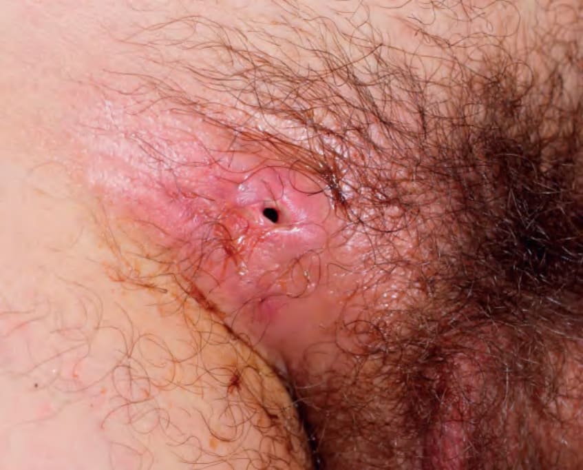
Fig. 82.24 Lymphogranuloma venereum. Inguinal bubo that has ruptured and drained.
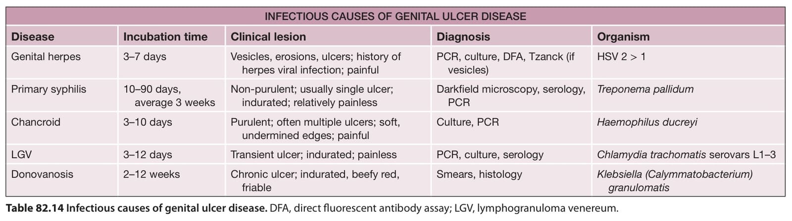
Table 82.14 Infectious causes of genital ulcer disease. DFA, direct fluorescent antibody assay; LGV, lymphogranuloma venereum.
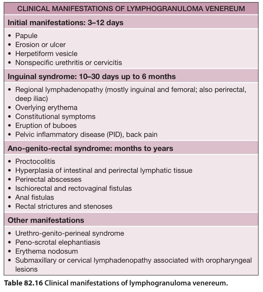
Table 82.16 Clinical manifestations of lymphogranuloma venereum.
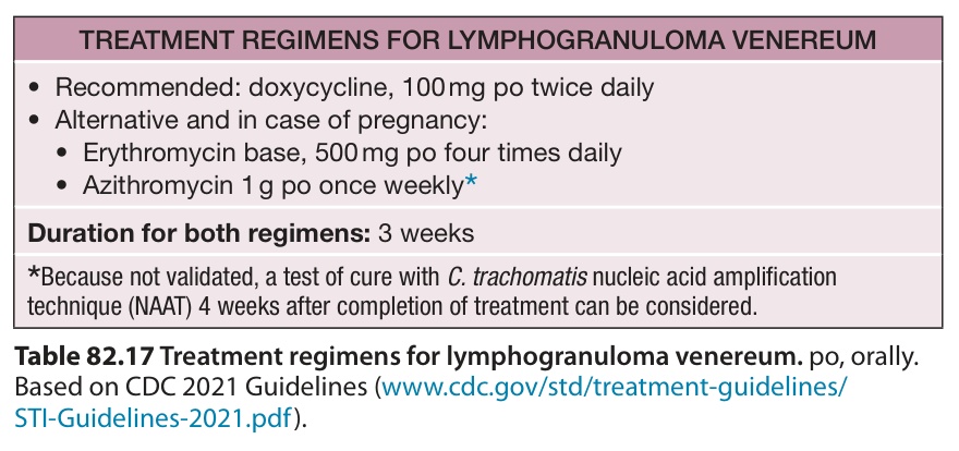
Table 82.17 Treatment regimens for lymphogranuloma venereum. po, orally. Based on CDC 2021 Guidelines (www. cdc. gov/std/treatment-guidelines/ STI-Guidelines-2021. pdf).