DONOVANOSIS (GRANULOMA INGUINALE)
Synonyms: Granuloma venereum Granuloma inguinale tropicum Granuloma venereum genitoinguinale Granuloma genitoinguinale Ulcerating sclerosing granuloma Ulcerating granuloma of the pudendum Serpiginous ulceration of the groin Lupoid form of groin ulceration
Key features
Donovanosis is a rare, chronic, progressive, ulcerative bacterial infection with Klebsiella granulomatis (previously known as Calymmatobacterium granulomatis), a Gram-negative bacillus
Ulcers occur primarily in the genital region
The responsible microorganisms are found within macrophages in smears or biopsy specimens (Donovan bodies)
History
The disease was initially described in India as ulcerations followed by elephantiasis of the genitalia. The microorganism was first identified in 1905 by Donovan, who noted the characteristic Donovan bodies in macrophages and epithelial cells of the stratum malpighii. In 1950, Marmell and Santora proposed the name “donovanosis”. Limited success in culturing the organism until the 1990s explains the slow progress in donovanosis research.
Epidemiology
In the pre-antibiotic era, donovanosis was distributed in both hemispheres, but now it is restricted primarily to a few low-income countries. A large number of cases, the majority in the 20- to 40-year-old age group, are reported from South Africa, India, Papua New Guinea, and some parts of Australia (Fig. 82.25).
Although donovanosis is listed among the group of STIs, the disease can also occur in individuals who are not sexually active, and a wide variation in the prevalence of the infection (from 0.5% to 50%) is reported in sexual partners. This is probably due to different diagnostic procedures and a long incubation period of up to 1 year. It is also not clear whether fecal contamination represents a route of non-sexual transmission.
Pathogenesis
Donovanosis is caused by Klebsiella granulomatis (previously known as Calymmatobacterium granulomatis), an intracellular Gram-negative bacillus. The primary lesion is a small cutaneous papule or nodule that contains mononuclear cells with cytoplasmic vacuoles that are filled with microorganisms. The cytoplasmic vacuoles can rupture and release bipolar Donovan bodies of coccoid, coccobacillary, and bacillary morphology.
Clinical Features
The average incubation period is thought to be ~17 days, but a range from 1 day to 1 year has been reported. A small papule or nodule initially appears and eventually ulcerates. The lesions of donovanosis are usually painless or mildly painful and slowly expand over a period of weeks to months. They are often highly vascular, with a beefy red appearance and a tendency to bleed (Fig. 82.26). Extensive tissue destruction may result, and a foul-smelling exudate is characteristic of necrotic donovanosis. Inguinal lesions occur in up to 20% of patients and are often combined with genital involvement. The most frequent sites of lesions in men are the prepuce, glans penis, frenulum, and coronal sulcus. In women, the most common location is the vulvar area, where large ulcers as well as granulomatous or verrucous papules may be observed.
Extragenital lesions due to autoinoculation or secondary to dissemination have been observed in the skin, bones, abdominal cavity, and oral cavity; they can involve any organ, with the bones most commonly affected. In some patients, primary extragenital lesions are seen.
Pathology
Histologically, ulceration with exuberant granulation tissue is evident. Pseudoepitheliomatous hyperplasia may be present at the ulcer edge. A diffuse infiltrate of histiocytes, plasma cells, and a few lymphocytes is present in the dermis, sometimes with small neutrophilic abscesses. The organisms are easier to find in smears than in histologic sections.
They are 1–2 microns in diameter and are within histiocytes, as is also characteristic of leishmaniasis, rhinoscleroma, and histoplasmosis (see Table 77.18). Bipolar staining is found at two ends of the organisms (Donovan bodies), giving them a “safety pin” appearance.
Diagnosis
The diagnosis of donovanosis is usually based on the demonstration of Donovan bodies in smears from active lesions by Giemsa, Wright, or Leishman stains. The smears are prepared from tissue scrapings or touch preparations of biopsies taken from the dermis or subcutis of lesions.
There are no serologic tests available for the diagnosis of donovanosis. Nucleic acid amplification assays such as PCR are under investigation, but they are not yet commercially available. Although successful culture of K. granulomatis has been reported in human peripheral blood mononuclear cells and Hep2 cells, attempts to grow this organism in fertilized eggs or on routinely available artificial media have been unsuccessful.
Differential Diagnosis
Donovanosis is frequently confused with other diseases of the anogenital region. Other infectious causes of genital ulcer disease have to be considered (see Table 82.14); early lesions of secondary syphilis, especially condylomata lata, often have a clinical appearance similar to donovanosis. Carcinoma, amebiasis, tuberculosis, dimorphic fungal infections, blastomycosis-like pyoderma, Crohn disease, and pyoderma gangrenosum may represent additional diagnostic possibilities.
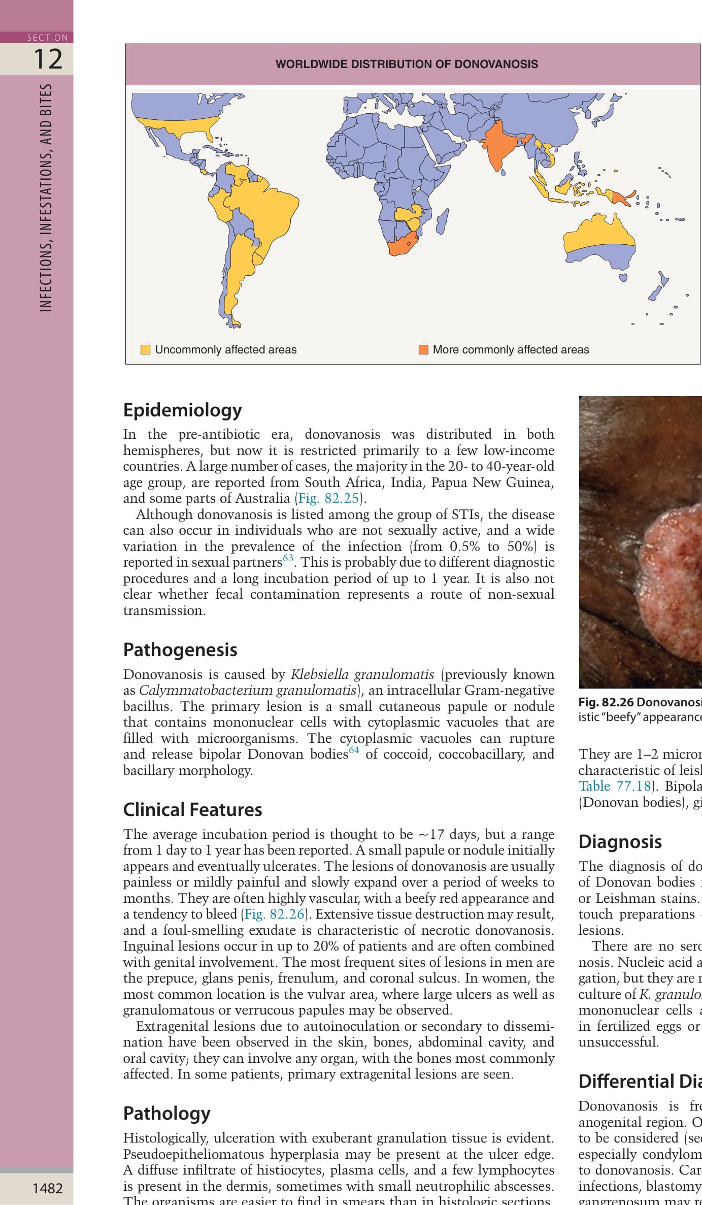
Fig. 82.25 Worldwide distribution of donovanosis.
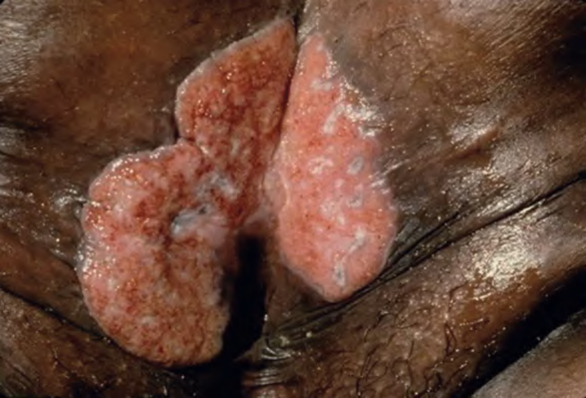
Fig. 82.26 Donovanosis (granuloma inguinale). Large ulcers with a characteristic “beefy” appearance. Courtesy Joyce Rico, MD.
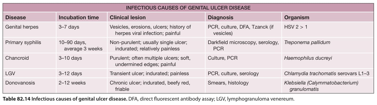
Table 82.14 Infectious causes of genital ulcer disease. DFA, direct fluorescent antibody assay; LGV, lymphogranuloma venereum.
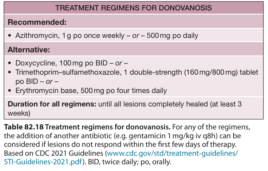
Table 82.18 Treatment regimens for donovanosis. For any of the regimens, the addition of another antibiotic (e. g. gentamicin 1 mg/kg iv q8h) can be considered if lesions do not respond within the first few days of therapy. Based on CDC 2021 Guidelines (www. cdc. gov/std/treatment-guidelines/ STI-Guidelines-2021. pdf). BID, twice daily; po, orally.
The recommended treatment is azithromycin, but treatment regimens in different geographic areas reflect the availability of antibiotics; for example, in Papua New Guinea, chloramphenicol is used. Doxycycline, trimethoprim–sulfamethoxazole, quinolones, and erythromycin (recommended during pregnancy) have also been shown to be effective in the treatment of donovanosis (Table 82.18). For azithromycin, successful treatment with a daily dose of 500 mg for 1 week has been reported. However, the CDC 2021 guidelines recommend 1 g weekly or 500 mg daily for at least 3 weeks and until all lesions have completely healed.
Examination of sexual partners is recommended if there has been sexual contact during the previous 60 days or if the partner is symptomatic. Relapses can occur 6–18 months later, despite effective initial treatment.
Ansteckungsstoff des weichen Schankers und über die Bubonen. Monatshr Prakt Dermatol. 1889;9:387–405.48. Ronald A. Chancroid. In: Mandell GL, (editor-in-chief),
Holmes KK, Sparling PF, Stamm WE, et al. , editors. Sexually Transmitted Diseases. New York: McGraw-Hill; 2008:607–626.33. Handsfield HH, Lipman TO, Harnisch JP, et al.
Rein MF, editor. Atlas of Infectious Diseases, vol. 5. Sexually Transmitted Diseases. New York: Current Medicine; 1995:16. 1–10.49. González-Beiras C, Marks M, Chen CY, et al. Epidemiology
in the adult. In: Holmes KK, Sparling PF, Stamm WE, et al. , editors. Sexually Transmitted Diseases. New York: McGraw-Hill; 2008:627–645.37. Stary A, Ching SF, Teodorowicz L, Lee H. Comparison
Holmes KK, Sparling PF, Stamm WE, et al. , editors. Sexually Transmitted Diseases. New York: McGraw-Hill; 2008:661–684.19. Merritt HH, Adams RD, Solomon HC. Neurosyphilis. New
Table 82.18 Treatment regimens for donovanosis. For any of the regimens, the addition of another antibiotic (e. g. gentamicin 1 mg/kg iv q8h) can be considered if lesions do not respond within the first few days of therapy. Based on CDC 2021 Guidelines (www. cdc. gov/std/treatment-guidelines/ STI-Guidelines-2021. pdf). BID, twice daily; po, orally.