LEISHMANIASIS
Synonyms: Old World cutaneous leishmaniasis: oriental sore, Delhi boil, Baghdad boil New World cutaneous and mucocutaneous leishmaniasis: American tegumentary leishmaniasis, chiclero ulcer (Mexico), uta and espundia (Peru), ulcera de Bauru (Brazil), bush or forest yaws, pian boi (Guyanas) Diffuse cutaneous leishmaniasis: anergic, lepromatous, or pseudolepromatous leishmaniasis Visceral leishmaniasis: kala-azar, Dumdum fever
Key features
Chronic parasitic (protozoan) disease in which organisms are found within phagolysosomes of mononuclear phagocytes
The clinical manifestations of leishmaniasis depend on the host’s cell-mediated immune response and the species of Leishmania involved; clinical subtypes include cutaneous, disseminated cutaneous, diffuse cutaneous, mucocutaneous/mucosal, and visceral leishmaniasis
The most common cutaneous finding is a papule at the site of inoculation that classically evolves into an ulcer
The vector is a sandfly infected with promastigotes
The disease has a worldwide distribution but is endemic in Latin
America, the Mediterranean basin, and parts of Asia and Africa
Introduction
Leishmaniasis encompasses a spectrum of chronic infections in humans and several animal species. It is caused by over 20 species of Leishmania (Table 83.1), flagellated protozoans belonging to the order Kinetoplastidae. Transmission is via the bite of infected female sandflies from the genera Phlebotomus and Lutzomyia.
The disease has a worldwide distribution, affecting millions of people in South America, the Mediterranean basin, and parts of Asia and Africa (Figs. 83.1 & 83.2). There are four major clinical patterns: (1) cutaneous, which is restricted to the skin and is seen more often in the Old World; (2) mucocutaneous, which affects both the skin and mucosal surfaces and occurs almost exclusively in the New World; (3) diffuse cutaneous, which occurs mainly in the New World; and (4) visceral, which affects the organs of the mononuclear phagocyte system, e.g. liver, spleen.
Epidemiology
According to the World Health Organization, more than a million new cases of cutaneous leishmaniasis and ~30 000 new cases of visceral leishmaniasis occur annually. Over 90% of cutaneous infections with Leishmania occur in the Middle East (Afghanistan, Algeria, Iran, Iraq, Saudi Arabia, Syria) and South America (Brazil, Peru, Colombia) (Table 83.2), but the disease is also found in the Mediterranean basin, sub-Saharan Africa, Central Asia, and India (see Fig. 83.1). In the US, climate change has led to the expansion of sandfly vectors to northern Texas. One study found that 59% (41/69) of novel cases of cutaneous leishmaniasis reported to laboratories in Texas from 2007–2017 were autochthonous (occurring in patients with no history of travel outside the US), and all 22 of such cases that were confirmed by PCR were due to L. mexicana.
Mucocutaneous leishmaniasis is endemic in several countries in Central and South America. Although the visceral form of leishmaniasis has a worldwide distribution, it is seen most frequently in Africa and Asia (see Fig. 83.2).
Old World cutaneous leishmaniasis is usually due to L. major or L. tropica, and less often L. infantum (Mediterranean basin) or L. aethiopica (Ethiopia and Kenya) (see Table 83.1 & Fig. 83.1). In the New World, cutaneous leishmaniasis is caused primarily by subspecies of L. mexicana and the L. braziliensis complex, whereas mucocutaneous/mucosal disease is associated with the latter organisms. Diffuse cutaneous leishmaniasis is most closely associated with L. amazonensis. Infections with L. donovani (e.g. India, Bangladesh, Sudan) and L. infantum (especially in the setting of HIV infection) as well as the subspecies L. infantum chagasi (e.g. Central and South America; previously known as L. chagasi) are the major causes of visceral leishmaniasis (Table 83.3 & Fig. 83.2).
The species of Leishmania can also vary within a particular geographic region. For example, in the Middle East, L. major is found in rural areas where the primary animal reservoirs are desert rodents, whereas L. tropica is endemic in urban areas.
Canine and rodent species act as the main reservoirs for Leishmania (see Table 83.1). Transmission between these species (as well as to humans) is via sandflies, primarily of the genera Phlebotomus (Old World) and Lutzomyia (New World). Human-to-human spread of L. tropica can also occur. In general, humans contract leishmaniasis as accidental hosts when they intrude into the sandfly’s habitat. Overall, the disease is most common in men between 20 and 40 years of age, but Old World cutaneous leishmaniasis occurs more often in children.
Epidemics of cutaneous leishmaniasis have occurred in populations affected by natural disasters, forced migration, and civil unrest as well as in foreign military personnel serving in endemic areas of the Middle East (e.g. Iran, Afghanistan, Syria, Iraq). Whereas the incidence of visceral leishmaniasis seems to be decreasing in endemic countries such as India, Bangladesh, and Nepal, new areas of epidemic visceral leishmaniasis are appearing in Sudan and South Sudan. Migration from rural areas to peri-urban centers has also contributed to an increasing incidence of visceral leishmaniasis in Brazil. Solid organ and hematopoietic stem cell transplant recipients and other individuals receiving immunosuppressive medications (e.g. tumor necrosis factor [TNF] inhibitors) represent additional groups at increased risk for visceral leishmaniasis.
Pathogenesis
Leishmania spp. are obligate intracellular parasites that exist in two forms: promastigote and amastigote. In the gut of the sandfly, the organisms multiply as extracellular flagellated promastigotes. Following migration to the proboscis, the parasites are inoculated (in the promastigote form) via the sandfly bite. Once inoculated into the skin, the parasites are rapidly engulfed by the host’s mononuclear phagocytes, where they transform into amastigotes and multiply via binary fission (Fig. 83.3). The incubation time from infection until the first clinical manifestation can vary from ≤2 weeks for cutaneous lesions (typically several weeks to 2 months) to >2 years for mucocutaneous lesions or visceral involvement (typically 3 to 9 months).
The clinical manifestations of leishmaniasis depend on the species of Leishmania (see Table 83.1), the host’s cell-mediated response, and the ability of the parasite to evade host defense mechanisms. A robust Th1 response with production of interleukin-2 (IL-2) and interferon-γ (IFN-γ) is associated with quicker resolution, whereas a lack of a Th1 response and/ or the development of a Th2-type response with production of IL-4 and IL-10 is associated with disease progression. Depending on the type of immune response, l-arginine metabolism within infected macrophages occurs via two alternative pathways: (1) Th2 cytokines promote its catabolism by arginase to produce l-ornithine, which favors parasitic growth; or (2) Th1 cytokines promote nitric oxide synthase-induced generation of l-citrulline plus nitric oxide, and the latter increases host resistance via NLRP3 (NLR family pyrin domain-containing-3) inflammasome-derived IL-1β. Resistance of L. amazonensis and L. braziliensis promastigotes to nitric oxide in vitro correlates with poorer clinical outcomes.
Mucocutaneous disease is associated with the infection of Leishmania organisms with Leishmania RNA virus-1 (LRV1); this virus is recognized by human Toll-like receptor 3, leading to a proinflammatory cascade that subverts the immune response to Leishmania, and allows disease progression. L. donovani has been shown to induce epigenetic changes via macrophage DNA methylation that downregulate host defense mechanisms, allowing parasitic replication and survival. Human genetic factors include HLA variants that contribute to leishmaniasis risk and IL2RA variants that reduce IL-2-dependent responses and thereby increase susceptibility to cutaneous and visceral leishmaniasis.
IFN-γ is the most potent cytokine involved in the induction of activity against organisms residing within macrophage phagolysosomes. It is produced by Th1 cells and leads to the production of oxygen species. The differentiation of naive CD4+ cells into Th1 cells is aided by IL-12 (see Fig. 4.9), which also stimulates natural killer (NK) cells to produce IFN-γ. TNF produced by macrophages and NK cells amplifies the IFN-γ-driven macrophage activation. Consequently, use of TNF inhibitors increases the risk of developing leishmaniasis, especially the visceral form. More recent studies have also highlighted the role of TNF-dependent IL-32γ in stimulating the production of proinflammatory cytokines and microbicidal molecules in human macrophages infected with New World Leishmania spp.. In addition, Th17 cells can potentially contribute to both host resistance and tissue damage in patients with leishmaniasis.
Animals that have recovered from L. tropica or L. donovani infections acquire immunity against reinfection from the same species of Leishmania, but not against other species. Similarly, humans with New World cutaneous leishmaniasis have been inoculated successfully with L. tropica. However, it has been reported that surviving an episode of visceral leishmaniasis confers lifelong immunity against all types of leishmaniasis.
Clinical Features
Cutaneous leishmaniasis
Cutaneous leishmaniasis is divided into two subsets based on the geographic region where the infection is acquired: Old World and New World. These two groups differ with regard to the causative organisms, vectors, reservoirs, clinical presentation, and prognosis. Both Old World and New World cutaneous leishmaniasis usually begin as a small, well-circumscribed papule at the inoculation site. This lesion may slowly enlarge over several weeks into a nodule or plaque and then become ulcerated or verrucous (Figs. 83.4–83.6). Exposed sites such as the face, neck, arms, and legs are most commonly involved. For example, the chiclero ulcer results from infection on the ear in forest workers who collect gum from the chicle tree in Mexico and Central America (see Fig. 83.4D). The lesions of leishmaniasis are often solitary but may be multiple, with the formation of satellites or lymphatic spread (sporotrichoid pattern; Fig. 83.7). Unusual clinical presentations include erysipeloid, zosteriform, psoriasiform, and eczematous. Most acute cutaneous infections resolve spontaneously within several months, often with scarring (cicatricial stage). A minority of patients develop chronic or disseminated disease.
Several clinical forms of Old World cutaneous leishmaniasis have been described, including zoonotic, anthroponotic, recidivans, and
This patient presented with an infiltrated hyperpigmented plaque with papules at its periphery. Cutaneous leishmaniasis due to L. aethiopica often affects the face and can evolve to mucocutaneous disease. Courtesy Edward Cowen, MD.
lupoid leishmaniasis. Zoonotic cutaneous leishmaniasis (rural, moist, or early ulcerative form) usually has a mild, rapid course and is caused by L. major. Anthroponotic cutaneous leishmaniasis (urban, dry, or late ulcerative form), has a more chronic course and is caused by L. tropica. Leishmaniasis recidivans is a chronic, destructive form that is characterized by recurrence at the site of an original ulcer, generally within 2 years and often at the edge of the scar. Anthroponotic or, less frequently, zoonotic cutaneous leishmaniasis occasionally develops into chronic lupoid leishmaniasis, which clinically and histologically (epithelioid granulomas surrounded by lymphocytes) resembles the lupus vulgaris form of cutaneous tuberculosis. Amastigotes are scarce and difficult to find in lupoid leishmaniasis, making the diagnosis more challenging.
New World cutaneous leishmaniasis has a wide clinical spectrum, including plaque-type, sporotrichoid, pustular, impetigo-like,
Montenegro test. This expanding ulcer could be misdiagnosed as pyoderma gangrenosum. Courtesy Omar P. Sangüeza, MD.
eczematoid, sarcoid-like, lupoid, erysipeloid, papulotuberous, verrucous, disseminated, and diffuse presentations. There is also the potential for developing leishmaniasis recidivans.
Disseminated cutaneous leishmaniasis, which most often results from L. braziliensis or L. amazonensis infection, is characterized by multiple (10 to >300) secondary lesions, either in proximity to or distant from the primary site. The lesions resemble those of classic cutaneous leishmaniasis, including papules and small nodules that may have an acneiform appearance, and ulceration is common (Fig. 83.8A). Up to 25% of affected individuals have concomitant mucosal disease, and patients may have systemic symptoms such as fever and malaise. Disseminated skin disease most likely results from early hematologic spread, and it is associated with decreased production of IFN-γ and TNF.
Diffuse cutaneous leishmaniasis is a rare presentation that develops in the setting of reduced cell-mediated immunity, analogous to the lepromatous form of Hansen disease. L. aethiopica (in Africa) and L. amazonensis (in the Americas) are the most common pathogens (see Table 83.1). Multiple nodular and keloid-like lesions on the face and limbs are usually observed (Fig. 83.8B); ulceration is uncommon but occasionally occurs secondary to trauma. Nasal infiltration and mucosal ulceration may develop due to spread from nearby skin lesions, but destruction of the nasal septum is rare. There may also be laryngeal and pharyngeal involvement.
Mucocutaneous/mucosal leishmaniasis
After a variable time period ranging from a few months to more than 20 years, mucocutaneous/mucosal disease develops in some patients
infected with Leishmania spp. in the Viannia subgenus (known as the L. braziliensis complex), most commonly L. braziliensis and occasionally L. panamensis, L. guyanensis, or hybrid genotypes. Mucosal lesions range from edema of the lips and nose to perforation of the nasal septum or (less often) the laryngeal cartilage or palate. Infiltration and/or ulceration of the mucosal surfaces of the nose, lips, and oropharynx are typical features (Figs. 83.9 & 83.10); ocular or genital involvement is rare. In some patients, there is extensive loss of tissue in both the mouth and nose, causing a characteristic “tapir face” known as espundia. Hoarseness may result from vocal cord involvement.
Ulceration and induration of the nasal vestibule extends onto the cutaneous lip. Courtesy Kalman Watsky, MD.
Visceral leishmaniasis (kala-azar)
Visceral leishmaniasis, or kala-azar, occurs when the parasite spreads to the bone marrow, spleen, and liver. It is primarily caused by L. donovani in adults and L. infantum or L. infantum chagasi in children (see Fig. 83.2). The incubation period ranges from 1 to 36 months. Fever, wasting, cough, lymphadenopathy, and hepatosplenomegaly are the most common systemic findings (Table 83.4). There may be an abrupt onset or slow progression, and fever may be continuous or inter-mittent. Additional complications include enteritis, oronasal or gastrointestinal hemorrhage, pneumonia, and nephritis, which may lead to death. Cutaneous manifestations may be disease-specific papules, nodules, or ulcers at infected sites as well as nonspecific findings such as purpura, hyperpigmentation, xerosis, and kwashiorkor-like hair discoloration.
Post-kala-azar dermal leishmaniasis develops as a sequela of untreated or treated visceral leishmaniasis (Fig. 83.11). This form of cutaneous leishmaniasis is most commonly seen in Sudan and India, where it occurs in 50% and 10% of patients cured of visceral leishmaniasis, respectively. Onset may be up to 20 years after treatment. Skin
findings include hypopigmented macules, malar erythema, skin-colored nodules, and verrucous papules.
Leishmaniasis and HIV
Co-infection of HIV and leishmaniasis can alter the progression and presentation of leishmaniasis, especially the visceral form (see Ch. 78). HIV infection increases the risk of developing visceral leishmaniasis 100- to over 2000-fold, with the prevalence of visceral leishmaniasis ranging from 4% to >15% among HIV-infected patients who live in Leishmania-endemic areas. Co-infection often results in an atypical clinical presentation, reduced therapeutic response, and increased mortality. Chronic immune activation from the Leishmania infection may lead to increased HIV load and accelerated progression of AIDS, while the immunosuppressed state from the HIV infection creates an ideal environment for parasitic growth.
Pathology
Histologically, cutaneous lesions typically show ulceration, pseudoepitheliomatous hyperplasia, and a mixed inflammatory infiltrate composed of histiocytes, lymphocytes, plasma cells, and neutrophils. Amastigotes present within dermal macrophages, especially those in the papillary dermis (Fig. 83.12), are evident in ∼50% of skin biopsies. Over time, lesions develop an increasing number of giant cells and fewer parasites; in longstanding cutaneous leishmaniasis, tuberculoid granulomas with caseation necrosis may be observed. In the cicatricial stage, the epidermis becomes flattened and hyperpigmented in areas with dermal fibrosis. Identical changes are seen in mucosal lesions. In diffuse cutaneous leishmaniasis, numerous amastigotes are present within foamy histiocytes; in contrast, the disseminated form features a primarily lymphoplasmacytic infiltrate with few amastigotes. The parasite can be detected in the lymph nodes, bone marrow, and spleen in patients with visceral leishmaniasis.
Diagnosis
The diagnosis of cutaneous leishmaniasis can be confirmed by demonstrating the presence of amastigotes in dermal macrophages within skin biopsy specimens, tissue impression smears (touch preparations), and smears obtained by dermal scraping or needle aspiration of skin lesions. Although routine hematoxylin and eosin staining allows the visualization of the amastigotes, Giemsa, Wright, or Feulgen stains can help to identify the organisms in smears and tissue: the cytoplasm appears blue, the nucleus pink, and the kinetoplast a deep red. CD1a immunostaining can also highlight the amastigotes, but its positivity may vary depending on the species (Fig. 83.12, inset). The edge of a relatively new ulcer is the location of choice for obtaining dermal scrapings, a biopsy specimen, or a needle aspirate; the latter two types of samples may be used for culture and all three types for polymerase chain reaction (PCR)-based assays (see below).
In longer-standing lesions of leishmaniasis, parasites may be scarce. In this context, the delayed skin reaction test (Montenegro skin test or
Leishman reaction), which uses leishmanial antigens to induce a cellmediated response, has traditionally been an important diagnostic tool. A phenolated suspension of killed promastigotes is injected intradermally, usually on the volar aspect of the forearm. The test is considered positive if a papule >5 mm in diameter forms at the site of inoculation after 48–72 hours (see Fig. 83.6). It is positive in up to 90% of patients with cutaneous and mucocutaneous leishmaniasis of over 3 months duration and is invariably negative in patients with diffuse cutaneous (anergic) leishmaniasis. However, this test (which is not approved by the US Food and Drug Administration [FDA]) cannot distinguish between past and present infection, so it is most useful in patients who do not reside in areas where leishmaniasis is endemic; positivity develops while the skin lesions are still active, and the test remains positive long after spontaneous cure. The test is usually negative during the febrile phase of visceral leishmaniasis, but it often becomes positive after cure.
For culturing Leishmania, specialized media are required, e.g. Nicolle– Novy–MacNeal (NNM) media or chick embryo media. Cultures are positive in ~40% of cases. Serologic and immunologic tests are also available, e.g. indirect immunofluorescence, ELISA, immunoprecipitation, and isoenzyme electrophoresis. Serologic testing is most useful for visceral and occasionally mucocutaneous disease, although it is not specific due to cross-reactivity (e.g. with Chagas disease antibodies). PCR-based methods represent the most sensitive and specific diagnostic tests, and their availability is increasing. The combination of PCR (heat shock protein 70, mini-exon) with histopathology increases sensitivity to >90%. Of note, an FDA-approved real-time PCR assay for the diagnosis of leishmaniasis is available through the Centers for Disease Control and Prevention (CDC).
Differential Diagnosis
The differential diagnosis of cutaneous leishmaniasis includes persistent arthropod bite reaction, basal cell carcinoma, tuberculosis, non-tuberculous mycobacterial infections, and subcutaneous mycoses; other infectious causes of lesions in a lymphocutaneous pattern are listed in Table 77.17. Mucocutaneous leishmaniasis can resemble paracoccidioidomycosis and tertiary syphilis. In addition, granulomatosis with polyangiitis and angiocentric NK/T cell lymphoma should be considered when ulcerative mucocutaneous lesions affect the central part of the face. Cocaine-induced nasal ulcerations can also mimic the mucosal involvement of leishmaniasis. Histologic examination allows distinction among these diseases. The histologic differential diagnosis includes other infections characterized by parasitized macrophages (see Table 77.18) and neoplasms such as squamous cell carcinoma when pseudoepitheliomatous hyperplasia is present. The morphology and
distribution of diffuse cutaneous leishmaniasis can mimic the lepromatous form of Hansen disease; however, in leishmaniasis the eyebrows are spared and the lesions are usually less infiltrative.
Treatment
Factors to consider in the treatment of leishmaniasis include the region of the world in which the infection was acquired, the species of Leishmania, the site(s) and severity of the infection, and host factors such as immune status and age (Table 83.5). The benefits of therapy need to be balanced with the goal of minimizing drug toxicity. Without treatment, Old World cutaneous leishmaniasis typically resolves within 2–4 months (L. major) or 6–15 months (L. tropica). Indications for systemic treatment of Old World cutaneous leishmaniasis include: (1) an immunocompromised host; (2) ≥5 lesions of substantial size (e.g. >1 cm) or individual lesion(s) measuring ≥5 cm; (3) enlarged regional lymph nodes; and (4) involvement of the mucosa, face, ears, genitalia, fingers, toes, or skin overlying a joint. For patients who do not meet these criteria, either observation, if the lesions are healing spontaneously within 6 months, or local therapy are options.
New World cutaneous leishmaniasis caused by L. mexicana resolves within 3 months in >75% of cases. In contrast, cutaneous disease caused by L. braziliensis and L. panamensis spontaneously heals in less than 10% and 35% of cases, respectively. Therefore, systemic treatment is indicated when L. braziliensis complex infection is suspected in order to accelerate healing, decrease scarring (especially in cosmetically sensitive sites), and prevent dissemination, relapse, or the development of mucosal or diffuse disease (see Table 83.5).
Both parenteral pentavalent antimonials and miltefosine are firstline systemic treatments for cutaneous and mucocutaneous/mucosal leishmaniasis. While miltefosine is a less toxic therapy, its use can be limited by cost. Liposomal amphotericin B is the treatment of choice for visceral leishmaniasis. Table 83.6 summarizes treatment regimens for cutaneous and mucocutaneous/mucosal leishmaniasis. Table 83.7 lists frequent adverse reactions to drugs used for the treatment of leishmaniasis. The addition of oral pentoxifylline to antimonial therapy has been utilized for refractory mucocutaneous leishmaniasis and is included as an alternative treatment by the Pan American Health Organization, although RCTs have not consistently shown a benefit. Additional interventions that have demonstrated some efficacy for cutaneous or mucocutaneous/mucosal leishmaniasis (in combination with other agents) include thermotherapy, cryotherapy, photodynamic therapy, and allopurinol.
Drugs or vaccines that completely prevent leishmanial infection have not yet been developed. The best form of protection is to avoid the bite of the sandfly and eliminate animal reservoirs. Personal protection includes applying DEET-based repellants, bed netting, permethrintreated clothing, and sleeping in minimal-risk areas. People are at increased risk of bites in the early morning and late evening.
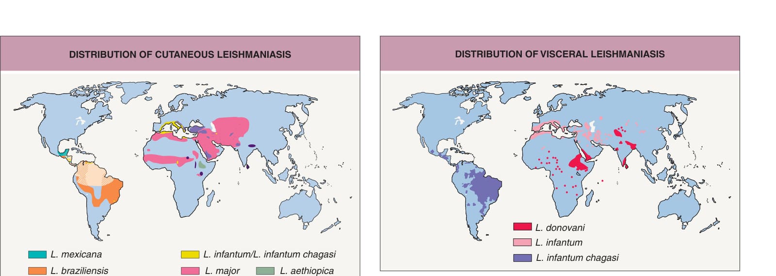
Fig. 83.1 Distribution of cutaneous leishmaniasis. Climate change is expected to lead to expansion of these distributions. For example, autochthonous cases of cutaneous leishmaniasis due to L. mexicana have reported as far north as Texas. Modified from Aronson N, Herwaldt BL, Libman M, et al. Diagnosis and Treatment of Leishmaniasis: Clinical practice guidelines by the Infectious Diseases Society of America (IDSA) and the American Society of Tropical Medicine and Hygiene (ASTMH). Clin Infect Dis 2016;63:1539–57 and Cohen J, Powderly WG, Opal SM (eds). Infectious Diseases, 4th edition. Edinburgh: Elsevier, 2016.
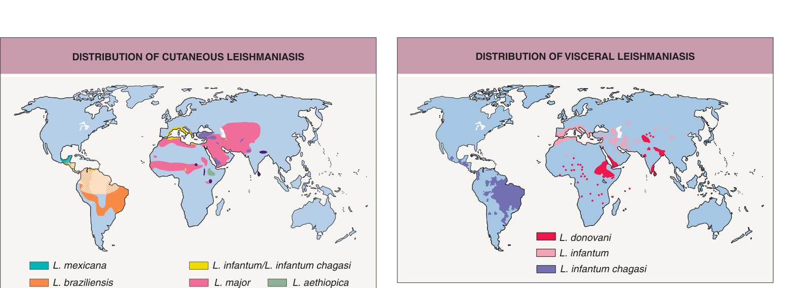
Fig. 83.2 Distribution of visceral leishmaniasis. These organisms, in particular L. infantum and L. donovani, can also cause cutaneous disease. Modified from Aronson N, Herwaldt BL, Libman M, et al. Diagnosis and Treatment of Leishmaniasis: Clinical practice guidelines by the Infectious Diseases Society of America (IDSA) and the American Society of Tropical Medicine and Hygiene (ASTMH). Clin Infect Dis 2016;63:1539–57 and Cohen J, Powderly WG, Opal SM (eds). Infectious Diseases, 4th edition. Edinburgh: Elsevier, 2016.
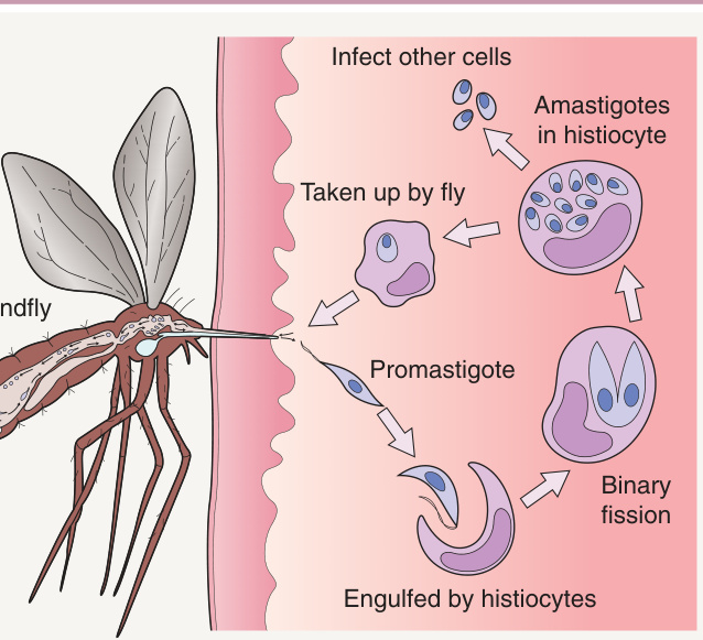
Fig. 83.3 Life cycle of Leishmania species. Promastigotes develop within the gut of the sandfly and then migrate to the proboscis.
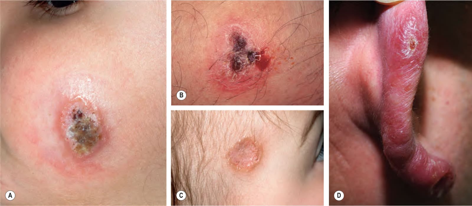
Fig. 83.4 Variable presentations of cutaneous leishmaniasis.A, B Ulcerated plaques with rolled border (A) and central crusting (A, B). C Plaque with translucent borders containing telangiectasias and central scarring. D New World cutaneous leishmaniasis of the ear (chiclero ulcer). Cutaneous leishmaniasis is sometimes mistaken for a basal cell carcinoma. A, C, Courtesy Julie V. Schaffer, MD; D, Courtesy Kalman Watsky, MD.
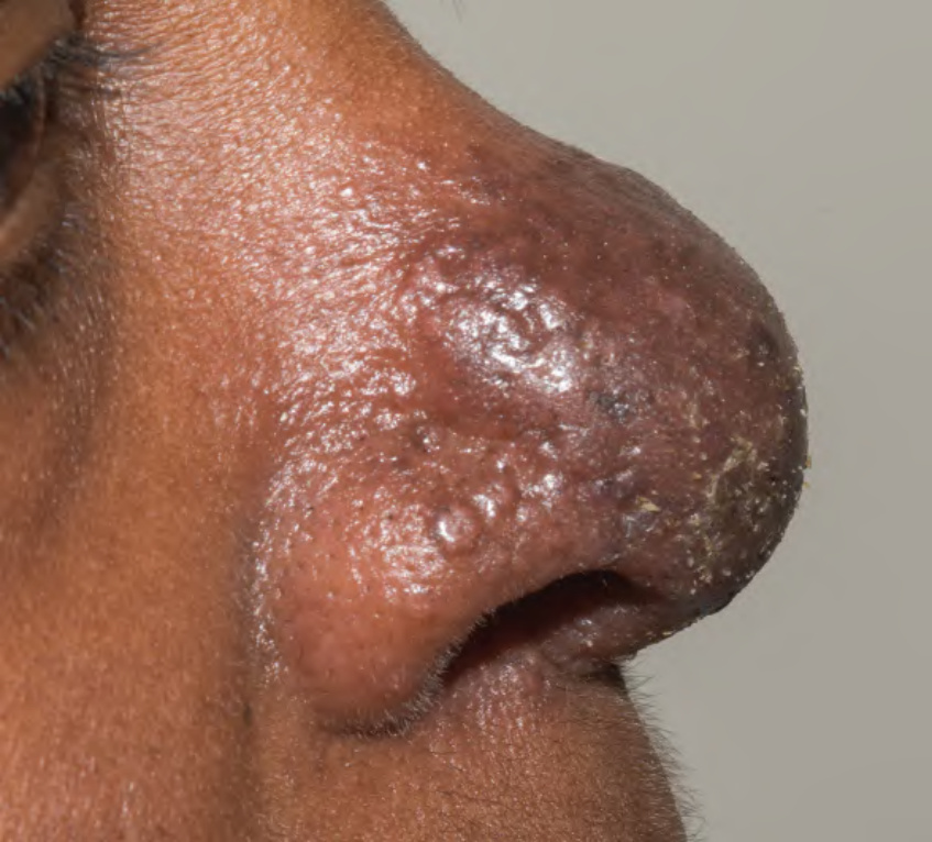
Fig. 83.5 Cutaneous leishmaniasis of the nose due to Leishmania aethiopica.
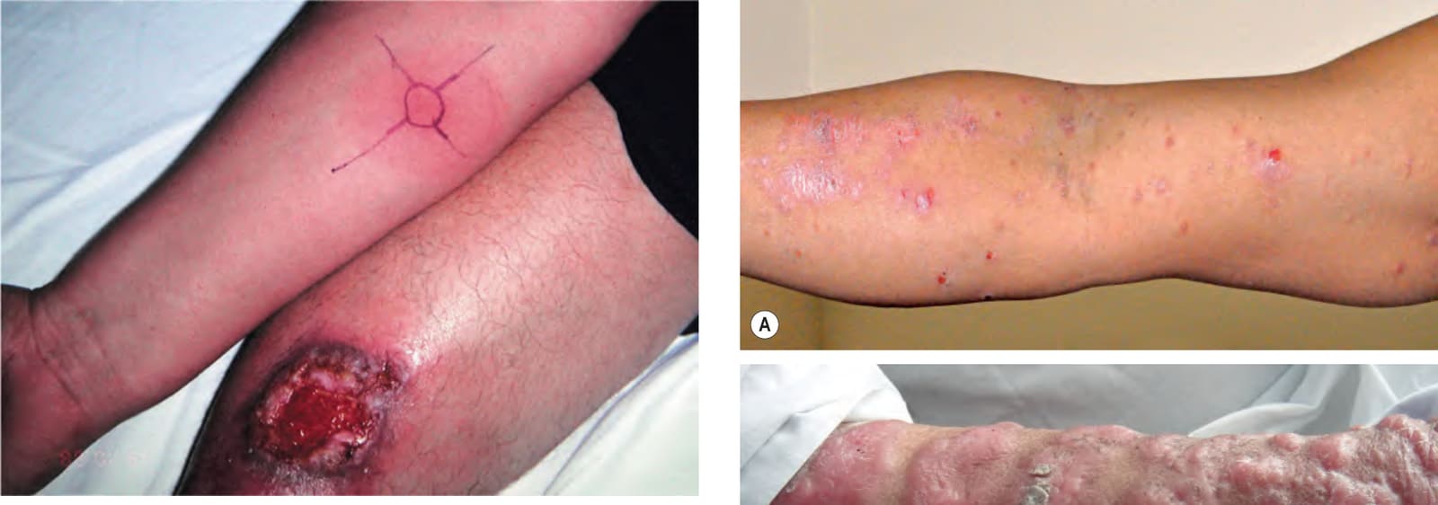
Fig. 83.6 Ulcerative cutaneous leishmaniasis on the leg and a positive
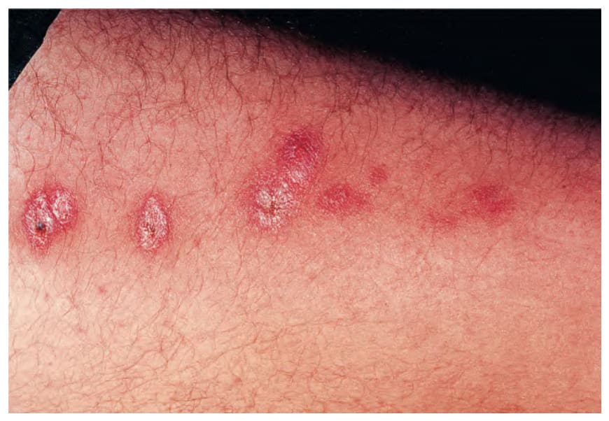
Fig. 83.7 Sporotrichoid form of cutaneous leishmaniasis.
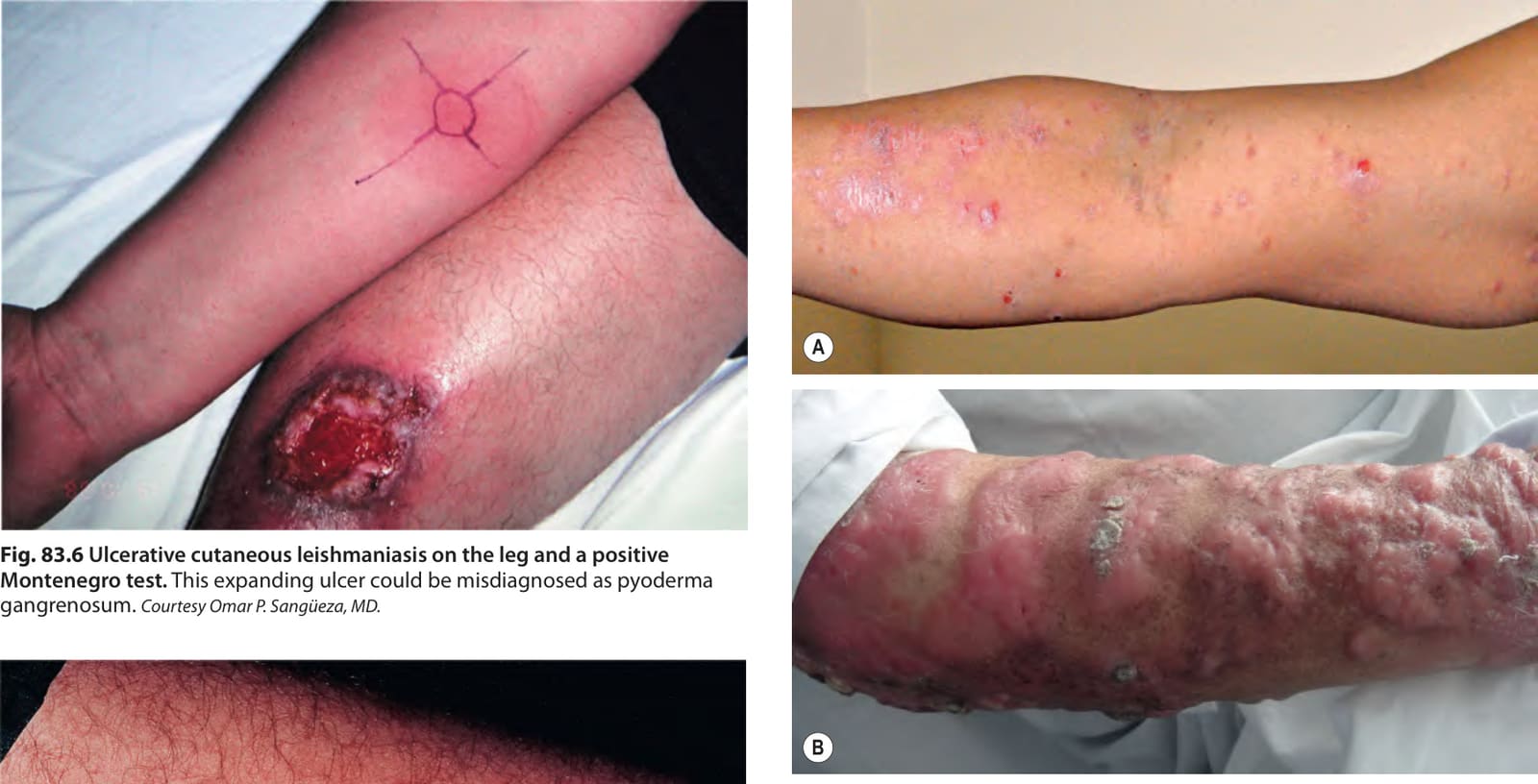
Fig. 83.8 Diffuse and disseminated cutaneous leishmaniasis.A Small papules in disseminated cutaneous leishmaniasis. B Diffuse cutaneous leishmaniasis presenting with large nodules that resemble the lepromatous form of Hansen disease.
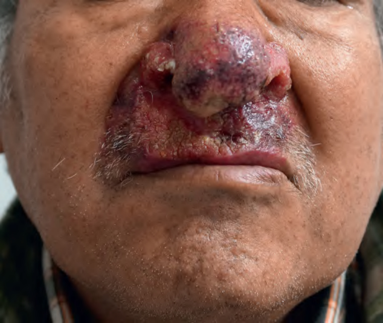
Fig. 83.9 Mucocutaneous leishmaniasis. Infiltration of the nose and upper lip is evident. Note the destruction of the nasal alae and crusting of the upper lip. Courtesy Jaqueline Luque, MD.
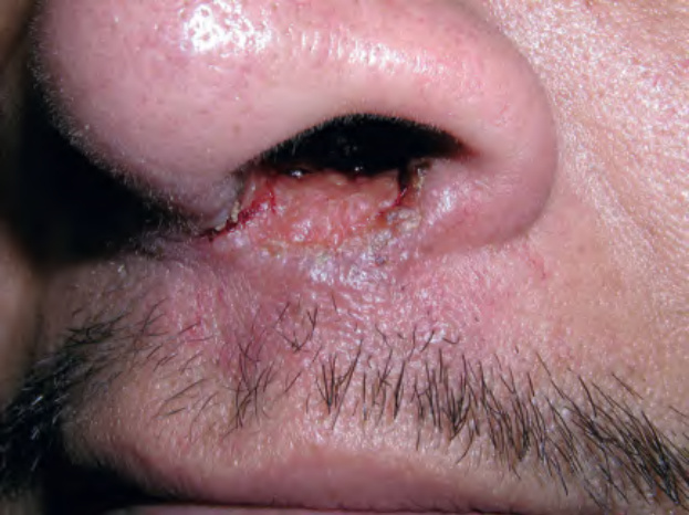
Fig. 83.10 Mucocutaneous leishmaniasis due to Leishmania braziliensis.
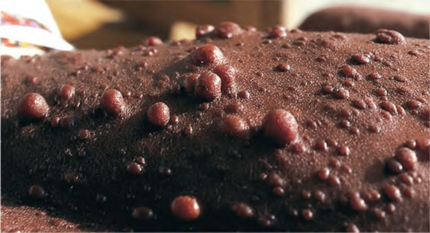
Fig. 83.11 Post-kala-azar dermal leishmaniasis. Nodules of various sizes, some pedunculated, are seen in this patient who had been treated for kala-azar over a period of 6 months, 20 years previously. With permission from Peters W, Pasvol G. Tropical Medicine and Parasitology, 6th edition. London: Mosby, 2007.
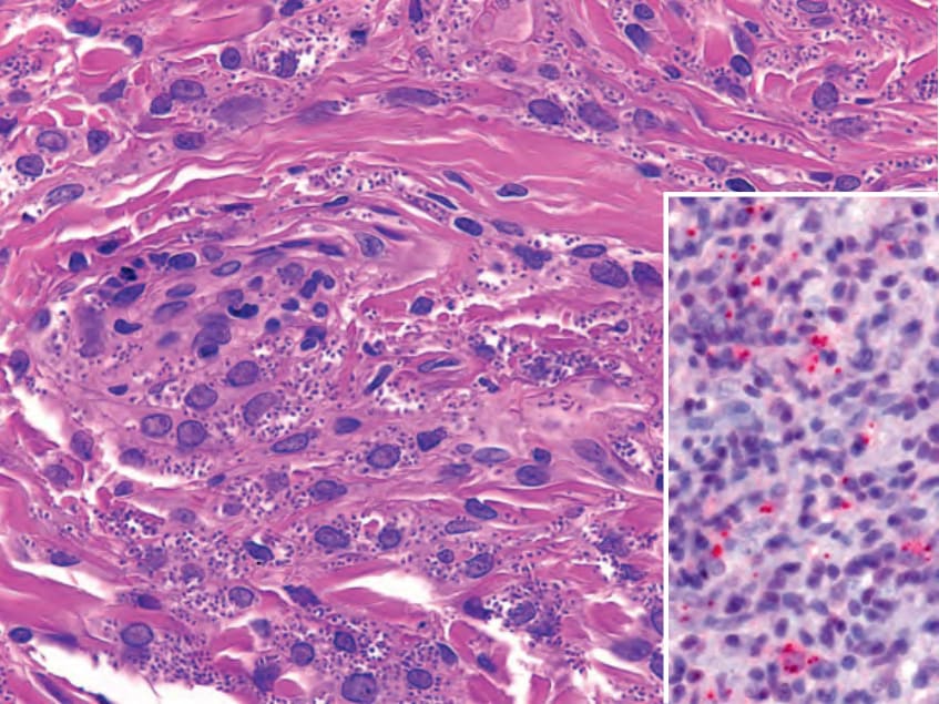
Fig. 83.12 Histopathology of leishmaniasis. Parasitized macrophages that contain amastigotes are seen in the dermis and highlighted by CD1a immuno staining (inset). Each amastigote has a nucleus and kinetoplast (not always visible) at opposite poles. Courtesy Luis Requena, MD; inset, courtesy Lorenzo Cerroni, MD.
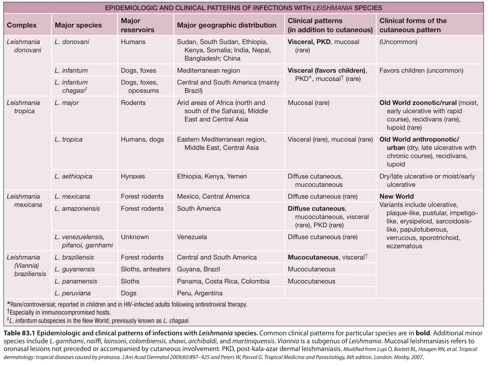
Table 83.1 Epidemiologic and clinical patterns of infections with Leishmania species. Common clinical patterns for particular species are in bold. Additional minor species include L. garnhami, naiffi, lainsoni, colombiensis, shawi, archibaldi, and martiniquensis. Viannia is a subgenus of Leishmania. Mucosal leishmaniasis refers to oronasal lesions not preceded or accompanied by cutaneous involvement. PKD, post-kala-azar dermal leishmaniasis. Modified from Lupi O, Barlett BL, Haugen RN, et al. Tropical dermatology: tropical diseases caused by protozoa. J Am Acad Dermatol 2009;60:897–925 and Peters W, Pasvol G. Tropical Medicine and Parasitology, 6th edition. London: Mosby, 2007.
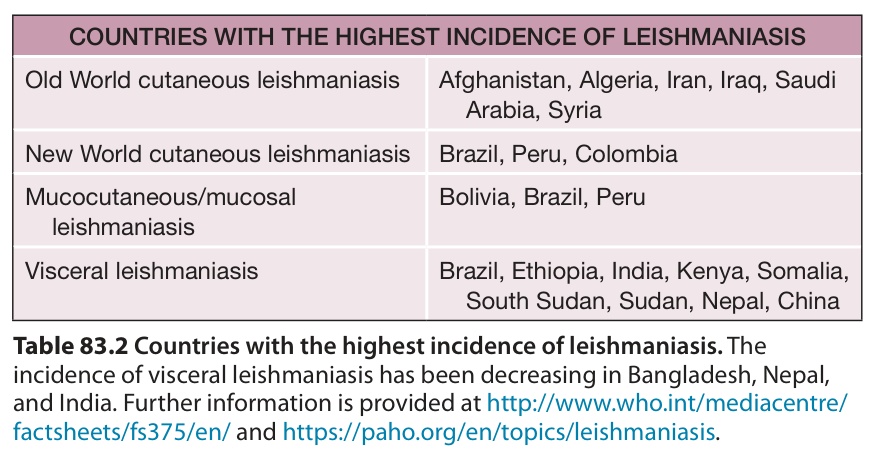
Table 83.2 Countries with the highest incidence of leishmaniasis. The incidence of visceral leishmaniasis has been decreasing in Bangladesh, Nepal, and India. Further information is provided at http://www.who.int/mediacentre/ factsheets/fs375/en/ and https://paho.org/en/topics/leishmaniasis.
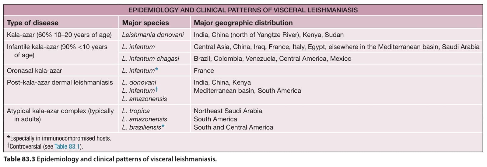
Table 83.3 Epidemiology and clinical patterns of visceral leishmaniasis.
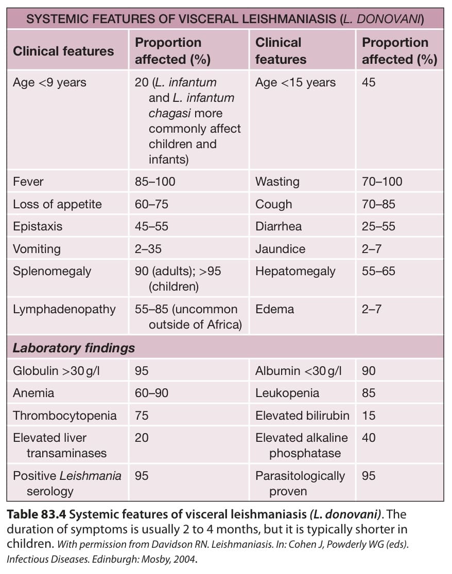
Table 83.4 Systemic features of visceral leishmaniasis (L. donovani). The duration of symptoms is usually 2 to 4 months, but it is typically shorter in children. With permission from Davidson RN. Leishmaniasis. In: Cohen J, Powderly WG (eds). Infectious Diseases. Edinburgh: Mosby, 2004.
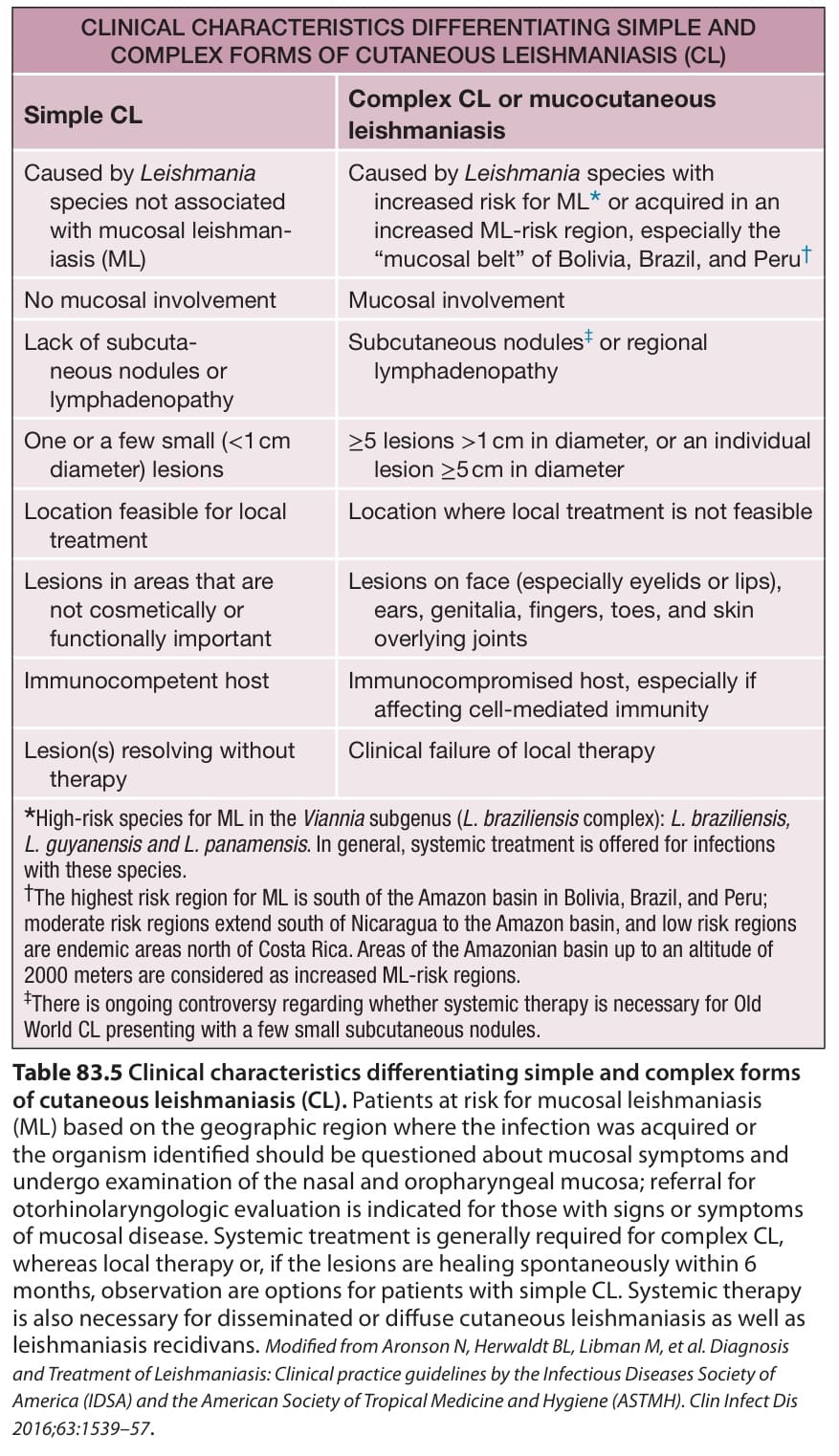
Table 83.5 Clinical characteristics differentiating simple and complex forms of cutaneous leishmaniasis (CL). Patients at risk for mucosal leishmaniasis (ML) based on the geographic region where the infection was acquired or the organism identified should be questioned about mucosal symptoms and undergo examination of the nasal and oropharyngeal mucosa; referral for otorhinolaryngologic evaluation is indicated for those with signs or symptoms of mucosal disease. Systemic treatment is generally required for complex CL, whereas local therapy or, if the lesions are healing spontaneously within 6 months, observation are options for patients with simple CL. Systemic therapy is also necessary for disseminated or diffuse cutaneous leishmaniasis as well as leishmaniasis recidivans. Modified from Aronson N, Herwaldt BL, Libman M, et al. Diagnosis and Treatment of Leishmaniasis: Clinical practice guidelines by the Infectious Diseases Society of America (IDSA) and the American Society of Tropical Medicine and Hygiene (ASTMH). Clin Infect Dis 2016;63:1539–57.
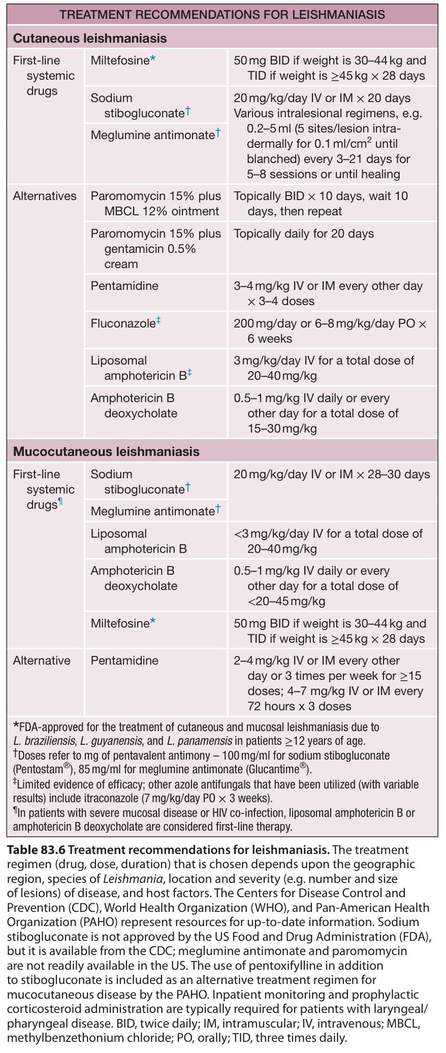
Table 83.6 Treatment recommendations for leishmaniasis. The treatment regimen (drug, dose, duration) that is chosen depends upon the geographic region, species of Leishmania, location and severity (e.g. number and size of lesions) of disease, and host factors. The Centers for Disease Control and Prevention (CDC), World Health Organization (WHO), and Pan-American Health Organization (PAHO) represent resources for up-to-date information. Sodium stibogluconate is not approved by the US Food and Drug Administration (FDA), but it is available from the CDC; meglumine antimonate and paromomycin are not readily available in the US. The use of pentoxifylline in addition to stibogluconate is included as an alternative treatment regimen for mucocutaneous disease by the PAHO. Inpatient monitoring and prophylactic corticosteroid administration are typically required for patients with laryngeal/ pharyngeal disease. BID, twice daily; IM, intramuscular; IV, intravenous; MBCL, methylbenzethonium chloride; PO, orally; TID, three times daily.
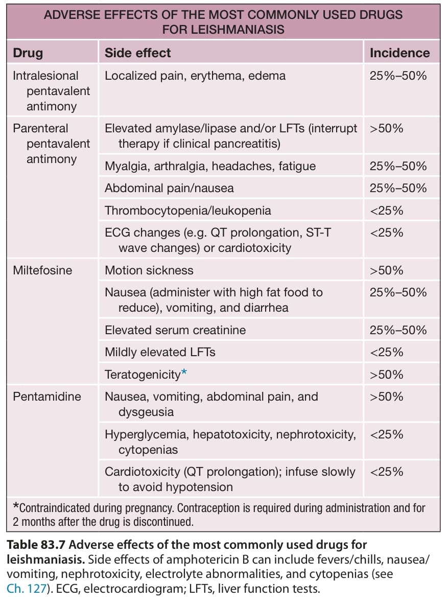
Table 83.7 Adverse effects of the most commonly used drugs for leishmaniasis. Side effects of amphotericin B can include fevers/chills, nausea/ vomiting, nephrotoxicity, electrolyte abnormalities, and cytopenias (see Ch. 127). ECG, electrocardiogram; LFTs, liver function tests.