CUTANEOUS LARVA MIGRANS
Synonym: Creeping eruption
Key features
Due to larvae of animal hookworms (intestinal nematodes)
Skin lesions are serpiginous and pruritic, representing the paths of migration of organisms within the epidermis
The rate of migration is 1–2 cm per day
Self-limited disease
Cutaneous larva migrans is a serpiginous cutaneous eruption caused by the accidental penetration and migration of animal hookworm larvae through the epidermis. The infection has a worldwide distribution and occurs most frequently in warmer climates. The skin lesions are usually self-limited.
Epidemiology and Pathogenesis
Cutaneous larva migrans is caused by the larvae of hookworms that infect domestic dogs and cats, most often Ancylostoma braziliense or A. caninum and occasionally Uncinaria stenocephala or Bunostomum phlebotomum. Although cutaneous larva migrans has a worldwide distribution, it is most commonly seen in warm climates, such as the southeastern US, Central and South America, Africa, and other tropical areas. The infection is usually acquired by walking barefoot on ground contaminated with animal feces, but the buttock or other body sites can become infected via contact with contaminated soil or sand. The larvae enter the skin and begin a prolonged process of migration within the epidermis. With rare exceptions, the parasite remains confined to the epidermis, producing visible tracts and intense pruritus. The parasite lacks collagenase, which is necessary to disrupt the basement membrane and enter the dermis.
Clinical Features
Patients have intense localized pruritus that begins shortly after the hookworm penetrates the skin. Several days later, the pruritus is associated with small vesicles and/or one or more edematous, serpiginous
tracts (Fig. 83.27). Each larva produces one tract and migrates at a rate of 1 to 2 cm per day (“creeping eruption”). The most frequent location is the distal lower extremities or buttocks. Additional sites of involvement include the hands, thighs, and rarely perianal area. In severe infections, hundreds of such lesions may develop. If untreated, a single larval tract may progress, disappear for a few days, reappear, advance further, and so on, for weeks or months; spontaneous resolution occurs after a mean of 2 to 4 weeks.
Unlike human hookworm disease and strongyloidiasis, the larvae rarely progress beyond the skin, and systemic manifestations such as migratory pulmonary infiltrates and peripheral eosinophilia (Loeffler syndrome) are rarely seen. The only common systemic finding is a
moderate peripheral blood eosinophilia. Due to intense pruritus and scratching, superimposed bacterial infections may complicate the clinical picture. Vesicles and bullae may develop in previously sensitized patients (see Fig. 83.27).
Pathology
Although the diagnosis is usually made clinically, based on the characteristic lesions and history of recent exposure (e.g. walking barefoot), biopsies are sometimes performed. It is unusual to see the parasite in biopsy specimens, but occasionally the larva can be identified within the epidermis. More commonly, cavities left by the parasite are located within the upper epidermis and are associated with spongiosis. In the dermis, there is a mixed inflammatory infiltrate composed of lymphocytes, histiocytes, and numerous eosinophils. Occasionally, collections of eosinophils may be present in the epidermis and within hair follicles.
Differential Diagnosis
The cutaneous eruption is characteristic, and the diagnosis is usually straightforward. The human species of intestinal hookworms (A. duodenale and Necator americanus) penetrate the skin prior to possible migration into venules (see Fig. 83.22), causing a nonspecific pruritic localized rash lasting 1–2 weeks that is referred to as “ground itch”. Unlike cutaneous larva migrans, A. duodenale and N. americanus infections can progress to systemic hookworm disease, which manifests with anemia, malnutrition, and pulmonary and gastrointestinal symptoms (see Table 83.10). With these human species, autoinfection from larvae in the feces can occur, sometimes years after the initial infection.
The differential diagnosis may also include allergic contact dermatitis, impetigo, inflammatory tinea, scabies, myiasis, and other nematode infections such as the superficial form of gnathostomiasis and strongyloidiasis. Larva currens is a localized or occasionally more widespread urticarial eruption that is caused by autoinfection with Strongyloides stercoralis in immunocompetent individuals (see Table 83.10); it typically begins in the perianal area and then rapidly extends, as quickly as 5–10 cm per day, onto the buttocks, thighs, and abdomen. The lesions are relatively short-lived, lasting hours to days, but often recur.
Treatment
Although cutaneous larva migrans is self-limited, the intense pruritus and prolonged course often necessitate treatment. A single ivermectin dose of 12 mg in adults or 150–200 mcg/kg in children has 80%–100% efficacy. Administration of a single 400 mg oral dose of albendazole to adults and children >2 years of age produces cure rates of 45%–100%, but a dose of 400–800 mg/day in adults or 10–15 mg/kg/day (maximum of 800 mg/day) in children for 3–5 days results in more consistent cure rates of 80%–100%. In the largest series to date, application of ivermectin 1% cream daily for two weeks led to improvement in only 1 of 12 patients with larva migrans involving primarily the feet/ ankles; however, a few case reports have described benefit when lesions involved the genitals or thighs, where cutaneous penetration may be higher. Topical 10%–15% thiabendazole solution or ointment (not available in the US) may have benefit for localized disease, but it requires application three times daily for at least 15 days. Oral thiabendazole is also effective but is less well tolerated than either ivermectin or albendazole.
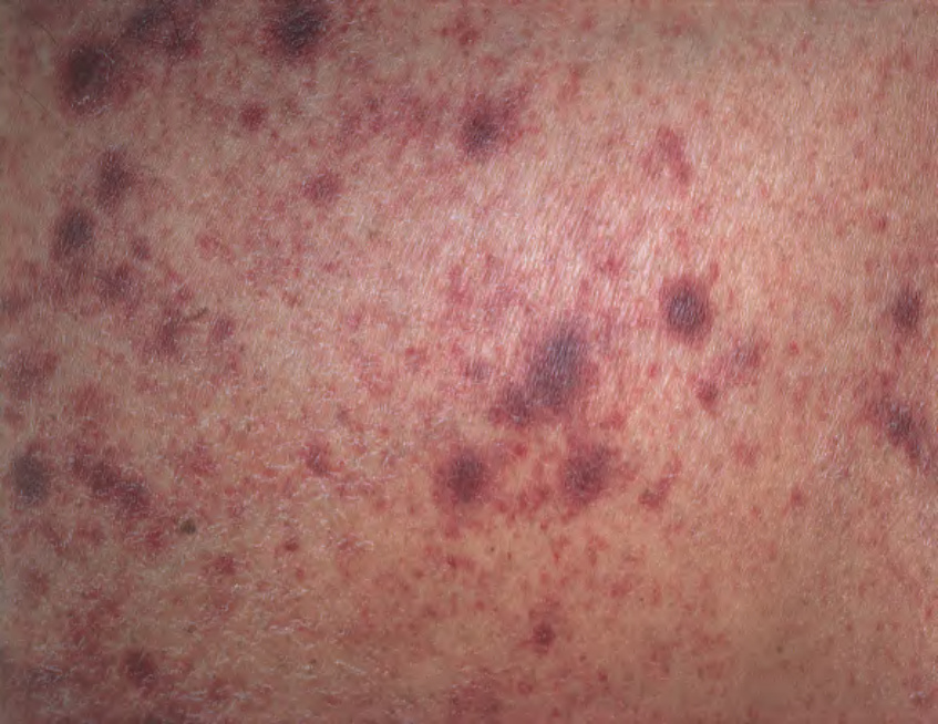
Fig. 83.21 Strongyloidiasis hyperinfection. Multiple purpuric lesions (sometimes referred to as “thumbprint” purpura) on the abdomen of an immunocompromised patient. Courtesy Jean L. Bolognia, MD.
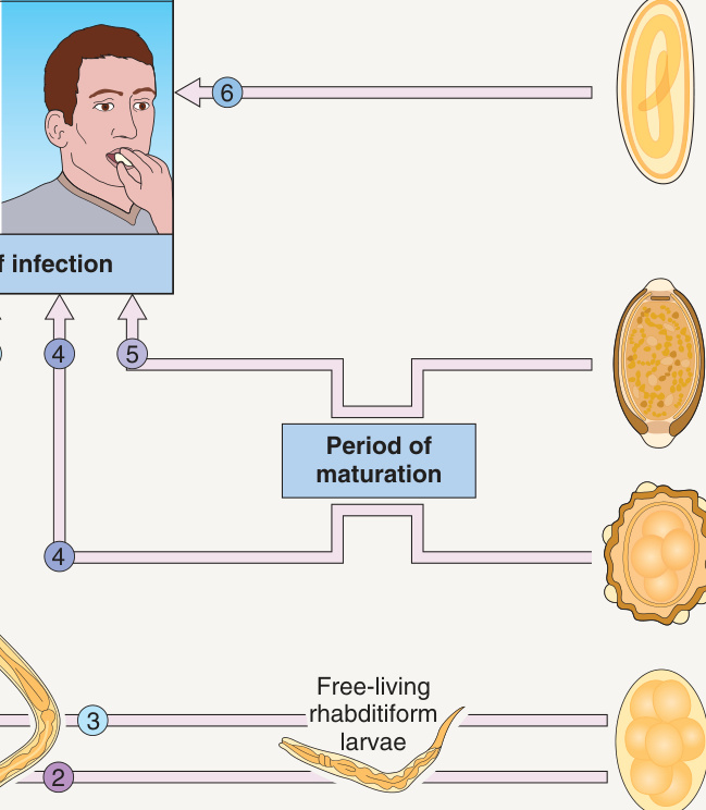
*Fig. 83.22 Life cycles of important human roundworms: adults living in the intestines. Larvae can migrate from venules into the right heart and lungs, then via alveoli into the trachea; eventually they are swallowed. With permission from Cross JH. Helminths. In Cohen J, Powderly W (eds). Infectious Diseases, 2nd edition. London: Mosby, 2004.
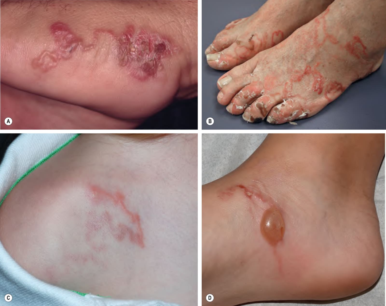
Fig. 83.27 Cutaneous larva migrans. Note the characteristic serpiginous erythematous tracts on the lateral foot (A), both feet (B), the shoulder (C), and the ankle (D). Vesiculation and crusting (A,B) or even bullae (D) are sometimes seen. B, Courtesy Peter Klein, MD; C, D Courtesy Julie V. Schaffer, MD.

Table 83.10 Major parasitic worms that cause diseases and cutaneous manifestations in humans. Continued