GNATHOSTOMIASIS
Synonyms: Nodular migratory eosinophilic panniculitis Shanghai rheumatism Tuao chid Yangtze edema
Key features
Typically acquired via consumption of raw freshwater fish
Clinical presentations include nodular migratory panniculitis, a superficial type that mimics cutaneous larva migrans, and furuncular lesions; peripheral eosinophilia is often present
Introduction
Gnathostomiasis is caused by the larvae of nematodes of the genus Gnathostoma and the usual definitive hosts are cats, dogs, and pigs. However, humans can become infected by eating raw freshwater fish, which represent an intermediate host during the parasite’s life cycle (see Fig. 83.25). The most common clinical presentation is a migratory panniculitis, associated with tissue and peripheral eosinophilia.
Epidemiology
Gnathostomiasis is endemic in Southeast Asia and Japan; it also occurs elsewhere in Asia as well as in Mexico, Central and South America, and South Africa. At least two cases of autochthonous gnathostomiasis have been reported in the US. Local and imported dishes containing raw freshwater fish, eels (e.g. Asian swamp), frogs, birds, and reptiles are the source of infection, and travelers may develop the disease after returning home.
Pathogenesis
G. spinigerum is the most common cause of human gnathostomiasis. Other pathogenic species include G. hispidum, G. doloresi, G. nipponicum, G. malaysiae, and G. binucleatum.
The adult worms are enteric parasites of cats, dogs, pigs, wild felines, weasels, and rats (see Fig. 83.25). Gnathostoma eggs are passed in the feces of these infected hosts and hatch when they reach the water, e.g. in a nearby river. The first-stage larva is ingested by a copepod (Cyclops) and develops into a second-stage larva. The copepod is then ingested by a fish or other aquatic intermediate host, and the larva reaches a third stage in its flesh, which is eaten either by a definitive host or human. The larva penetrates the human gastrointestinal mucosa and starts to migrate, first to the peritoneal cavity and subsequently to the subcutaneous tissues and internal organs.
Clinical Features
Abdominal pain, other gastrointestinal symptoms, and fever may occur during early infection. Cutaneous manifestations typically develop 2–4 weeks after ingestion of the larva. The classic presentation is migratory panniculitis characterized by a single, deep-seated, pruritic or tender inflammatory nodule that disappears spontaneously after a few days to weeks and then reappears a few centimeters away or in a different body region. Sites of involvement may develop a peau d’orange appearance. Occasionally, the parasite’s migratory pattern becomes more superficial
and mimics that of cutaneous larva migrans. Both patterns may be seen simultaneously in the mixed form (Fig. 83.28A). Following treatment, the parasite tends to be located near the skin surface, inducing the formation of a vesicle or pustule overlying a papulonodule; this has been called furuncular gnathostomiasis. Rarely, the parasite reaches the surface and becomes visible.
Migration of the larva into deeper tissues can result in pulmonary, gastrointestinal, renal, or ocular involvement. Invasion of the CNS is associated with a mortality rate of up to 25% and may lead to radiculomyelitis, eosinophilic meningitis, and subarachnoid hemorrhage. Neurologic symptoms often wax and wane, reflecting the migratory pattern of infection, and an altered state of consciousness or coma can occur with cerebral involvement.
Pathology
Nodular lesions demonstrate an eosinophilic lobular panniculitis with linear hemorrhage. In superficial lesions, an eosinophilic infiltrate is present in the mid and deep dermis. The parasite itself may be seen, especially in the furuncular type. The worm has a cylindrical shape, measuring 0.2–0.4 mm in diameter, with an external cuticle and central intestine (Fig. 83.28B).
Diagnosis and Differential Diagnosis
Gnathostomiasis should be suspected in patients with a migratory panniculitis or dermatitis plus recent consumption of raw fish or another potentially contaminated raw food. The diagnosis is supported by the presence of peripheral eosinophilia (≤50% of cases) as well as tissue eosinophilia. Worldwide serologic testing via immunoblot based on a specific L3 antigen is available in specialized research centers. Development of a rapid filtration-based immunoassay is currently under investigation.
The differential diagnosis includes other nematode infections with migratory skin lesions, including cutaneous larva migrans, strongyloidiasis, and toxocariasis. Similar inflammatory nodules may also be seen in sparganosis, fascioliasis, and myiasis.
Treatment
Recommended therapy is either albendazole 400–800 mg/day for 21 days or ivermectin 200 mcg/kg/day for 1–2 days. Relapses often occur.
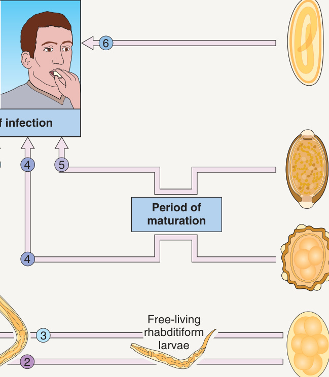
*Fig. 83.22 Life cycles of important human roundworms: adults living in the intestines. Larvae can migrate from venules into the right heart and lungs, then via alveoli into the trachea; eventually they are swallowed. With permission from Cross JH. Helminths. In Cohen J, Powderly W (eds). Infectious Diseases, 2nd edition. London: Mosby, 2004.
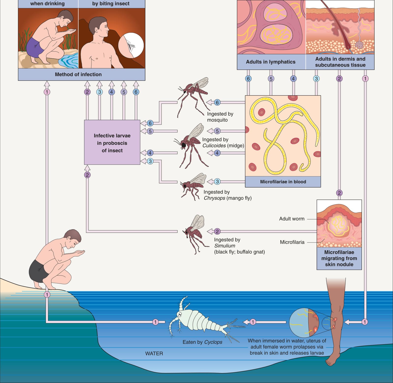
Fig. 83.23 Life cycles of important human roundworms: adults living in tissues.With permission from Cross JH. Helminths. In Cohen J, Powderly W (eds). Infectious Diseases, 2nd edition. London: Mosby, 2004.
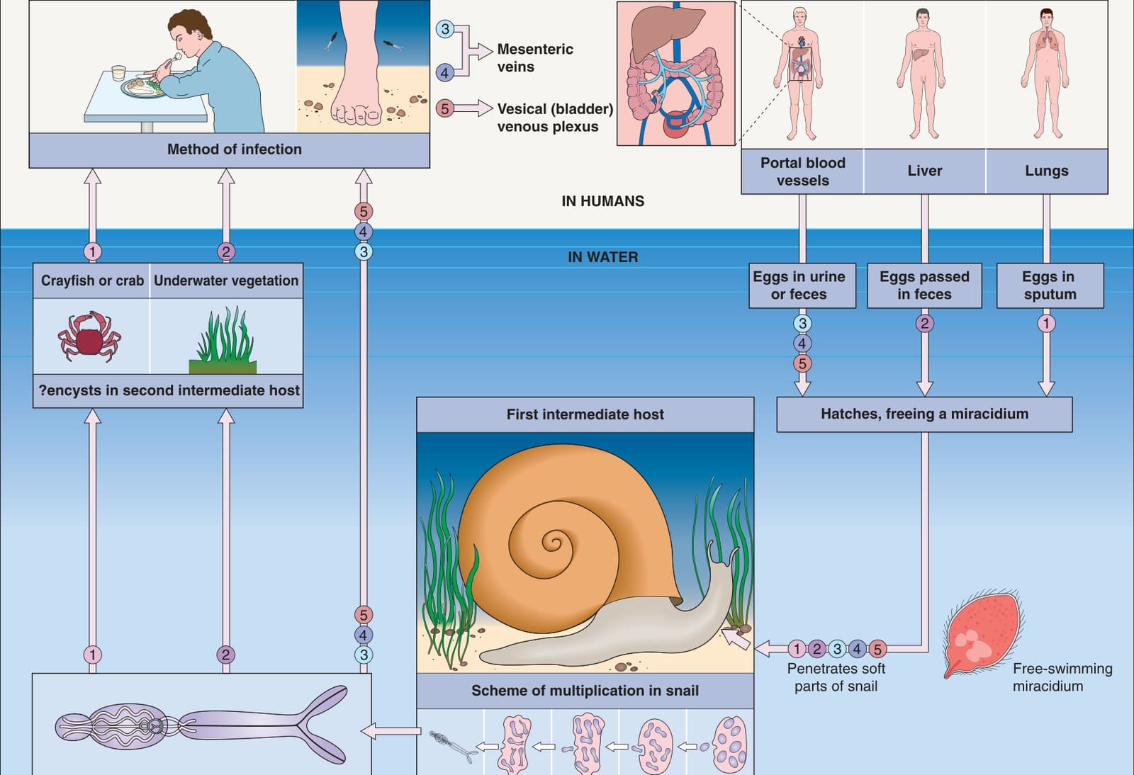
Fig. 83.24 Life cycles of important human flukes: adults living in the liver, lungs, and blood.Adapted with permission from Cross JH. Helminths. In Cohen J, Powderly W (eds). Infectious Diseases, 2nd edition. London: Mosby, 2004.
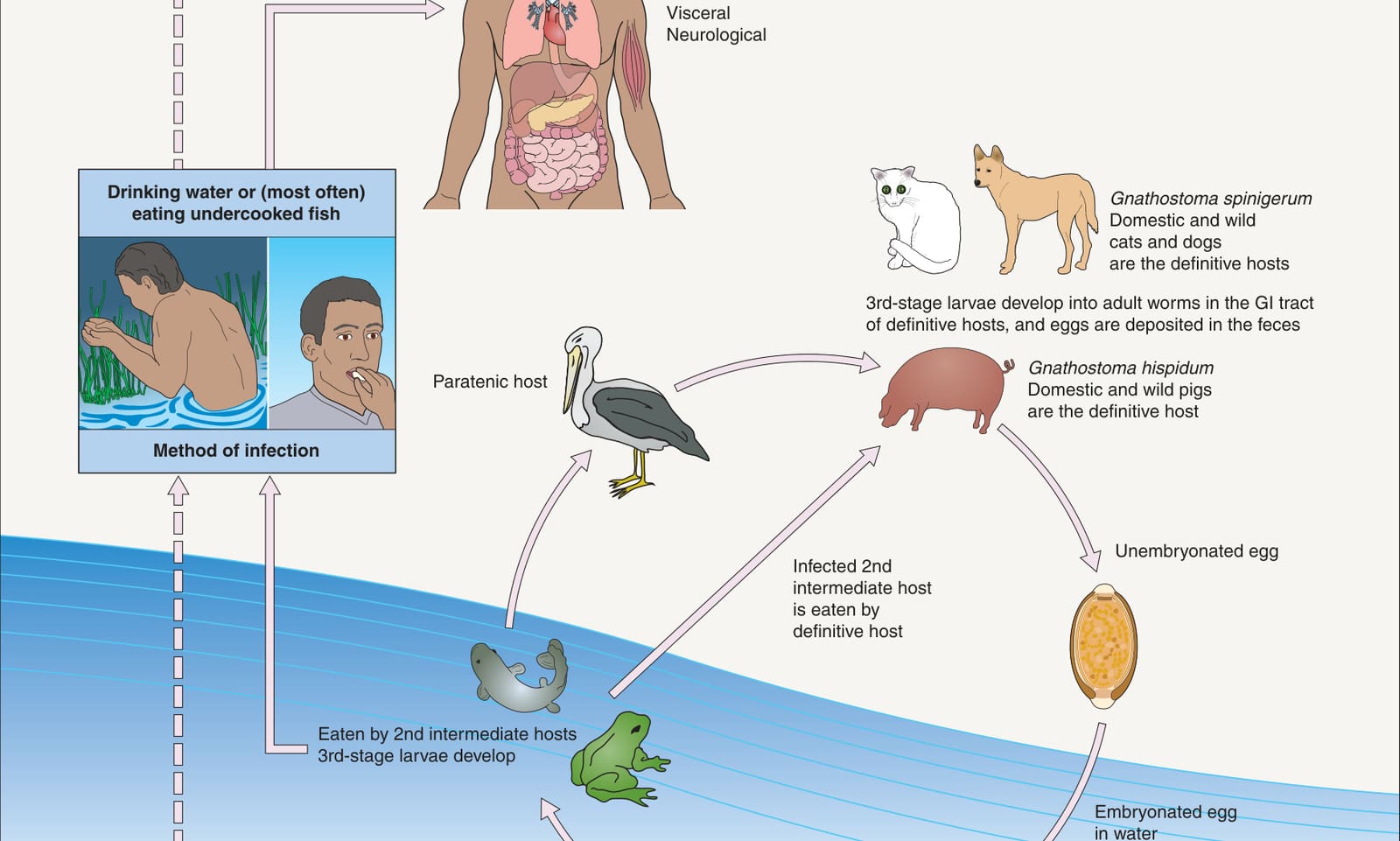
Fig. 83.25 Life cycle of Gnathostoma species. Transmission to humans via copepods in drinking water has been postulated but not confirmed. Modified from www.cdc. gov/parasites/gnathostoma/biology.html.