ONCHOCERCIASIS
Synonyms: River blindness Enfermedad de Robles Erysipelas (erisipela) de la costa Craw-craw
Key features
Onchocerca volvulus is a tissue-dwelling nematode that is mainly found in Africa and transmitted by black flies Most clinical manifestations are caused by microfilariae in the skin and eyes Cutaneous manifestations include subcutaneous nodules that contain adult worms, a pruritic papular dermatitis, lichenification, and leukoderma The diagnosis can be established by identification of microfilariae via microscopic examination of a skin sample (skin snip) or slit-lamp examination of the eyes
Introduction
Onchocerciasis, or river blindness, predominantly affects cutaneous and ocular tissue and is caused by Onchocerca volvulus, a filarial nematode. Onchocerciasis most commonly occurs in inhabitants of tropical Africa and occasionally develops in residents of tropical areas in South America and the Middle East. In the early stage of the disease, cutaneous manifestations include subcutaneous nodules and pruritic papular dermatitis, while skin thickening and leukoderma are later findings. Progressive sclerosing keratitis can lead to blindness.
Epidemiology
Onchocerciasis is a debilitating disease that worldwide affects ~16 million people, in particular those living for prolonged periods of time near fast-flowing rivers in endemic areas. The larvae of O. volvulus have an obligatory aquatic stage during which they require high oxygen tension, thus explaining the significant role well-oxygenated water plays in this disease. Severe disease requires repeated exposures over many years. Approximately 99% of all cases are acquired in equatorial Africa, but some endemic foci remain in the Middle East, Venezuela, and Brazil (Fig. 83.29). As of 2016, onchocerciasis was considered eradicated in Colombia, Ecuador, Mexico, and Guatemala; in addition, Niger is currently undergoing verification to become the first African nation to eliminate onchocerciasis.
Pathogenesis
Onchocerciasis is transmitted by the bite of an infected black fly (Simulium spp.). Because these flies often assume a “hump-backed” stance, they are sometimes referred to as “buffalo gnats”. Black flies feed by “rasping” (abrading) the surface of the skin until a pool of blood forms, which they lap up. Microfilariae previously residing in the dermis are present in this blood, and they penetrate the gut wall of the insect and migrate to its thoracic muscles, where they develop into larvae. The larvae then migrate to the proboscis, from which they will be transmitted to another human host during a blood meal (see Fig. 83.23). Wolbachia bacterial endosymbionts are required for normal development of O. volvulus larvae and embryos, are thought to support the survival of adult worms, and drive immune responses that contribute to disease pathogenesis.
Once in their human host, larvae take almost a year to reach maturity. The adult worms are large, measuring up to 50 cm in length. They are encapsulated within fibrous tissue and reside in nodules close to the surface of the skin or near joints. These nodules are known as onchocercomas (Fig. 83.30). Each female worm produces hundreds of microfilariae, which live for 1–2 years and migrate into the skin, connective tissue, eyes, and lymph nodes. It is the microfilariae that cause most of the serious clinical features of onchocerciasis. There is little inflammation until the microfilariae degenerate, which triggers a hypersensitivity reaction. In the skin, this results in intense pruritus and dermatitis.
Clinical Features
The primary manifestations of onchocerciasis involve the eye and the skin. Cutaneous findings are classified into the following groups: acute papular onchodermatitis, chronic papular onchodermatitis, lichenified onchodermatitis, onchocercal atrophy, and depigmentation (Fig. 83.31). Patients first present with subcutaneous nodules that are usually located over bony prominences; they favor the upper part of the body in patients living in South America and the lower part of the body in those from Africa. The subcutaneous nodules represent fibrous tissue surrounding adult worms. An intermittent, intensely pruritic acute papular onchodermatitis follows, in which small papules form around the microfilariae (called “craw-craw” in Africa).
In chronic onchocerciasis, the skin becomes thickened and wrinkled; this is accompanied by lichenification and hyperpigmentation and is sometimes referred to as “lizard” or “elephant” skin or “sowda”. The back, thighs, and lower trunk are frequently affected, and hyperpigmentation may develop on one lower extremity in association with inguinal lymphadenopathy (see Fig. 83.31). Atrophy and a loss of pigment with sparing of perifollicular skin are also seen in late stage onchocerciasis. The latter changes, sometimes referred to as “leopard” skin, are most often evident on the shins (see Fig. 83.31 and Ch. 66). Chronic lymphatic obstruction and involvement of the inguinal lymph nodes can lead to hanging groin or elephantiasis of the genitalia (see Fig. 83.31).
In Central America, young patients who are heavily infested may develop erythema on the face or upper trunk (erisipela de la costa).
Older patients may have violaceous papules or plaques (mal morado), which can lead to leonine facies.
In addition to skin lesions, microfilariae can also be found in the conjunctivae; from there they can move through the cornea into the anterior and posterior chambers of the eye, leading to conjunctivitis, sclerosing keratitis, uveitis, chorioretinal lesions, optic atrophy, and glaucoma. Blindness occurs in the most severe cases. Dead microfilariae in the cornea can induce a tissue reaction that produces characteristic “snowflake” opacities.
Pathology
In onchodermatitis, microfilariae are present in all levels of the skin but are most concentrated in the dermal papillae, where they can infect black flies during feeding. Microfilariae are ~250 microns long with two or three pairs of anterior nuclei. Initially, they are surrounded by an infiltrate with numerous eosinophils. Lymphocytes, macrophages, plasma cells, and mast cells may become more prominent at later stages, and epidermal hyperplasia (secondary to rubbing) may develop. The final outcome is fibrosis surrounding the microfilariae with minimal inflammation.
The adult female worms are found in a subcutaneous nodule. They are initially surrounded by an infiltrate containing many eosinophils, with progressive development of fibrosis.
Diagnosis
The clinical diagnosis of onchocerciasis is usually not difficult in endemic areas. It is confirmed by microscopic visualization of microfilariae as they emerge from multiple skin snips placed in saline; these small, thin discs (including superficial dermis) are taken from skin near a nodule and over the shins, iliac crests, and/or scapulae. Onchocerciasis can also be diagnosed by the identification of the adult worm in an excised nodule. In heavily infected individuals, microfilariae can be found in the blood, sputum, and urine. By slit-lamp examination, microfilariae (when present) are easily visualized within the anterior chamber of the eye.
When skin snips are negative, the “Mazzotti test” may be helpful in diagnosing onchocerciasis: 50 mg of diethylcarbamazine (DEC) is administered orally, and a pruritic eruption develops within 15 minutes (as the microfilariae die) if the patient is infected. In the “Mazzotti patch test”, DEC is applied topically (e.g. in a 10% lotion under occlusion) to a small area of skin, which is examined 24 hours later for the development of papules and edema; this diagnostic method is not as sensitive as oral administration of DEC but less likely to produce a systemic reaction. A dipstick test to detect Onchocerca volvulus antigen in the urine or tears has a sensitivity of >90% and a specificity of 100%. Highly sensitive and specific PCR-based tests have also been developed. Tests based on antibody detection cannot distinguish between active and past infections.
Photos: left, depigmentation with perifollicular sparing (“leopard” skin); middle, hanging groin due to chronic lymphatic obstruction and lymph node involvement; right, lichenified onchodermatitis (“sowda”). Insets, courtesy David O. Freeman, MD.
Differential Diagnosis
Early lesions of onchocercal dermatitis must be distinguished from insect bites, scabies, and atopic or allergic contact dermatitis. The chronic skin lesions may be mistaken for chronic eczema with postinflammatory pigmentary changes, Hansen disease (leprosy), or the leukoderma of scleroderma. The differential diagnosis of elephantiasis includes other types of filarial infestation (see Fig. 83.23), and a falsepositive skin snip can occur if the tissue is contaminated with blood filariae, especially Mansonella perstans or Loa loa.
Treatment
Therapy for onchocerciasis has drastically improved as a result of the widespread use of oral ivermectin since 1987. This drug is effective in rapidly killing microfilariae and preventing their escape from gravid females. Ivermectin causes few or no adverse reactions and no Mazzotti reaction, whereas older drugs, e.g. diethylcarbamazine and suramin, are associated with severe hypersensitivity or dangerous toxic reactions. Ivermectin is administered as a single dose of 150 mcg/kg every 3 to 12 months. After treatment, microfilariae usually disappear from the skin within 1 week and from the eye within 3 months. Although multiple doses of oral ivermectin can kill adult worms, treatment is typically continued for the worm’s lifetime (10–15 years).
Moxidectin is a macrocyclic lactone in the milbemycin family that was recently FDA-approved for the treatment of onchocerciasis. Compared to ivermectin (a macrocyclic lactone in the avermectin family), moxidectin has a longer half-life and greater potency against microfilariae of O. volvulus as well as other nematodes such as Dirofilaria immitis. In addition, moxidectin has potential as an alternative treatment for scabies. Administration of doxycycline (100–200 mg/day for 6 weeks) to target Wolbachia endosymbionts represents another potential therapeutic approach that can markedly reduce or eliminate O. volvulus microfilariae for >18 months. Nodulectomy is also a popular treatment in countries where nodules on the head are common (see Fig. 83.31); their removal reduces the chance of ocular disease.
Preventive measures such as spraying black fly breeding sites with larvicides have not significantly changed the epidemiology of the disease. In contrast, mass treatment of endemic populations with ivermectin once or twice yearly has resulted in significant progress toward elimination of the disease in both the Americas and Africa. In addition to onchocerciasis and scabies, mass drug administration of ivermectin or moxidectin targets strongyloidiasis, pediculosis, and cutaneous larva migrans.
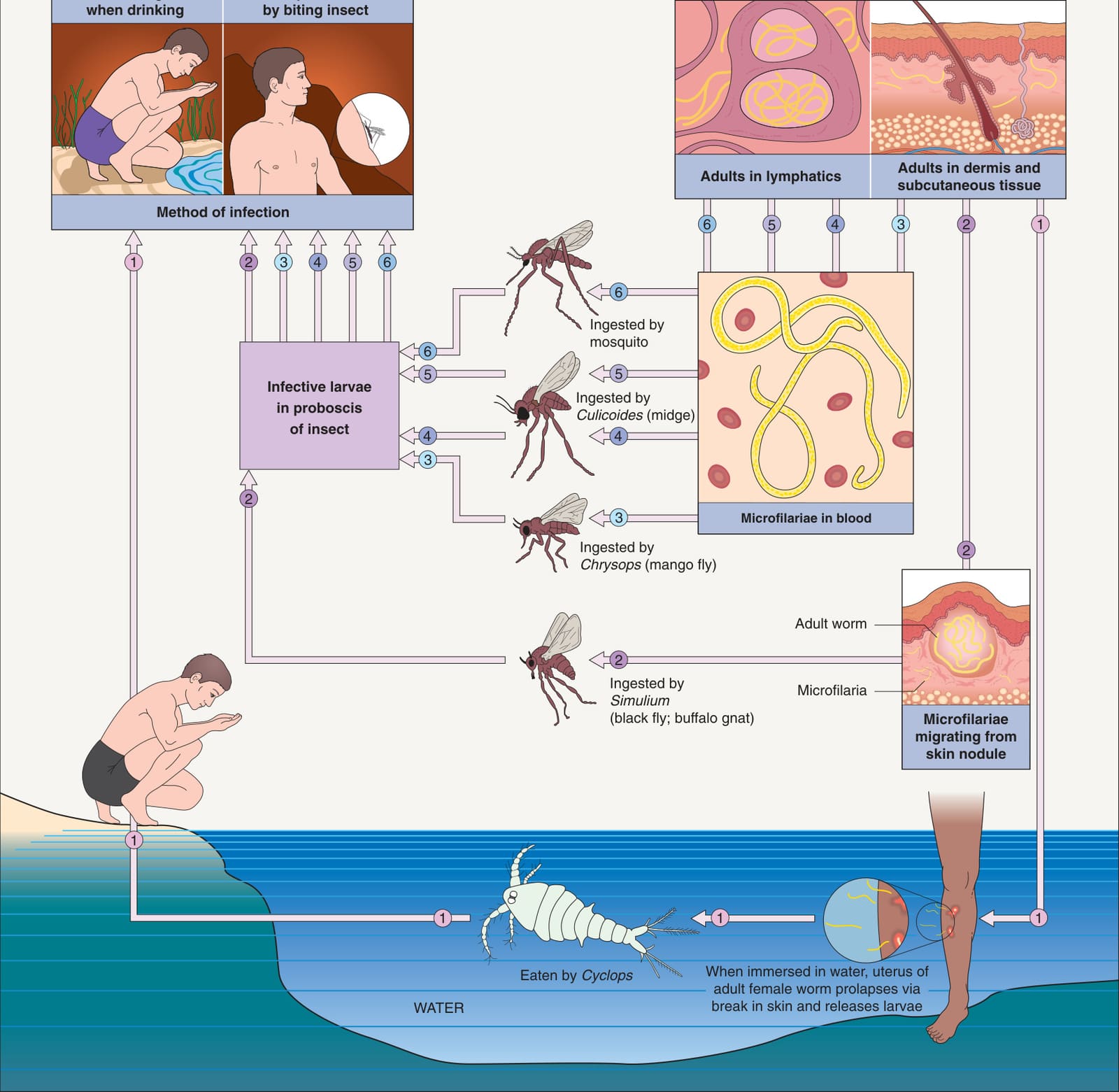
Fig. 83.23 Life cycles of important human roundworms: adults living in tissues.With permission from Cross JH. Helminths. In Cohen J, Powderly W (eds). Infectious Diseases, 2nd edition. London: Mosby, 2004.
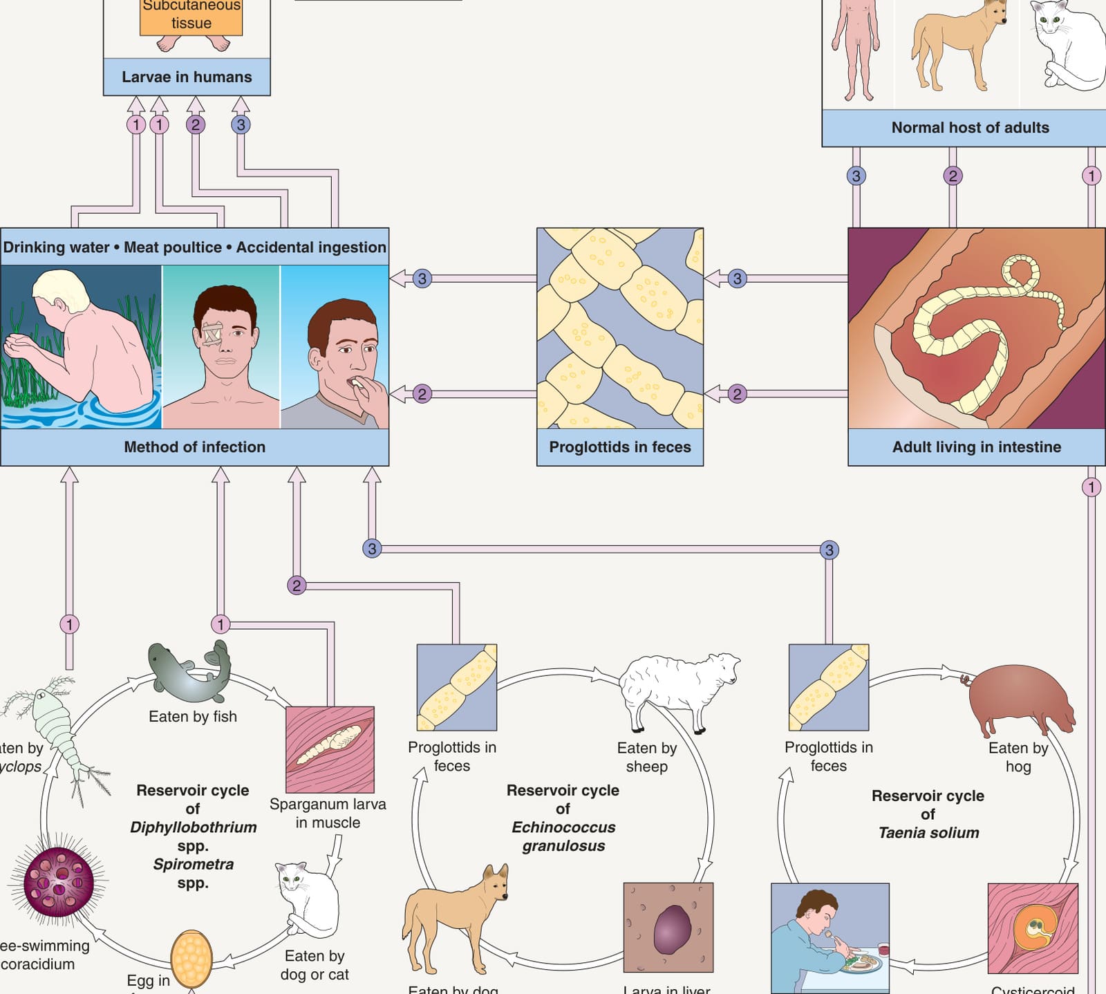
Fig. 83.26 Life cycles of important human tapeworms: adults living in tissues, intestines, and blood.Adapted with permission from Cross JH. Helminths. In Cohen J, Powderly W (eds). Infectious Diseases, 2nd edition. London: Mosby, 2004.
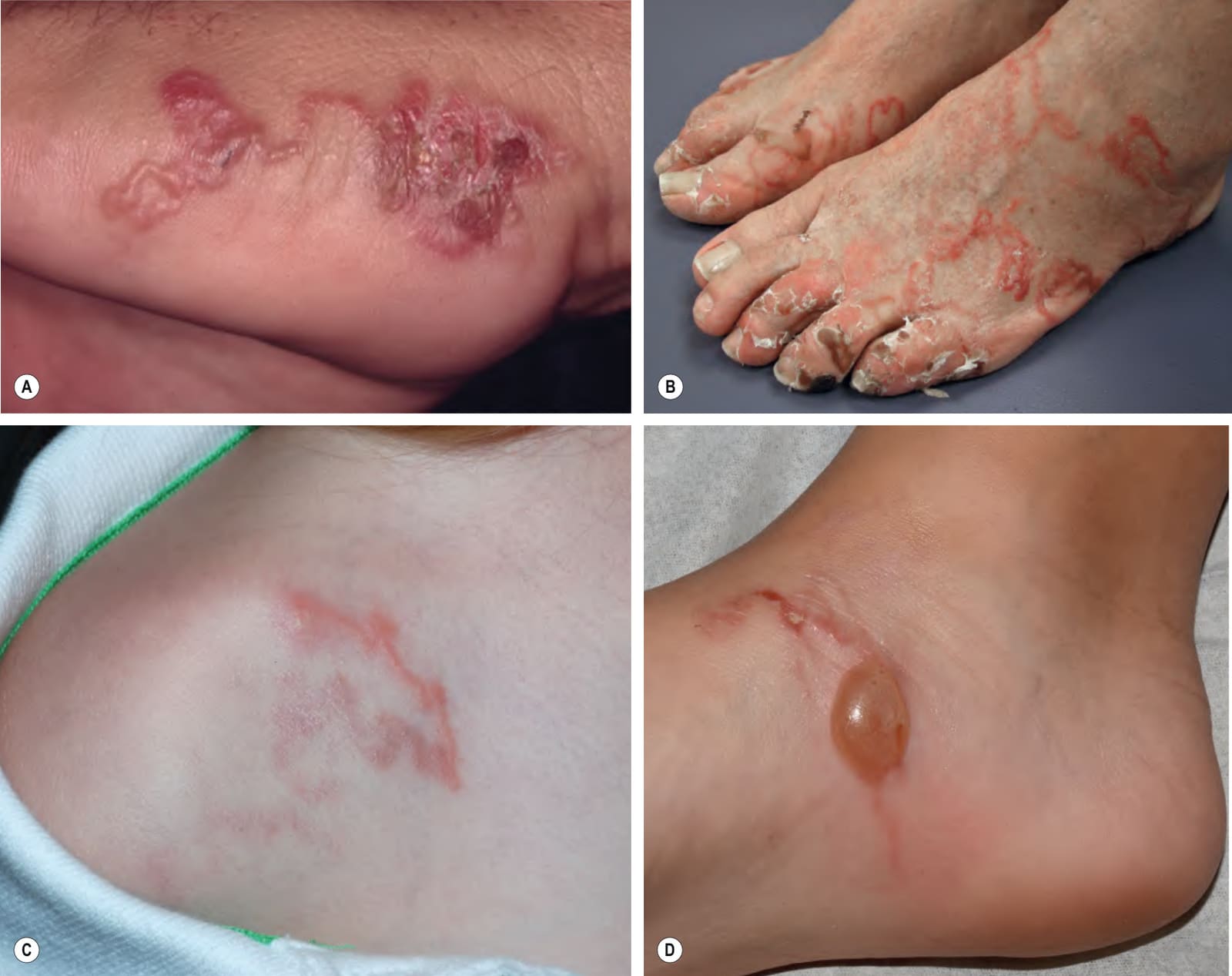
Fig. 83.27 Cutaneous larva migrans. Note the characteristic serpiginous erythematous tracts on the lateral foot (A), both feet (B), the shoulder (C), and the ankle (D). Vesiculation and crusting (A,B) or even bullae (D) are sometimes seen. B, Courtesy Peter Klein, MD; C, D Courtesy Julie V. Schaffer, MD.
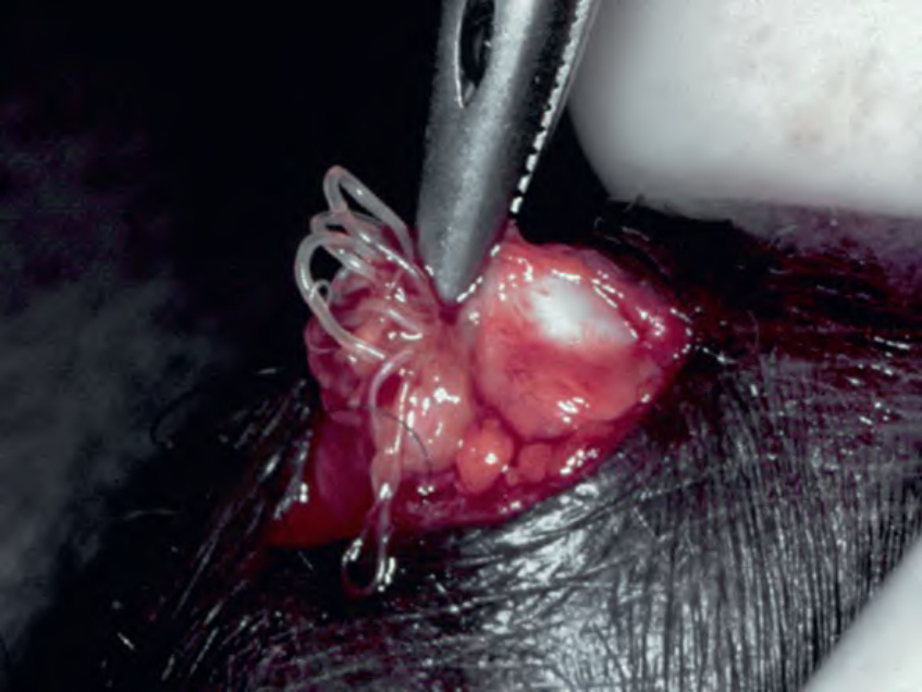
Fig. 83.28 Gnathostomiasis.A In addition to a serpiginous pattern on the lower back that is similar to larva migrans, there is an indurated pink plaque with a peau d’orange appearance. B A skin biopsy specimen occasionally reveals the Gnathostoma third-stage larva. A, Courtesy Edward Cowen, MD.
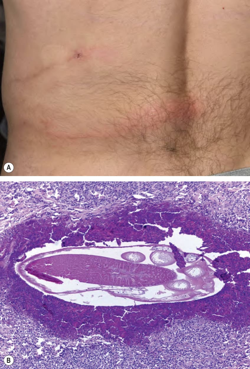
Fig. 83.29 Distribution of onchocerciasis. The disease is found primarily in Africa and focal areas of the Amazon region in South America.
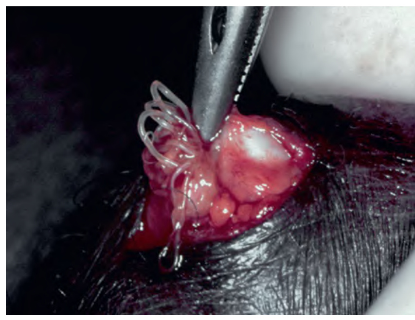
Fig. 83.30 Onchocerciasis. Extraction of adult worms from an onchocercal nodule on the buttocks.
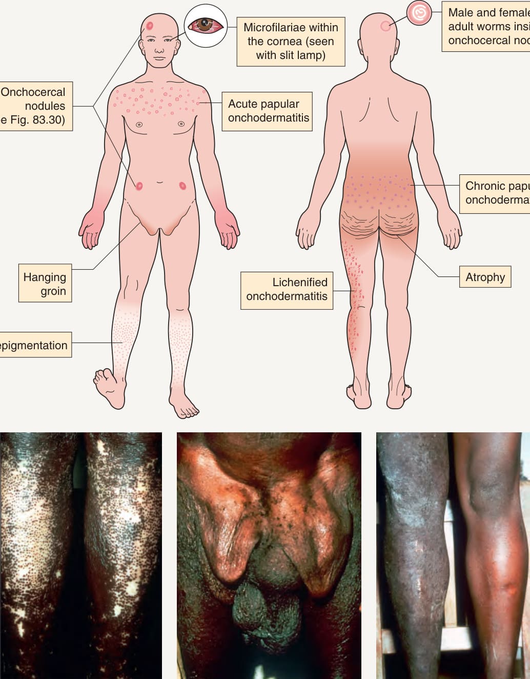
Fig. 83.31 Cutaneous findings in onchocerciasis.