FILARIASIS
Synonyms: Lymphatic filariasis, elephantiasis Elephantiasis
Key features
Filariasis is an infection of the lymphatic system by tissue nematodes (roundworms) acquired via mosquito vectors
There are acute, chronic, and asymptomatic forms of filariasis
Manifestations of the acute phase include lymphangitis and orchitis
Chronic disease is characterized by the sequelae of lymphatic obstruction, e.g. lymphedema, elephantiasis, hydroceles, and chyluria; associated skin findings include hyperkeratosis, verrucous changes, soft tissue hypertrophy, and fibrosis
Introduction
Lymphatic filariasis is an infection of the lymphatic system by two genera of filarial nematodes (roundworms). Several species of mosquitoes act as vectors (see below). Cutaneous findings during the acute and chronic stages include lymphangitis and lymphedema accompanied by changes of elephantiasis.
Epidemiology
There are two major forms of filariasis, Bancroftian (due to Wuchereria bancrofti), causing 90% of infections, and Malayan (due to Brugia malayi and B. timori). The disease affects ~120 million people living in ~50 countries located primarily within the tropical and subtropical regions of South America, Africa, Asia, and the Pacific Islands. W. bancrofti is found worldwide, whereas B. malayi is restricted to South and East Asia. The distribution of B. timori is limited to islands in the Indonesian archipelago.
A number of mosquito species act as vectors for filarial nematodes. The most important ones for W. bancrofti are Culex quinquefasciatus, Anopheles gambiae, Anopheles funestus, Aedes polynesiensis, Aedes scapularis, and Aedes pseudoscutellaris. Both B. malayi and B. timori are transmitted by Anopheles barbirostris. B. malayi can also be transmitted by several species of Aedes and Mansonia mosquitoes.
Pathogenesis
Multiple bites from infected mosquitoes seem to be required to produce symptomatic infections, and the incubation period varies from 2 to 18 months. Larvae enter lymphatic vessels and then, once mature, the adult worms mate within the lymphatics or lymph nodes and release sheathed microfilariae into the bloodstream. The mosquito then acquires microfilariae during a blood meal. Over several days, the microfilariae develop into larvae within the mosquito, which can then transmit infection to other humans during subsequent blood meals (see Fig. 83.23).
The clinical manifestations of lymphatic filariasis reflect a complex interplay of the pathogenic potential of the parasite, the immune response of the host, and secondary bacterial and fungal infections. Maturing adult worms provoke an eosinophilic and chronic inflammatory cell infiltrate around lymphatic vessels, leading to dilation and valvular damage with eventual scarring and obstruction of lymphatic flow. The death of worms within lymphatic vessels and lymph nodes results in an intense inflammatory reaction with granulomatous changes and necrosis. Wolbachia bacterial endosymbionts are required for the worms’ development, embryogenesis, and survival; Wolbachia also stimulates an innate and adaptive immune response that leads to expression of vascular endothelial growth factors (VEGFs) that promote lymphangiogenesis, lymphatic endothelial proliferation, and dilation of lymphatic vessels.
Clinical Features
There are acute, chronic, and asymptomatic forms of filariasis. Acute episodes of retrograde adenolymphangitis typically last up to one week and usually recur 1 to 10 times per year; the inguinal lymph nodes are most often involved. Early on, the affected body part appears clinically normal in between these episodes. Men with W. bancrofti infection often present with orchitis and epididymitis. Another disease manifestation is acute dermatolymphangioadenitis characterized by cutaneous or subcutaneous inflammatory plaques associated with ascending lymphangitis, regional lymphadenitis, and fever; this may result from a bacterial or fungal superinfection, often with an interdigital entry point. Acute symptoms, which are thought to reflect a lack of tolerance to filarial antigens, are most common in recently infected young adults and older patients. Children residing in endemic areas are often asymptomatic until they reach puberty, when they typically develop chronic manifestations.
After 10 to 15 years of infection, the clinical features of chronic disease, i.e. the sequelae of lymphatic obstruction due to both adult worms and granulomatous inflammation, are noted. These include lymphedema, elephantiasis, hydroceles, and chyluria. In some communities in endemic regions, 40%–60% of adult men have hydroceles. In sites of lymphedema and elephantiasis, the skin can be hyperkeratotic, verrucous, and fibrotic with soft tissue hypertrophy and redundant folds. Fissures, ulceration, and gangrene may also develop. Secondary bacterial and fungal infections are common.
There are two major reasons for the predilection of filariasis for the lower extremities and genitalia: (1) the female mosquito flies near the ground, thus it is more likely to bite the lower limbs, which is followed by retrograde involvement of the inguinal region; and (2) due to gravitational forces, the legs are at greater risk for venous hypertension and valvular incompetency and therefore more susceptible to the development of chronic lymphedema.
Tropical pulmonary eosinophilia occurs in some patients with filarial infections. Clinical findings include cough, wheezing, dyspnea, chest pain, and fever accompanied by marked eosinophilia and pulmonary infiltrates. This may progress to restrictive pulmonary disease.
Travelers to endemic areas who become infected do not manifest the classic findings of filariasis. Instead, they have a hyperresponsive clinical presentation, known as “expatriate syndrome”, with more intense inflammatory reactions to the filarial parasites. Clinical findings include lymphangitis, lymphadenitis, and genital pain from the associated lymphatic inflammation, as well as urticaria, nonspecific cutaneous eruptions, and peripheral eosinophilia.
Diagnosis
Detection of circulating filarial antigen is now the preferred method for diagnosis of Bancroftian filariasis; immunochromatographic card tests or test strips are typically utilized. Similar tests are not currently available for Malayan filariasis, which can be diagnosed using a dipstick test for IgG4 antibody specific for the Brugia antigen BmR1. Alternatively, the diagnosis can be established by the demonstration of microfilariae in the blood, urine, or other body fluids and tissues, or by the identification of adult worms (Fig. 83.32). Visualization
of motile adult worms may be possible using ultrasound to evaluate the infected lymphatics. As the parasite exhibits a nocturnal periodicity in humans, 10 p.m. to 2 a.m. is the optimal time period for collection and direct smear of blood or other fluids. The use of filters with a pore size of 3 microns has improved the rate of detection of microfilariae in the blood and urine. However, parasite detection is not a particularly reliable method because many symptomatic patients are amicrofilaremic. Species-specific PCR-based assays have been developed and are utilized in research studies on filariasis.
Peripheral eosinophilia and elevated serum IgE are very common laboratory findings. In addition, more than 50% of microfilaremic patients with W. bancrofti infections have hematuria and/or proteinuria.
Differential Diagnosis
The differential diagnosis depends upon the stage of the infection. Acute infection may be misdiagnosed as a bacterial lymphangitis. Lymphedema and elephantiasis have multiple possible etiologies, including recurrent bacterial lymphangitis, severe venous hypertension, lymph node dissections, podoconiosis, Milroy disease, and Klippel– Trenaunay syndrome (see Ch. 105).
Treatment
The drug of choice is diethylcarbamazine, which is active against microfilariae but has a limited effect on adult worms. Filariasis is usually treated with a single 6 mg/kg dose of diethylcarbamazine; a 14–21-day course is recommended for patients with tropical pulmonary eosinophilia.
Mass drug administration programs with the goal of eradicating filariasis from endemic countries initially provided at-risk individuals yearly single doses of diethylcarbamazine. Current programs utilize annual administration of a triple-drug regimen including albendazole, ivermectin, and diethylcarbamazine, which has resulted in increased microfilarial clearance. In areas co-endemic for loiasis or onchocerciasis, diethylcarbamzine is held (see below), with albendazole administered alone or together with ivermectin, respectively.
Doxycycline (200 mg/day for 4–8 weeks) targeting the Wolbachia endosymbiont can also eliminate microfilariae, kill adult worms, and improve lymphatic drainage. During the treatment period, antihistamines and corticosteroids may be useful in decreasing the allergic reaction that may result from massive disintegration of microfilariae. Because severe reactions may follow the administration of diethylcarbamazine in patients who have loiasis or onchocerciasis, it is important to exclude other helminthic diseases before instituting therapy. Other management strategies include elevation of the affected body part, compression stockings, other anti-inflammatory drugs, treatment of secondary infections, and protection of the affected area from trauma.
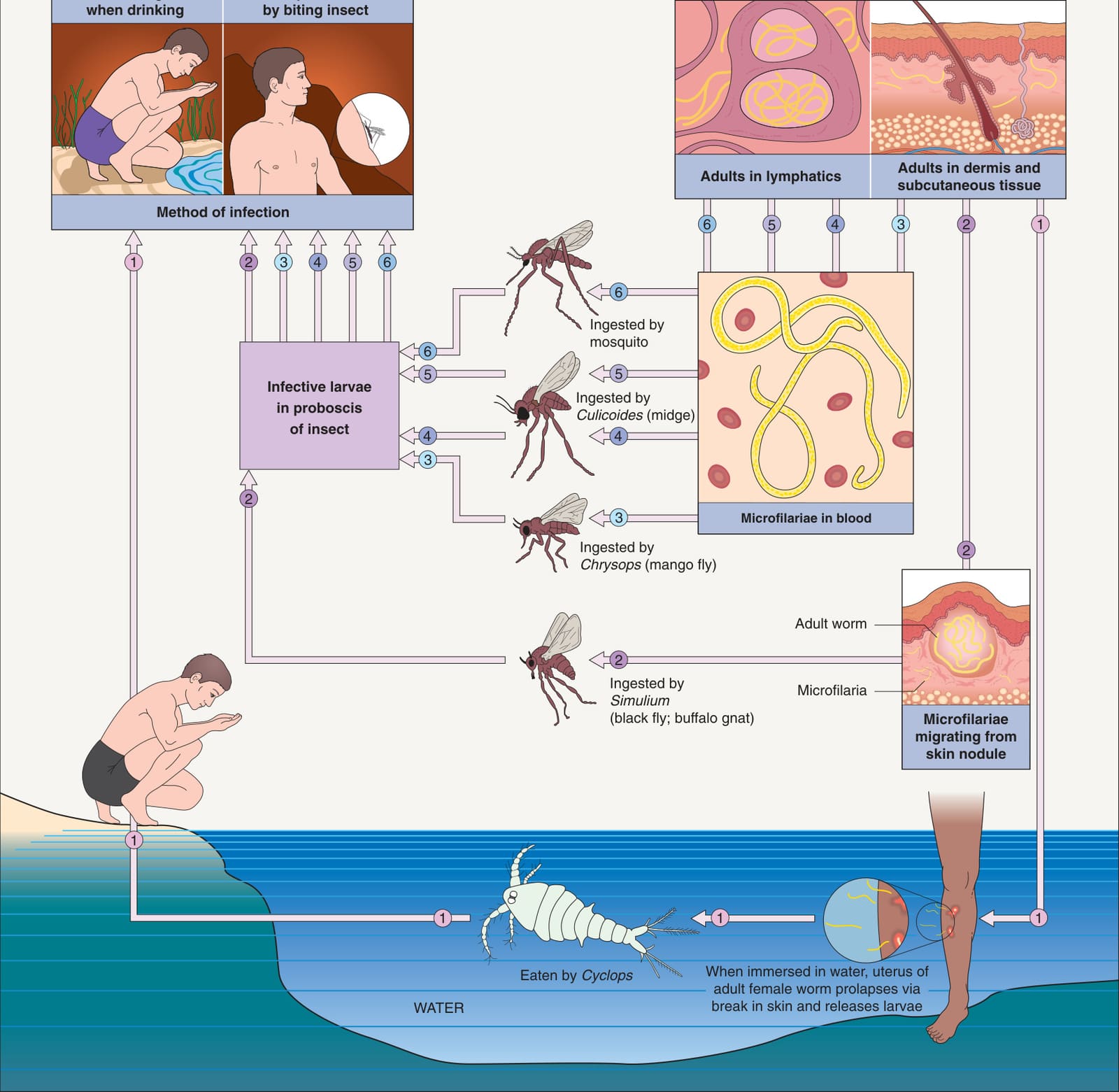
Fig. 83.23 Life cycles of important human roundworms: adults living in tissues.With permission from Cross JH. Helminths. In Cohen J, Powderly W (eds). Infectious Diseases, 2nd edition. London: Mosby, 2004.
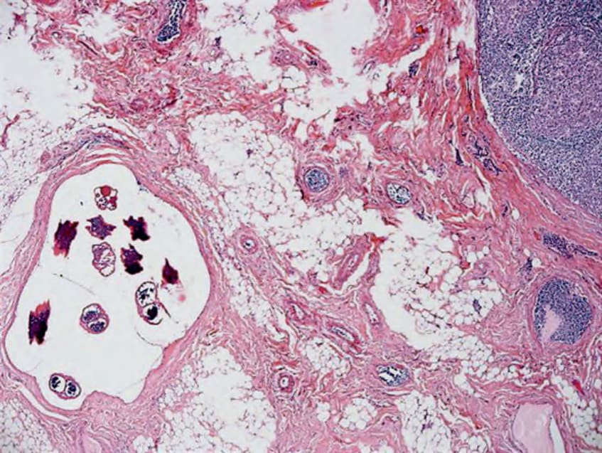
Fig. 83.32 Histopathology of filariasis. Dilated lymphatic with a mature adult worm and surrounding enlarged inguinal lymph nodes.