CYSTICERCOSIS AND ECHINOCOCCOSIS
Synonyms: Echinococcosis: hydatidosis, hydatid disease
Key features
Cestodes (tapeworms) that commonly affect animals (e.g. Taenia solium, Echinococcus granulosus) can also infect humans
Cutaneous cysticercosis is characterized by papules and nodules that are usually multiple and asymptomatic; the brain (neurocysticercosis), eye, heart, other muscles, and peritoneal cavity may also be affected
In echinococcosis, the liver and lungs are the major sites of involvement, with development of one or more hydatid cysts. Allergic symptoms, such as urticaria, asthma and anaphylaxis, are uncommon
Introduction
In the two stages of their life cycle (adult and larval), cestodes (tapeworms) can cause disease in humans. The adult worms reside in the definitive host’s intestines (including humans) and are often associated with minimal clinical manifestations (see Fig. 83.26). In intermediate hosts, the larval stage can lead to clinical disease, e.g. cysticercosis or echinococcosis in humans.
Epidemiology
The four most common human cestodes (tapeworms) are Taenia solium (pork tapeworm), Taenia saginata (beef tapeworm), Diphyllobothrium latum (fish tapeworm), and Hymenolepis nana. In humans, they cause primarily gastrointestinal symptoms, but the larval stage of T. solium also causes cysticercosis. Larvae of T. solium and T. saginata, respectively, can cause cysticercosis in pigs and cattle. Cysticercosis has a worldwide distribution, affecting more than 50 million people, and it is more common in rural areas. Endemic regions include Central and South America, sub-Saharan Africa, India, and East Asia.
Echinococcosis is a zoonosis caused by Echinococcus species, the definitive hosts of which are dogs and other members of the family Canidae. Humans are only infected by the larval stage, i.e. they are intermediate hosts, and develop cysts within viscera. Most human disease is due to larval stages of E. granulosus and E. multilocularis. Echinococcosis occurs in tropical and subtropical countries worldwide, primarily in sheep- and cattle-raising areas. Endemic regions include the southern province of Brazil, the Middle East, parts of Asia, and Northern and East Africa. It is particularly common in Lebanon and Greece.
Pathogenesis
The adult tapeworm can reach a length of up to 30 feet in the intestinal tract of its definitive host. Its body has two main parts: a single head (scolex) for attachment to the host and many proglottids, each of which has male and female reproductive organs. Fertilized eggs accumulate in large numbers inside the uterine horns of the proglottids; the eggs can be released directly into the host’s intestine or the entire gravid terminal proglottid can be excreted by the host. Both eggs and proglottids are found in the host’s stool (see Fig. 83.26).
When eggs or proglottids are ingested by a susceptible intermediate host, they develop into larvae called oncospheres. In humans, oncospheres can develop into encysted forms (cysticerci in cysticercosis) or generate germinal tissue with a cyst (hydatid in echinococcosis). Subsequent ingestion of cyst-containing tissues by a susceptible definitive host allows the development of the larvae into adults and completion of the life cycle.
Humans are the only definitive host for T. saginata (beef tapeworm) and T. solium (pork tapeworm), becoming infected with the adult form after eating poorly cooked muscle of a diseased animal (see Fig. 83.26). Humans are also intermediate hosts of T. solium, developing cysticercosis as a result of ingestion of food or water contaminated with infected human feces or via autoinfection from anus to mouth. In contrast, humans are only intermediate hosts of E. granulosus (as are sheep and cattle) as a result of ingesting contaminated dog feces. Dogs are the definitive host of E. granulosus, becoming infected with adult tapeworms after eating infected beef or lamb.
When humans are infected by the adult stage of T. solium (i.e. are the definitive host), the tapeworms within the intestine usually cause no morphologically identifiable change in the mucosa or submucosa. Some patients may develop moderate peripheral eosinophilia. Immunity plays only a limited role during infections with the adult stage and has little effect on the duration of infection or on the susceptibility to reinfection. However, immunity plays an important role when humans are the intermediate host, i.e. develop cysticercosis. After penetrating the host’s intestinal wall and disseminating, the oncosphere matures rapidly and by 10 weeks is fully developed. The cyst that forms is usually 0.5–1.0 cm in diameter and remains viable for 3 to 5 years before degenerating. At this time, the inflammatory response is markedly intensified, and calcification also often takes place. The signs and symptoms triggered by these reactions depend on the location of the cysts. For example, if there are cysts at the base of the brain, a cysticercic meningitis may ensue. In brain tissue itself, cysts that calcify may cause seizures.
When humans ingest the eggs of E. granulosus, oncospheres penetrate the mesenteric vessels. While the majority are trapped by the liver sinusoids, some bypass the liver and are carried in the bloodstream to other organs and tissues. Within hours, an inflammatory response may be seen. If the larvae survive, they develop into hydatid cysts within 5 days. Cell-mediated immunity controls dissemination during the early phase of infection.
Clinical Features
In cysticercosis, cysts can develop in almost any organ or tissue in the body. Skin involvement presents as subcutaneous papulonodules that are more often palpable than visible. They are usually multiple and asymptomatic. The brain, eye, heart, other muscles, and peritoneal cavity may also be affected. Subcutaneous involvement is recognized in <1%–20% of patients, and it is said to be more common in Asia and Africa than in Latin America. However, in a Brazilian study of 30 patients with cysticercosis, 90% had cysts in the subcutaneous tissue, skeletal muscle, or mucous membranes, while the CNS was affected in <10%.
In echinococcosis, the cysts may be single or multiple and they affect primarily the liver (50%–75%) and lungs (20%). The skin is involved in fewer than 2% of the patients. Cysts increase in size at the rate of ~1 cm/year. The clinical features are highly variable and reflect the location and size of the cysts. Most symptoms are due to mechanical pressure by the cyst mass or an allergic reaction (urticaria, wheezing, anaphylaxis) to foreign antigens in leaking cyst fluid.
Soft tissue cysts may present as firm subcutaneous nodules or as masses that are fluctuant and nontender. E. multilocularis has been noted to cause multiple supraumbilical dermal and subcutaneous nodules with associated ulceration, local inflammation, and painless cutaneous fistulas.
Diagnosis
The diagnosis of cysticercosis is often made by X-ray examination of the head or extremities, which reveals the calcified cysts. CT or MRI can also demonstrate lesions. ELISA and hemagglutination tests utilizing cyst vesicular fluid as antigen are 80%–95% sensitive and specific. An enzyme-linked immunoblot assay is the serologic test of choice, as its specificity and sensitivity approach 100% and 94%, respectively. Tests that detect circulating cysticercal antigens and PCR-based assays have also been developed.
The diagnosis of echinococcosis is based on detection of cysts via ultrasound, CT, or MRI. Examination of the contents of the cyst and identification of the parasite provides confirmation. Peripheral eosinophilia is present in one-third of patients. Western blot and enzyme immunoassays are also available, which have a high degree of
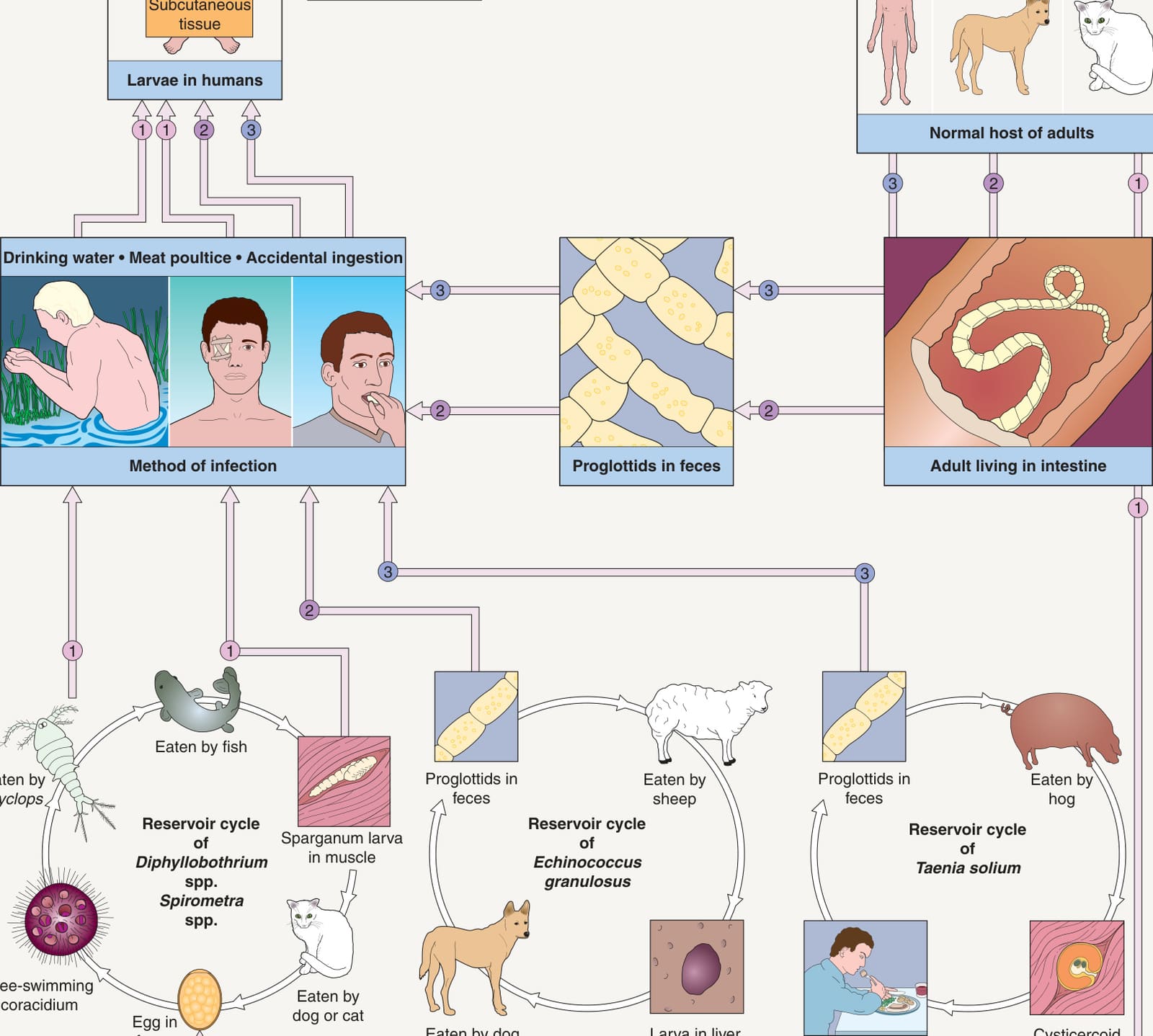
Fig. 83.26 Life cycles of important human tapeworms: adults living in tissues, intestines, and blood.Adapted with permission from Cross JH. Helminths. In Cohen J, Powderly W (eds). Infectious Diseases, 2nd edition. London: Mosby, 2004.
cross-reactivity with assays for cysticercosis. The assays have decreased sensitivity when the lesions are calcified, only one cyst is present, or the echinococcal cysts are not located in the liver.
“Old”, inactive lesions of cutaneous cysticercosis are treated surgically. Patients with active disease may be treated with albendazole (15 mg/kg/ day with a maximum of 800 mg, for ≥8 days) or praziquantel (50–100 mg/ kg/day in three divided doses for 2 weeks). Albendazole is cheaper and has 20% more cysticidal activity than praziquantel. Evaluation for neurocysticercosis is mandatory in patients with cutaneous cysticercosis. Antiparasitic treatment in individuals with multiple CNS cysts may induce cerebral inflammation and edema that can exacerbate neurologic symptoms; concurrent administration of a systemic corticosteroid and close inpatient monitoring are recommended.
Surgical removal of the intact cyst is the preferred form of therapy for echinococcosis. Albendazole has been used in doses of 10 mg/kg/day for 8 weeks for the treatment of inoperable disease and as preoperative therapy for 4 days.
Control of the deposition of human feces is important for the prevention of cysticercosis. In addition, meat inspection for T. solium decreases its transmission. Proper disposal of the carcasses and entrails of sheep, cattle, and hogs can help to prevent infection of dogs and thereby break the cycle of Echinococcus spp. transmission.
Additional figures and tables on Guidelines for cutaneous and mucocutaneous leishmaniasis, and Comparison between West African and East African trypanosomiasis, available in our eBook (see inside front cover for access code).
Connor DH, Chandler FW, Schwartz DA, eds. Pathology of Infectious Diseases. Vol. II. Stamford: Appleton & Lange; 1997:1205–1221.2. Samady JA, Schwartz RA. Old World cutaneous leish-
Available from: https://www.who.int/health-topics/ leishmaniasis
S, Lupi O, Hengge U, eds. Tropical Dermatology. 2nd ed. London: Elsevier; 2017:42–49.21. Turetz ML, Machado PR, Ko AI, et al. Disseminated
5th ed. London: Mosby; 2001.7. Aronson NE. Addressing a clinical challenge: guidelines
Leishmaniasis mucosa. In: Atlas Interactivo de Leishmaniasis en las Américas: Aspectos Clínicos y Diagnósticos Diferenciales. Washington D.C.: Organización Panamericana de la Salud, 2020.40. Reithinger R, Mohsen M, Wahid M, et al. Efficacy of guide for specimen collection and reference diagnosis of leishmaniasis. Available from: www.cdc.gov/parasites/ leishmaniasis/resources/pdf/cdc_diagnosis_guide_leishmaniasis.pdf.
variants reduce IL-2-dependent responses and histolytica: studies of adherence, secreted toxin