SCABIES
Synonyms: Itch mite infestation “Seven-year itch”
Key features
Human scabies is a pruritic condition caused by infestation with the host-specific mite Sarcoptes scabiei var. hominis, which lives its entire life within the epidermis
Although the scabies mite is not a known vector for any systemic disease, secondary bacterial infections with Streptococcus pyogenes or Staphylococcus aureus may develop
Transmission typically occurs via direct close contact with an infested person; fomite transmission is also possible, especially with the crusted variant
Permethrin 5% cream is generally the first-line treatment for classic scabies
Introduction
The scabies mite continues to plague all countries of the world. An estimated 150–200 million individuals are affected globally, with an overall worldwide prevalence of ~2.5%. In 2017, the World Health Organization (WHO) designated scabies as a neglected tropical disease to highlight the need for global control strategies. Pruritus associated with this infestation is usually severe, especially at night, and treatment typically requires prescription scabicidal therapy.
History
Scabies has been a common companion of the human species for over 2500 years.
Epidemiology
Scabies is a worldwide problem and all ages, races, and socioeconomic groups are susceptible. Environmental factors that promote its spread include overcrowding, institutional settings, delayed treatment, and lack of public awareness of the condition. There is considerable variation in the prevalence of scabies, with rates in low-income countries ranging from 4% to 70%. Higher incidences occur in tropical regions, and outbreaks may occur in the setting of natural disasters, wars, economic depression, and refugee displacement. Scabies can be transmitted directly by close personal contact, sexual or otherwise, or indirectly via fomites. Prevalence is higher in children and people who are sexually active, and spread of the infestation among family members and other close contacts is common. The scabies mite is not a known vector for systemic disease.
Crusted scabies (formerly called Norwegian scabies) is found in individuals with compromised immune systems, such as the elderly, people infected with HIV or human T cell lymphotropic virus type 1 (HTLV-1), and solid organ transplant recipients; it can also occur in those with decreased sensory functions and/or ability to scratch (e.g. patients with leprosy or paraplegia). These patients may experience minimal pruritus despite their infestation with a large number of mites and are highly contagious.
Pathogenesis
The species-specific, eight-legged mite Sarcoptes scabiei var. hominis causes human scabies (Fig. 84.1). The Sarcoptes mites that cause infestations in animals (e.g. S. scabiei var. canis in dogs) are not a source of human infestation, but they can produce bite reactions (see Ch. 85). The scabies mite is 0.35 × 0.3 mm in size and too small to be seen by the naked eye. The entire 30-day life cycle of these mites is completed within the epidermis (Fig. 84.2). Each day a female mite lays 2–3 eggs, which require approximately 10 days to mature. The number of mites living on an infested host can vary greatly, although there are usually fewer than a hundred and often no more than 10–15. However, patients with crusted scabies may have thousands of mites on their skin surface, and live mites can be recovered in debris from sheets, the floor, curtains, and chairs in the environment of affected individuals. Scabies mites usually live 3 days or fewer off a human host, but those from patients with crusted scabies may live up to 7 days by feeding on sloughed skin.
The incubation period before symptoms develop can range from days to months. In first-time infestations, it usually takes 2–6 weeks before the host’s immune system becomes sensitized to the mite or its by-products, resulting in pruritus and cutaneous lesions. In contrast, a subsequent infestation often becomes symptomatic within 24–48 hours. Asymptomatic scabies-infested individuals are not uncommon, and they can be considered “carriers”.
Clinical Features
The epidemiologic history (e.g. pruritus in household members or other close personal contacts), the distribution and types of lesions, and pruritus form the basis of the clinical diagnosis. The intense pruritus is classically accentuated at night and by a hot bath or shower. Pruritus may be present before any overt physical signs appear and is thought to be mediated by nonhistaminergic mechanisms. Cutaneous lesions are symmetrical, typically involving the interdigital web spaces of the hands, flexural aspect of the wrists, axillae, posterior auricular area, waist (including the umbilicus), ankles, feet, and buttocks. In men, penile and scrotal lesions are common, while in women, the areolae, nipples, and vulvar area are often affected. In infants, the elderly and immunocompromised hosts, all skin surfaces are susceptible, including the scalp and face.
Typically, small erythematous papules are present in association with a variable degree of excoriation (Fig. 84.3A–C). Vesicles, indurated nodules, and eczematous dermatitis are also common (Fig. 84.3B–F). The pathognomonic sign is the burrow, representing the tunnel that a female mite excavates while laying eggs. Clinically, the burrow is wavy, thread-like, grayish-white, and 1–10 mm in length (Fig. 84.3G). Many patients, however, do not have obvious burrows on inspection, especially in warm climates.
Acral vesiculopustules can represent a clue to the diagnosis of scabies in infants (see Fig. 84.3D). Crusted scabies often manifests with marked hyperkeratosis that favors acral sites including subungual areas,
but involvement may be widespread (Fig. 84.4). Areas of affected skin have been referred to as pachyderma.
Secondary bacterial infections with Staphylococcus aureus or Streptococcus pyogenes sometimes complicate scabies. In resourcepoor countries with endemic scabies, post-streptococcal glomerulonephritis is a significant issue. Peripheral eosinophilia can be the primary sign of scabies in patients with disorders of keratinization.
Confirmation of the diagnosis can be achieved by light microscopic examination of mineral oil preparations of skin scrapings (from infested areas) for adult mites, eggs, and/or fecal pellets (scybala; Fig. 84.5). A scalpel or curette may be used to obtain the skin sample. Microscopic examination of transparent adhesive tape following its application to infested areas of skin represents another diagnostic technique. Dermoscopy and confocal microscopy can prove useful for direct in vivo visualization of mites and eggs (see Fig. 84.5). Recent consensus criteria for a confirmed scabies diagnosis require visualization of mites, eggs, or feces either by light microscopy of skin samples or on an individual by a high-powered imaging device or dermoscopy. A skin biopsy may confirm the clinical diagnosis, but only if the specimen obtained happens to contain the mite or its eggs. Often, however, the diagnosis is a clinical one, resting on history and physical findings (e.g. burrows, genital lesions) as well as response to treatment. The diagnostic potential of PCR-based analysis
of skin scrapings or swabs and serologic testing to detect IgE specific for a recombinant S. scabiei antigen are under investigation.
Pathology
A patchy to diffuse infiltrate with prominent eosinophils as well as lymphocytes and histiocytes is noted in the reticular dermis. A transected scabies mite may occasionally be seen within the epidermis (Fig. 84.6). Pink “pigtail”-like structures attached to the stratum corneum, which represent fragments of the adult mite exoskeleton, can serve as a clue to the diagnosis of scabies when entire mites, scybala, and eggs are not identified.
Differential Diagnosis
Unless burrows or (via dermoscopy) mites and eggs are noted clinically, a wide variety of pruritic skin diseases should be considered in the differential diagnosis. These include atopic, allergic contact, autosensitization (“id” reaction), and nummular dermatitis as well as arthropod bites, pyoderma, dermatitis herpetiformis, and bullous pemphigoid. Occasionally, scabies can mimic Langerhans cell histiocytosis clinically and histologically, as a dense infiltrate of Langerhans cells may be present. The clinical and histologic features of infantile scabies may also resemble findings in the inflammatory stage of incontinentia pigmenti. Acropustulosis of infancy can both mimic scabies and follow scabies as a hypersensitivity phenomenon.
Treatment
Two treatments 1 week apart with an antiscabetic medication are typically recommended, with permethrin 5% cream a standard first-line therapy (Table 84.1). Topical preparations are applied to the entire body surface, from head to toe, in infants and the elderly. In other age groups, the face and scalp can be excluded from treatment. Special attention should be paid to the interdigital spaces, intergluteal cleft, umbilicus, and subungual areas. To reduce the potential for reinfestation by fomite transmission, at the time of each treatment, clothing, linens, and towels used within the previous week can be either washed in hot water and dried on high heat or stored in a bag for 7–10 days. The relatively common occurrence of asymptomatic mite carriers in households necessitates that all family members and other close contacts be treated simultaneously, even if they have not developed any pruritus or clinical signs. Pets cannot harbor human mites and do not have to be treated. Secondary bacterial infections should be treated with appropriate antibiotics.
Following successful treatment, pruritus and skin lesions can persist for 2–4 weeks or longer, especially for acral vesiculopustules in infants and nodules. This is referred to as “postscabetic” pruritus or dermatitis. Patients should be informed that such reactions do not imply treatment failure, but rather represent the body’s response to dead mites that are eventually sloughed off (within 2 weeks) along with normal epidermal exfoliation. Many patients, however, experience relief from pruritus within 3 days. The second treatment is performed in order to reduce the potential for reinfestation from fomites as well as to ensure killing of any nymphs that may have survived within the semi-protective environment of the egg and subsequently hatched (see Fig. 84.2).
Permethrin
Permethrin is a synthetic pyrethroid formulated in a 5% cream that is currently the standard topical scabicide4,12,14a. Like other pyrethroids, it inhibits sodium transport in arthropod neurons, thereby causing paralysis. Adverse reactions are rare and are usually related to brief stinging on application. Prolonged in vitro survival of S. scabiei var. hominis following permethrin exposure has been noted, but clinical resistance has not been documented.
Spinosad
In 2021, 0.9% spinosad suspension was FDA-approved for the treatment of scabies in patients ≥4 years of age. In a randomized clinical study (n=206), a single application of 0.9% spinosad suspension to the entire body (neck to toes; left on for ≥6 hours) led to a complete cure on day 28 in 78% of patients with scabies, compared to 40% for placebo (p<0.001)14b.
Lindane
Lindane (gamma-hexachlorocyclohexane), an organochlorine agent, is marketed in a 1% lotion and cream. CNS side effects may occur from increased percutaneous absorption through damaged skin, misuse, overuse, or accidental ingestion. Because of the potential for CNS toxicity, lindane is contraindicated in premature infants, individuals with crusted scabies or pre-existing skin conditions (e.g. extensive atopic dermatitis) that may increase its systemic absorption, as well as patients with an uncontrolled seizure disorder. Potential toxicity, relatively poor efficacy, widespread resistance, and issues related to environmental contamination make lindane an inferior treatment choice compared to permethrin or oral ivermectin. In 2003, the US Food and Drug Administration (FDA) issued a “black box” warning for lindane products, stating that they should be used with caution in infants, children, other individuals who weigh <110 pounds (50 kg), and the elderly due to an increased risk of neurotoxicity.
Crotamiton
An alternative scabicide is crotamiton, which is formulated in a 10% lotion and cream. Although it has an antipruritic effect, crotamiton can cause irritation of denuded skin and is less effective than the other prescription options.
Sulfur ointment
Another alternative therapy for scabies is three consecutive overnight applications of 5%–10% sulfur in a petrolatum base. A pharmacist must compound the medication, and toxicity studies have not been performed. Sulfur is messy, malodorous, and irritating to the skin and can stain clothing. However, its efficacy has been reported to be as high as 60%–96% with three consecutive 24-hour applications16a.
Benzyl benzoate
Benzyl benzoate is an ester of benzoic acid and benzyl alcohol that is neurotoxic to mites. It is available over-the-counter (OTC) in a 10%–25% lotion or emulsion (not directly sold in US pharmacies; lower concentrations used in children) that is left on for 24 hours. Regimens include: 2-3 applications over a 24-hour period; applications on 3 consecutive days; and applications on day 1 and day 8. Cure rates of ~90% have been reported, and the main side effect is skin irritation16b.
Ivermectin
Ivermectin is a macrocyclic lactone produced by Streptomyces avermitilis. Although not FDA-approved for scabies, ivermectin represents an effective treatment for this and other ectoparasitic infestations. By blocking transmission across nerve synapses that utilize glutamate or γ-aminobutyric acid (GABA), ivermectin causes paralysis of peripheral motor function in insects and acarines. Although GABA and glutamate are neurotransmitters within the human cerebral cortex, after early infancy, the blood–brain barrier prevents CNS penetration of the drug. However, neurotoxicity can occur in rare individuals with biallelic nonsense mutations in the ABCB1 (ATP-binding cassette subfamily B member 1) gene encoding the P-glycoprotein that functions in the blood–brain barrier. Ivermectin is not recommended for pregnant women or breastfeeding mothers because of the lack of safety data in these groups. Oral ivermectin treatment for scabies in infants and children weighing <33 pounds (15 kg) was well-tolerated in a recent large series, although traditionally not recommended in this group.
In clinical studies, oral ivermectin has proven to be extremely safe. Over 18 million people >5 years of age have been treated annually with ivermectin in the WHO Onchocerciasis Eradication Program. An increased death rate in elderly patients who received ivermectin in a single retrospective study was determined to be causally unrelated to the medication by the WHO, medical examiners, and the scientific community. A related medication with a longer half-life, moxidectin, was recently FDA-approved for onchocerciasis and is under investigation as a potential scabies treatment.
For treatment of scabies, an oral ivermectin dose of 200 mcg/kg is usually administered twice, 1–2 weeks apart. Although there is only one report to date of clinical resistance to this agent by the scabies mite, increased in vitro survival times of mites exposed to ivermectin have been described in communities with a high prevalence of scabies. In areas with endemic disease, mass administration of oral ivermectin or topical permethrin (± coadministration of azithromycin) can help to control scabies and reduce the prevalence of impetigo.
Topical ivermectin in a 1% concentration (FDA-approved for the treatment of rosacea) also appears to be effective for scabies, but further study is warranted before recommending its routine use for this indication. In patients with subungual disease, addition of a topical scabicide is recommended because a systemic medication will not penetrate into thickened keratotic debris. A combination of oral ivermectin and topical permethrin is often required to treat patients with crusted scabies (see Table 84.1), and use of a topical keratolytic agent (e.g. salicylic or lactic acid) can help to decrease the associated hyperkeratosis.
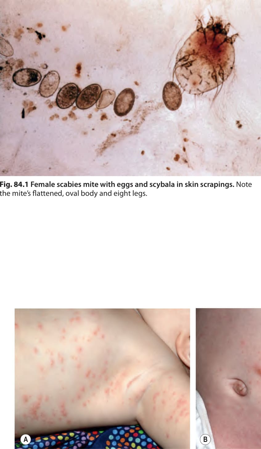
Fig. 84.1 Female scabies mite with eggs and scybala in skin scrapings. Note the mite’s flattened, oval body and eight legs.
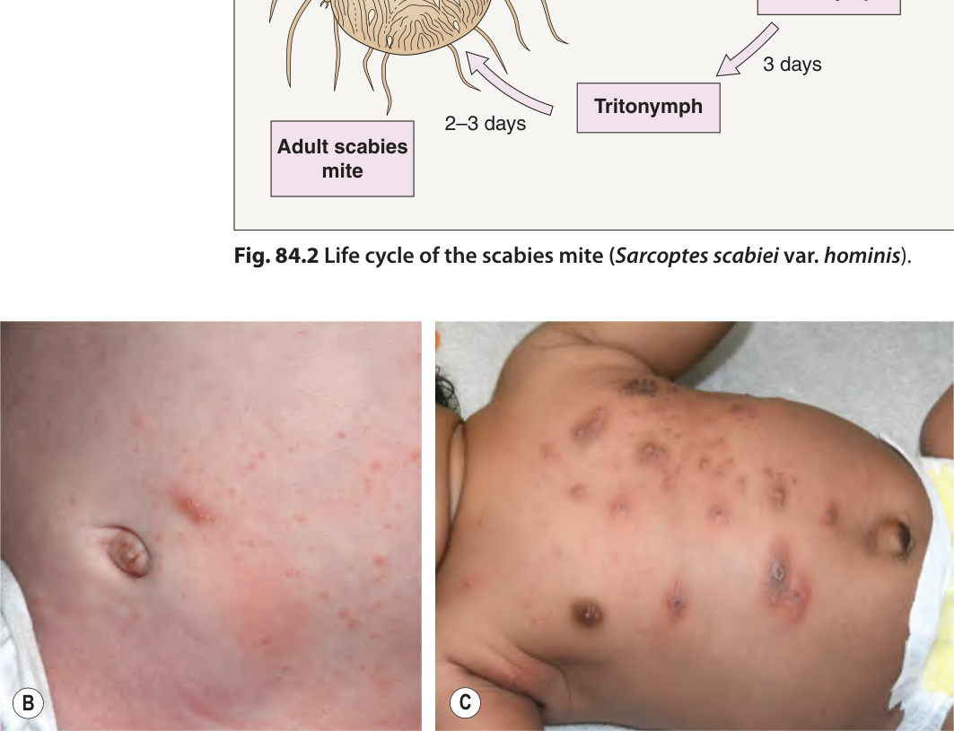
Fig. 84.2 Life cycle of the scabies mite (Sarcoptes scabiei var. hominis).
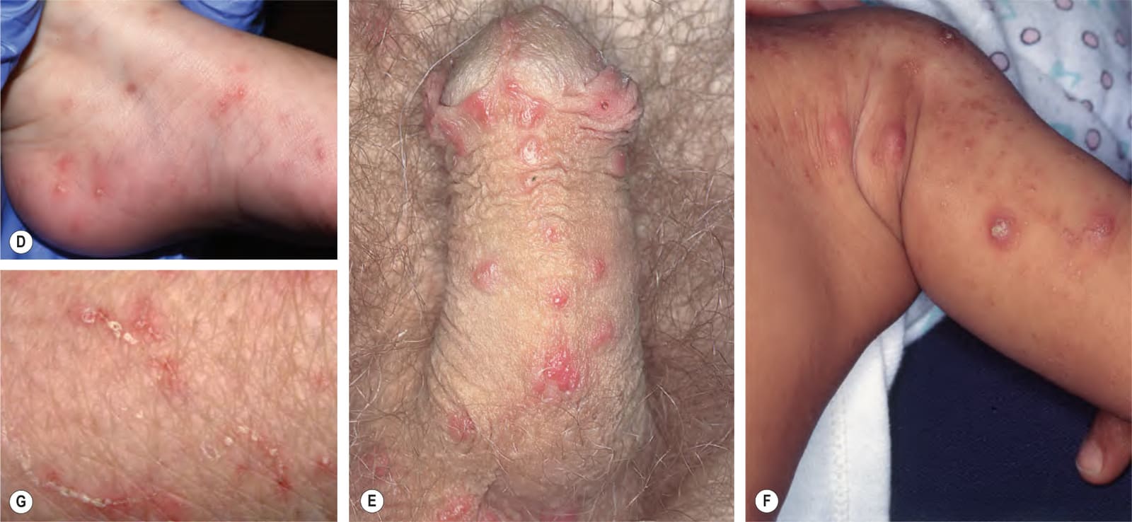
Fig. 84.3 Scabies.A–C Erythematous papules and oozing dermatitic plaques in infants with scabies. D Vesiculopustules on an infant’s foot. Similar findings are seen in acropustulosis of infancy, which can occur following successful scabies treatment. E Penile involvement with erythematous papules and nodules. F Nodular scabies in an infant. G Classic scabetic burrows. Burrows are of high yield for visualizing mites via dermoscopy and isolating mites via skin scraping. A–D, Courtesy Julie V. Schaffer, MD; F, Courtesy Kalman Watsky, MD.
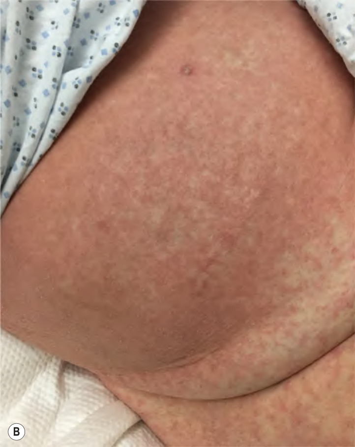
Fig. 84.4 Crusted scabies. Scabies incognito presenting as an asymptomatic hyperkeratotic “rash” on the hand in a patient with impaired sensory function (A) and on the abdomen of a hospitalized man (B). A, Courtesy Joyce Rico, MD; B, Courtesy Kalman Watsky, MD.
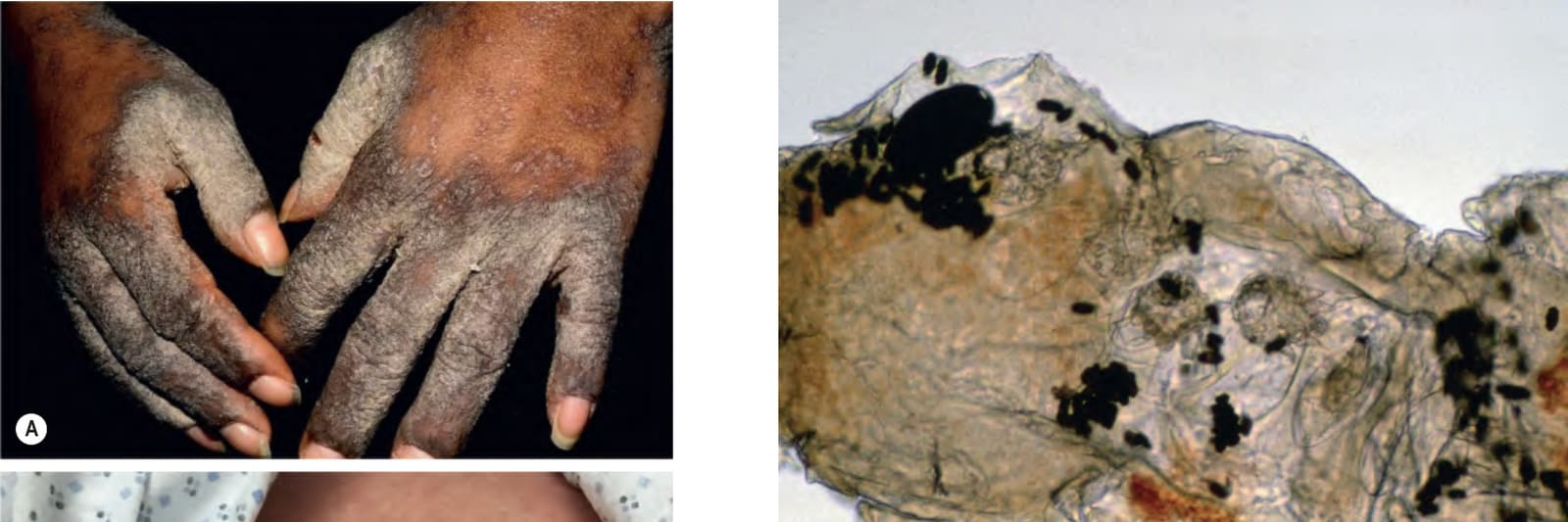
Fig. 84.5 Microscopy of a skin scraping from a patient with scabies. Four mites, eggs, and scybala are present. The mites blend in with the background scale, making them difficult to see.
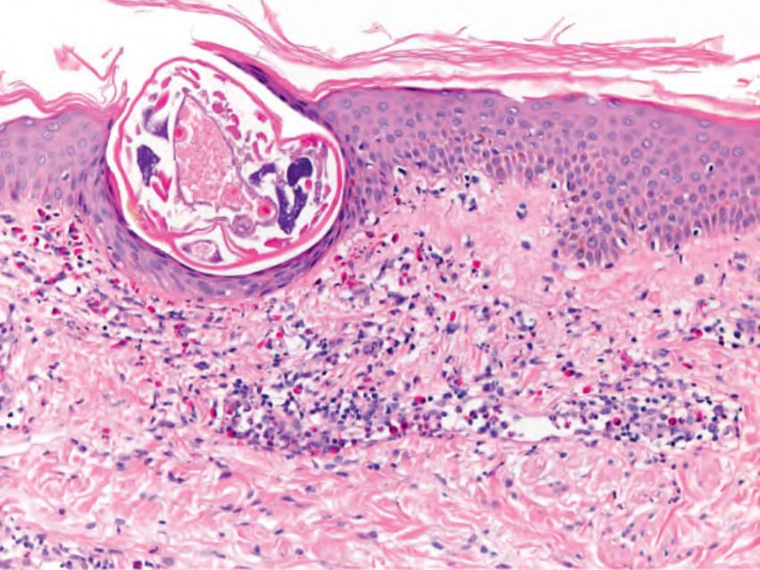
Fig. 84.6 Scabies – histologic features. A mite is evident in the stratum corneum and an eosinophil-rich dermal infiltrate is present. Courtesy Lorenzo Cerroni, MD.
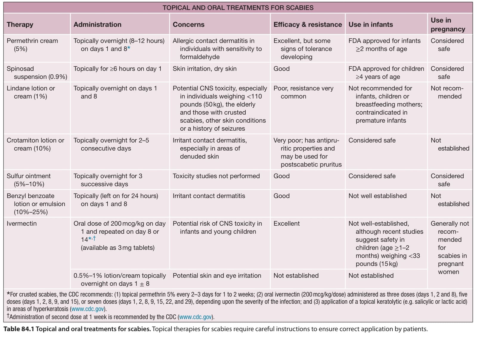
Table 84.1 Topical and oral treatments for scabies. Topical therapies for scabies require careful instructions to ensure correct application by patients.