CUTANEOUS MYIASIS
Synonym: Names for the various flies associated with this entity include human botflies, screwworms, blowflies, fleshflies, and tumbu flies
Key features
Infestation of the skin by fly larvae (maggots)
The larvae inhabit cutaneous wounds (wound myiasis) or burrow into the dermis causing boil-like lesions (furuncular myiasis)
Although they usually cause minimal morbidity, larvae can cause problems when they involve the nasal cavity and sinuses
Introduction
Cutaneous myiasis is an infestation of the skin by developing larvae (maggots) of a variety of fly species within the arthropod order Diptera. The two main clinical types are wound and furuncular myiasis.
History
Maggots were central to Francesco Redi’s experiment rejecting the theory of spontaneous generation. Wound debridement by maggots has been used for centuries, including during the American Civil War and by Napoleon’s troops.
Epidemiology
Myiasis is a worldwide infestation with seasonal variation whose prevalence is related to latitude and the life cycle of the various species of flies. Its incidence is higher in the tropics and subtropics of Africa and the Americas. The flies responsible prefer a warm and humid environment, thus they are restricted to the summer months in temperate zones, while living year-round in the tropics.
Pathogenesis
Myiasis is the infestation of living humans (or other vertebrates) with dipterous larvae that feed on the host’s tissues, liquid body substances, or ingested food. Myiasis can be caused by several species of arthropods
within the order Diptera, the two-winged true fly. The causative flies can be classified by: (1) taxonomic family; and (2) differences in pathogenic behavior of the fly species.
Worldwide, the most common flies that cause human infestation are Dermatobia hominis (human botfly) and Cordylobia anthropophaga (tumbu fly). The routes of transmission of fly larvae to human hosts differ among the fly species. For example, D. hominis lays its eggs on mosquitoes, which in turn deposit them on a warm-blooded mammal (Fig. 84.14). C. anthropophaga deposits its eggs on moist clothing and soiled blankets and in sand. The larva can live 15 days without feeding, but once it makes contact with a host, it penetrates the skin, which initiates further maturation. In endemic areas, people often iron their clothes after hanging them out to dry to kill the fly eggs. In wound myiasis, an open wound or orifice attracts flies to deposit their eggs. Any body area can be infested, and the most serious sequelae occur when the nasal cavity, sinuses, or scalp are involved. The incubation period for larvae to mature into adulthood depends on the fly and can range from 1 to 12 weeks.
Clinical Features
Furuncular cutaneous myiasis, which is caused by the human botfly and tumbu fly, causes boil-like lesions. Whereas myiasis from the tumbu fly typically occurs on the trunk, thighs and buttocks, botfly lesions are usually on exposed areas of the body, including the scalp, face, forearms and legs. A pruritic papule develops within 24 hours of penetration, enlarging to a 1–3 cm nodule with a 2–3 mm central punctum (Fig. 84.15A–C). These lesions can be painful or tender and may become crusted and purulent; there may be a sensation of movement.
In wound myiasis, the larvae are deposited in a suppurating wound or on decomposing flesh. Cochliomyia hominivorax, a screwworm, is the most common cause of this form of myiasis in the Americas, while Chrysomya bezziana is typically seen in Africa, Australia, and Asia. The diagnosis is obvious when larvae are visible on the surface of the wound (Fig. 84.15D) and more difficult when they have burrowed beneath the surface.
A–C Furuncular myiasis on the forearm presenting as a papulonodule with a central punctum (A) through which the larva’s posterior end, which contains respiratory spiracles, may protrude (B). Note the characteristic appearance of the extracted botfly larva, with parallel rows of dark spines and hooks (C). D Wound myiasis in an amputation stump. A–C, Courtesy Edward W. Cowen, MD; D, Courtesy Louis A. Fragola, Jr, MD.
Creeping (or migratory) cutaneous myiasis may be caused by Hypoderma bovis if there is exposure to infested cattle, or by Gasterophilus intestinalis in those who work with horses. This form of myiasis resembles cutaneous larva migrans, but the fly larvae migrate more slowly, persist for longer (often months), and are larger than helminth larvae.
Myiasis is a self-limiting infestation with minimal morbidity in the vast majority of cases. The major reasons for treatment are reduction of pain, cosmesis, and psychological relief. However, larvae such as C. hominivorax can infest around orifices of the head and may burrow into brain tissue.
Pathology
Histologically, an inflammatory response occurs in stages in which lymphocytes, giant cells, neutrophils, eosinophils, mast cells, and plasma cells participate. The larvae can be seen in cross-section.
Differential Diagnosis
Furuncular cutaneous myiasis should be differentiated from a ruptured epidermoid cyst, an abscess, furunculosis, a foreign body reaction, onchocerciasis, tungiasis, an exaggerated arthropod bite reaction, and lymphadenopathy.
Treatment
The larva in furuncular cutaneous myiasis should not be forcibly removed through the central punctum because its tapered shape with rows of spines and hooks prevents simple extrusion (see Fig. 84.15C). Surgical debridement under local anesthesia is curative, although a foreign body reaction can occur if parts of the larva remain. Occlusion/
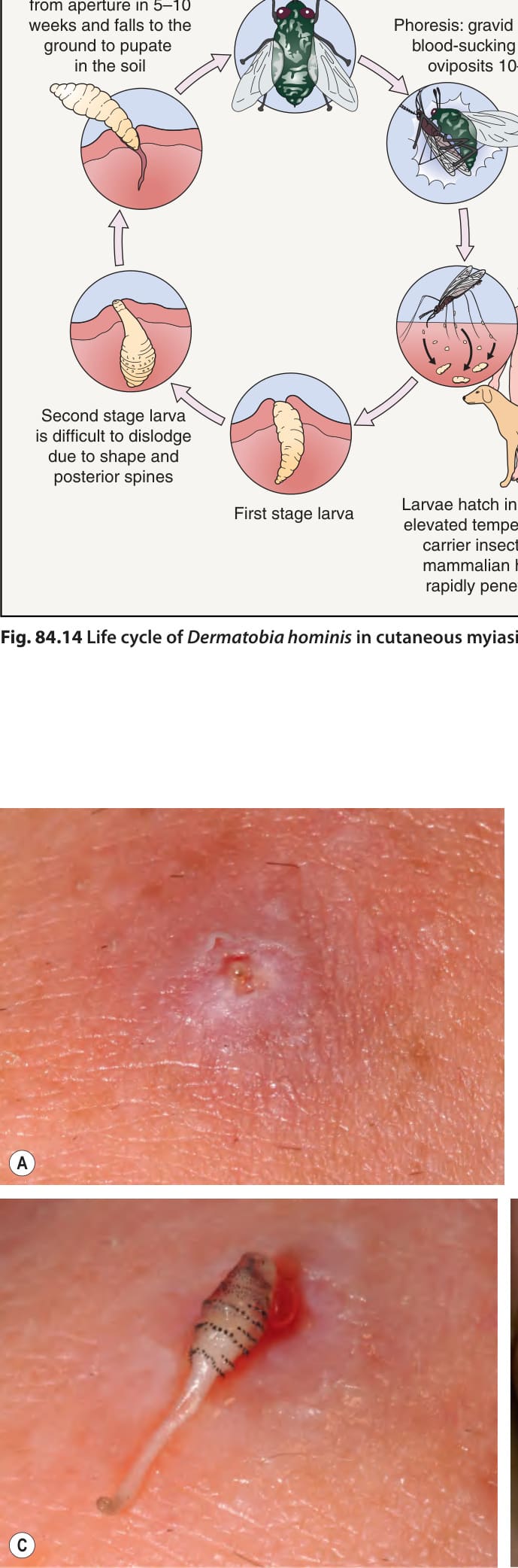
Fig. 84.14 Life cycle of Dermatobia hominis in cutaneous myiasis.
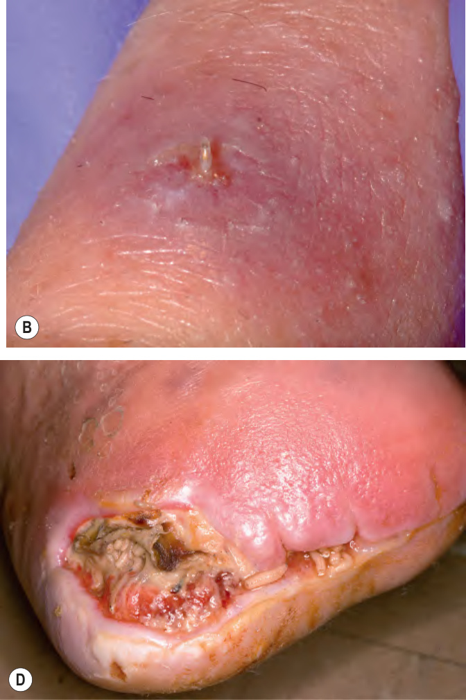
Fig. 84.15 Cutaneous myiasis.
suffocation approaches involve placement of petroleum jelly, liquid paraffin, beeswax, nail polish, heavy oil, lard, or strips of bacon over the central punctum. Such blockage forces the aerobic larva to surface for air over the course of several hours – at which time, forceps aid in their capture. Alternatively, ethyl chloride sprays, liquid nitrogen, chloroform in vegetable oil, or insecticides have been used alone or in combination. In addition, the larva can be forced to the surface by injecting lidocaine into the base of the tissue cavity or by using a suction-based venom extractor. After removal of larvae, antiseptic dressings are indicated, as are antibiotics if secondary infection is present.
Wound myiasis requires debridement with irrigation to eliminate the larvae from the wound, or surgical removal. If burrowing has occurred, the method of extraction is identical to that for furuncular cutaneous myiasis.
An alternative treatment for all types of myiasis is oral ivermectin, which has proven especially helpful with oral and orbital involvement. Surgical removal is not required unless requested by the patient, as the larvae are naturally sloughed within 2 weeks. Myiasis can be a portal of entry for Clostridium tetani, and vaccination of affected individuals should be considered.
In endemic areas, it is important to avoid activities that increase the risk of myiasis, such as wearing damp clothing or resting in sandy areas. The use of insect repellents (including on the scalp) can be useful in preventing mosquitoes that are harboring the larvae of the human botfly from depositing their living cargo.
Additional figures and tables on Diagnostic criteria for scabies, and Inpatient management of classic scabies and crusted scabies, available in our eBook (see inside front cover for access code).
diseases in dermatology: reassessment of scabies and pediculosis. In: James W, ed. Advances in Dermatology, vol. 15. St Louis: Mosby; 1999:67–108.2. Zhang W, Zhang Y, Luo L, et al. Trends in prevalence and