ARACHNIDS
Tick Bites (Class Arachnida: Subclass Acarina)
Key features
Hard ticks are common disease vectors
Ornithodoros soft ticks transmit borrelial relapsing fever
Dermacentor ticks hidden in the scalp can cause fatal tick paralysis
Tick bites may lead to galactose-α-1,3-galactose sensitization and red meat allergy
Tick control measures are important in preventing disease
Introduction
Patients with tick bites often present with the tick still attached. Scalp attachment is particularly common for Dermacentor ticks, and the patient may be unaware of a tick that is hidden by hair. This is of concern considering that Dermacentor ticks produce a neurotoxin that can cause paralysis when the tick is attached for ≥4 days. Symptoms resolve rapidly upon tick removal, and any child presenting with rapidly progressive ascending flaccid paralysis should be examined for ticks. In Australia, Ixodes ticks can cause paralysis, but this occurs primarily in dogs. Tick bites (especially Amblyomma spp.; see below) can result in sensitization to the carbohydrate antigen galactose-α-1,3- galactose associated with red meat allergy.
Correct identification of an attached tick (see Fig. 76.1) provides the physician with an opportunity to discuss tick control measures and symptoms of tick-borne disease. Most hard ticks have a threehost life cycle. Larval (seed) ticks have six legs and feed on small animals in nature. The nymphs and adults feed on larger animals.
Nymphs resemble small adult ticks, with eight legs, but are sexually immature. Because they attach for one long blood meal during each phase of their life cycle and search for a new host for each blood meal, ticks are efficient disease vectors. Hard ticks have a hard dorsal plate (scutum) and account for most medically important ticks, transmitting human diseases including Lyme borreliosis, ehrlichiosis, human granulocytic anaplasmosis, babesiosis, and a variety of rickettsial infections (including Rocky Mountain spotted fever [RMSF]) (see Table 76.1). Soft ticks such as Ornithodoros (the vector of borrelial relapsing fever) lack a scutum and have retroverted mouthparts. These ticks tend to live in close association with a single host.
Clinical features
Once the tick has been removed, hypersensitivity reactions at the site are common. Papular, nodular, and vesiculobullous lesions can occur and are typically pruritic. Hypersensitivity reactions presenting as erythematous plaques are commonly misinterpreted as erythema migrans. Nodules with pseudolymphomatous histology can also develop.
Treatment
Milder tick bite reactions may respond to application of a potent (class 1 or 2) topical corticosteroid; occlusion of these agents can increase efficacy. More intense reactions may require intralesional corticosteroid injection or surgical excision.
Most tick-borne diseases respond readily to tetracyclines. Suspected RMSF requires empiric therapy, which should not be delayed until confirmatory tests are available (see Ch. 76). Delayed treatment of RMSF has led to deaths in children. Doxycycline is the drug of choice for children as well as adults with RMSF. Staining of teeth is unlikely with a short course of doxycycline, and chloramphenicol is less effective for RMSF.
Environmental measures
Exclusion of animal hosts from communities and recreational areas can be helpful in control of tick-borne illness (Table 85.4), although this may be difficult to achieve for small animal hosts (e.g. mice, rabbits). There is evidence that communities located close to wildlife reserves have higher rates of tick-borne illnesses. Oral agents, such as the avermectins, can be placed in corn eaten by deer to kill ticks that feed on the deer. Injectable ivermectin is helpful in reducing tick infestation of livestock. Removal of leaf debris is a critical part of tick control efforts because it: (1) enables acaricides to reach the ticks; and (2) leads to a reduction in tick numbers via dehydration of adults (which require leaf debris to maintain hydration) and eggs (which require sufficient humidity to hatch).
Repellents
Permethrin has been marketed as a tick repellent. The spray formulation is most widely available. Once applied to clothing, permethrin is stable through several wash cycles. It is also suitable for treating sleeping bags, tents, and fabric used for insect screening. A liquid concentrate can be used to impregnate fabric for the life of the garment. Permethrin is particularly helpful in the prevention of tick and chigger bites, since these crawling arthropods must travel across the treated fabric, whereas flying insects tend to be attracted directly to exposed skin. The combination of a DEET-containing repellent and permethrin-treated clothing is generally highly effective against a wide range of biting arthropods.
Some North African camel ticks have demonstrated high-level resistance to permethrin. Instead of killing the tick, permethrin may produce a pheromone-like attachment response. This attachment phenomenon has not yet been described in North American ticks, but permethrin resistance does occur. High-level permethrin resistance in cattle ticks has been linked to outbreaks of bovine babesiosis and anaplasmosis in Mexico.
Methods to eliminate and prevent tick infestations in dogs include: topical application of products containing fipronil, permethrin, or fluralaner every month; oral administration of an isoxazoline (e.g. afoxolaner) every 1–3 months; and collars impregnated with imidacloprid/flumethrin, deltamethrin, or amitraz.
Tick removal
Ticks should be removed as completely as possible, and care should be taken not to squeeze the abdomen. Gentle traction is generally preferred, but a twisting motion can also be used; the latter technique may leave a small mouthpart. Inexpensive plastic tick removal devices (e.g. Tick Nipper™, Tick Twister®) can be utilized to grasp the tick just behind the mouthparts without severing them. A tick removal device can also be made by cutting a V-shaped notch in a plastic spoon or credit card with a sharp pair of scissors or hot blade. Petroleum jelly, fingernail polish, isopropyl alcohol, and hot matches are generally ineffective. Hands should be washed if they were in contact with the tick.
Antibiotic prophylaxis after tick bites
Antibiotic prophylaxis after tick bites remains controversial, and most authorities do not recommend its routine use, including for prevention of Lyme disease (see Ch. 19). In endemic areas (>20% of ticks infected), if a nymphal or adult I. scapularis has been attached for >36 hours and prophylaxis can be started within 72 hours of tick removal, a single 200 mg dose of doxycycline may decrease the risk of developing Lyme disease (from 3.2% in the placebo group to 0.4% in the treatment group in one study). A meta-analysis of antibiotic prophylaxis concluded that in order to prevent one case of Lyme disease, 50 people bitten by an
Ixodes tick would need to be treated – the risk in the placebo group was 2.2% compared with 0.2% in the antibiotic-treated group. Routine serologic testing for Lyme disease in individuals bitten by Ixodes ticks is not useful, and there is no evidence that molecular testing for Borrelia burgdorferi in a tick is helpful in predicting the risk of Lyme disease in a human bitten by that tick. Treatment regimens for erythema migrans are outlined in Table 19.4.
The utility of antibiotic prophylaxis in preventing other tick-borne illnesses has not been established. For example, in a guinea pig model of RMSF, a single dose of oxytetracycline was found to prevent disease if given shortly before the expected onset of illness. However, administration any earlier merely delayed the development of clinical symptoms, making correct timing of administration of a single dose difficult. Single-dose doxycycline for Lyme disease prophylaxis should be avoided in RMSF-endemic areas, since changing the course of RMSF could potentially delay definitive treatment.
Amblyomma Ticks
Synonym: Lone star tick
Key features
Ornate scutum (hard dorsal plate)
Single white dorsal spot on female Amblyomma americanum
Papular, nodular, and bullous lesions at attachment sites
Amblyomma ticks are vectors of ehrlichiosis, several rickettsial infec- tions, southern tick-associated rash illness (STARI), and tularemia
Amblyomma tick bites have been associated with the development of red meat allergy
Introduction
Amblyomma ticks are particularly common in the southern US. This, plus the characteristic white dorsal spot of the female A. americanum (Fig. 85.11; see Fig. 76.1), has given rise to the common name “lone star tick”. Males have ornate markings that often form pale “inverted horseshoes” on the posterior portion of their dark scutum. Amblyomma ticks have prominent eyes, festoons (grooves in the posterior edge of the scutum), and long anterior mouthparts.
A. americanum has a wide distribution, now ranging from Texas to Iowa and Maine (see Fig. 76.12), that is continuing to expand due to the explosion in the white-tailed deer population. Other species of Amblyomma ticks are common in Africa, the Caribbean, and Central and South America. Some Amblyomma ticks attach to birds, and migration of avian hosts has been implicated in dissemination of tickborne diseases.
Clinical features
Amblyomma tick attachments favor the legs, buttocks, and groin and often affect people who work outdoors or take part in outdoor recreation (e.g. construction workers, fishermen, hunters, hikers), especially when new development displaces the tick’s usual hosts. Patients usually become aware of Amblyomma tick bites while the tick is still attached. This is partly because the ticks often attach in large numbers. Once the tick has been removed, delayed-type hypersensitivity to tick antigens can lead to a reaction at the attachment site. Papular, nodular, and bullous lesions are common.
A. americanum is the major vector of Ehrlichia chaffeensis, the agent of human monocytic ehrlichiosis. This disease has been documented in more than 30 states, with systemic symptoms similar to those of RMSF (e.g. fever and headache) and variable cutaneous manifestations (see Ch. 76). A. americanum is also the primary vector for Ehrlichia ewingii infection.
Southern tick-associated rash illness (STARI; “Missouri Lyme disease”) was originally described in Missouri but can also occur in other southern and southeastern states. It presents with an expanding erythematous annular plaque 3–14 days after A. americanum attachment, often in association with fever, headache, myalgias, and arthralgias. Borrelia lonestari has been implicated as a potential cause.
Amblyomma ticks have been implicated in the transmission of RMSF (Rickettsia rickettsii; primarily in Central and South America, where it may be referred to as Brazilian spotted fever), American tick bite fever (Rickettsia parkeri), African tick bite fever (Rickettsia africae), and tularemia (see Chs. 74 & 76). In the midwestern and southern US, A. americanum is also the vector of Heartland virus disease and likely Bourbon virus infection, which are due to a phlebovirus and thogotovirus, respectively. A related phlebovirus causes severe fever with thrombocytopenia syndrome, an emerging infection in Asia transmitted by Haemaphysalis ticks, which have recently been identified in the eastern US.
Bites from Amblyomma ticks have been most strongly associated with allergy to beef, pork, and lamb via development of IgE antibodies specific to galactose-α-1,3-galactose, a blood group substance of non-primate mammals. Urticaria, angioedema, or anaphylaxis typically develops 3–5 hours after eating red meat. Of note, IgE against galactose-α-1,3- galactose has also been implicated in cetuximab-induced anaphylaxis.
Dermacentor Ticks
Synonyms:D. andersoni: Rocky Mountain wood tick D. variabilis: American dog tick
Key features
Large ticks
Ornate scutum (hard dorsal plate) with deep punctuations
The adult tick prefers to attach to the head, neck, and shoulder
D. variabilis is the major vector for RMSF in the United States, while
D. andersoni can transmit Colorado tick fever as well as RMSF
Introduction
Dermacentor ticks are commonly found in open areas with low bushy vegetation, but they are scarce in heavily wooded areas. Peak abundance of the tick is in April and May, declining by July. Dermacentor ticks have an ornate scutum, prominent eyes and festoons (grooves in the posterior scutum), small anterior mouthparts, and a rectangular basis capituli. Both D. variabilis (Fig. 85.12; see Fig. 76.1) and D. andersoni have brown legs, and coxa 1 (the attachment base for the first pair of legs) is bifid. Coxa 4 is greatly enlarged in the male, and males have no ventral plates. As in most hard ticks, the scutum in the female is smaller, leaving a portion of the abdomen exposed.
D. variabilis is found throughout the US, except for the Rocky Mountain states. In parts of Canada, its range overlaps with that of D. andersoni, which is generally confined to the Rocky Mountains. In Georgia and South Carolina, D. variabilis is the second most common tick found on humans (A. americanum being the most common).
This species is the major vector for Rocky Mountain spotted fever in the US.
Pathogenesis
During the course of engorgement, D. andersoni saliva downregulates local cell-mediated immunity. This phenomenon may explain the delayed onset of many nodular tick bite reactions. It might represent an adaptive mechanism, allowing the tick to feed unnoticed.
Clinical features
Dermacentor ticks preferentially attach to the head and neck region; in contrast, Amblyomma americanum prefers the lower legs, buttocks, and groin, and Ixodes scapularis shows less site preference but most often attaches to the trunk. Prolonged attachment of a Dermacentor tick is the most frequent cause of tick paralysis in the US (see above).
D. variabilis is the major vector for RMSF in the US. The highest incidence of RMSF is in the eastern states, especially in North Carolina (see Fig. 76.2). RMSF typically presents with fever and headache. An acral eruption of erythematous macules and/or petechiae may be evident but is commonly absent early in the disease process. As many as 40% of patients with RMSF are not aware of a recent tick bite. As the mortality rate in RMSF is heavily dependent on the interval between onset of symptoms and initiation of antibiotic therapy, the disease should be considered in every patient with fever and a headache in endemic areas.
D. andersoni is the major vector for RMSF in the Rocky Mountain states, and it is also the vector for the viral illness Colorado tick fever. Because the symptoms of Colorado tick fever overlap with those of RMSF, specific diagnostic tests such as paired serologic studies (acute and convalescent) and immunohistochemical staining, direct immunofluorescence, or PCR-based analysis of lesional skin biopsy specimens can help to confirm the diagnosis of RMSF; however, empiric treatment should not be delayed until the results are available. Additional diseases that can be transmitted by Dermacentor ticks include tularemia, rickettsial infections in Europe and Asia, Pacific Coast tick fever in California (Dermacentor occidentalis), and occasionally, human monocytic ehrlichiosis and Q fever.
Rhipicephalus Ticks
Synonym: Brown dog tick
Key features
Common dog ticks
Most are brown with brown legs, a teardrop shape and inornate scutum (hard dorsal plate)
Vectors for boutonneuse fever and (occasionally) RMSF in the southwestern US
Introduction
Rhipicephalus ticks (Fig. 85.13) are common dog ticks that are typically brown in color with an inornate scutum. They have prominent eyes and festoons (grooves in the posterior scutum), but small mouthparts attached at a hexagonal basis capituli.
Clinical features
Rhipicephalus ticks carry RMSF (primarily in the southwestern US and Mexico) and boutonneuse fever (see Ch. 76). They may also be vectors for Congo–Crimean hemorrhagic fever virus, although Hyalomma ticks usually transmit this condition.
Ixodes Ticks
Synonyms: Blacklegged tick Deer tick
Key features
Teardrop shape, often with dark legs
Inornate brown scutum (dorsal hard plate) that is small relative to the large abdomen in females
Vectors for Lyme disease, human granulocytic anaplasmosis, babesiosis, viral encephalitis (e.g. due to Powassan virus), and other borrelial and rickettsial infections
Introduction
Ixodes scapularis (formerly also known as I. dammini), the eastern blacklegged tick, is the best known of the Ixodes ticks. Adult females are typically found attached to the host in late fall and spring. Their small, inornate brown scutum is often overshadowed by a large, soft, cream-colored engorged abdomen (Fig. 85.14; see Fycks have short anterior mouthparts, prominent broad “antennae”, and an upside-down U-shaped ventral groove with its apex anterior to the anus.
Clinical features
Ixodes ticks are vectors for Lyme disease, babesiosis, and human granulocytic anaplasmosis (HGA). Babesiosis is a malaria-like illness, whereas HGA is an acute febrile illness characterized by leukopenia, thrombocytopenia, and increased serum hepatic transaminases. In
the western US, I. pacificus (the western blacklegged tick) is the major vector for Lyme disease (see Fig. 76.12). In the Great Lakes region and eastern US, I. scapularis is the major vector. I. ricinus and I. persulcatus are important vectors for Lyme disease in Europe and Eurasia, respectively. Because the larval and nymph stages of the tick often attach to birds in the spring and summer, avian migration (particularly of robins) may be responsible for the broad geographic distribution of the tick and of Lyme disease.
A single Ixodes tick may transmit more than one disease. In one study of a rural New Jersey county, 55% of adult I. scapularis ticks carried at least one disease (Lyme disease, 43%; babesiosis, 5%; HGA, 17%) and 10% carried more than one of the three diseases. Co-infection with more than one of these three diseases is also common in Wisconsin and Minnesota, and I. scapularis has been reported to carry both Lyme disease and HGA in New York City parks.
Ixodes ticks carry viral encephalitis in Europe and transmit rickettsial infections in Europe and Australia (see Table 76.1). Emerging infectious diseases that are transmitted by Ixodes ticks include a viral-like illness or relapsing fever due to the spirochete Borrelia miyamotoi (Asia, Europe, North America), Powassan encephalitis due to a flavivirus (northeastern US, Great Lakes region), ehrlichiosis due to E. muris eauclairensis (Minnesota and Wisconsin), and neoehrlichiosis due to Candidatus Neoehrlichia mikurensis (Europe and Asia).
Mites (Class Arachnida: Subclass Acarina)
Key features
Mites are identified by their mouthparts and legs; the shape of their body, dorsal plates, and shields (sternal, anal, and genitoventral); and the setae (bristles or hairs) on the latter structures
Trombiculid mites are typically larval (chiggers; six-legged) when attached to a host; they are vectors for scrub typhus
Cheyletiella mites are commonly found on pets and may produce papular or vesiculobullous lesions in the owner
Species that parasitize birds, mammals, and grains can produce dermatoses; the house mouse mite is a vector for rickettsialpox
Introduction
Mites are small arachnids that range from 0.1 to 2 mm in length. Tens of thousands of species exist worldwide (Table 85.5). The majority of mites
are free-living, but thousands of species parasitize animals or plants. Like fleas, most mites lack host specificity. Mites of medical importance may cause skin lesions, incite allergic reactions (especially in atopic individuals), or spread disease (Table 85.6). Like ticks, mites mature through larval, nymph, and adult stages. Typically, there
is a single larval stage (referred to as chiggers in trombiculid mites) and several nymphal stages. Most mites lay eggs, but some, such as the spiny rat mite (Laelaps echidninus), are viviparous, producing non-motile larvae. The straw itch mite (Pyemotes tritici) retains its young until they emerge as sexually mature mites. Sarcoptic mites are relatively species-specific and complete their entire life cycle on the host (see Ch. 84).
Clinical features
Cutaneous reactions to mites are common and include papular, papulovesicular, bullous, urticarial, and morbilliform eruptions (Fig. 85.15). Chigger bites most often affect the lower legs, skin at the edges of underwear, and the genital region. Larvae of trombiculid mites in Asia are vectors for scrub typhus, while house mouse mites transmit rickettsialpox (see Ch. 76). Eschars at the site of the bite are a clue to rickettsial disease, and serologic testing can be helpful. As with tickborne rickettsial diseases, biopsy of the eschar can be sent for immunohistochemical staining or PCR to distinguish typhus group organisms from the spotted fever group.
Differential diagnosis
Mite-induced dermatoses are often misdiagnosed. Bullous lesions are especially common, and false-positive direct immunofluorescence has been described. Histologic features resembling those of an insect bite may suggest the correct diagnosis, as may a clinical pattern of skin lesions corresponding to areas of exposure. However, eosinophilrich subepidermal bullae occur in both mite infestation and bullous pemphigoid, so a careful history is essential.
Mite-induced dermatitis can be associated with pets, rodents, straw bedding, bird nests, grains, and even onion bulbs (see Table 85.6). Avian mite dermatitis may also be associated with pet rodents, such as gerbils. Pets should be examined by a knowledgeable veterinarian; skin scraping, adhesive tape stripping, or biopsy may reveal the mite(s) on the animal. Vacuum cleaners can also be fitted with filters to collect mites (Table 85.7). Another helpful technique is to spray areas of dandruff or crust on a pet with an acaricidal agent recommended by a veterinarian. A fine-toothed comb or toothbrush is then used to remove the scale-crust, which is placed in a sealed bag. When alcohol is added to the bag, scalecrust and hair will sink, whereas mites will float.
Treatment
Some types of mites inflict a “bite and run” injury, while others remain attached for a prolonged meal. Scraping, tape stripping, or vigorous washing with soap and water are all effective in removing or killing attached mites. Scrapings, tape strippings, and biopsy specimens may be helpful diagnostically as well as therapeutically.
Definitive treatment requires avoidance or treatment of the infested source. Mite identification can be helpful in establishing the likely source. Management strategies differ for free-living, zoonotic, and grocery mites. An acarologist can be of immeasurable value in this setting, and the Medical and Veterinary Entomology text by Gary Mullen and Lance Durden is an excellent resource. As noted above, animals should be examined and treated by a veterinarian. In public parks and training areas, animal exclusion and acaricides may be helpful measures. Personal protection with permethrin-treated clothing is also of value.
Skin lesions in humans may be treated with topical antipruritics such as camphor and menthol, or with topical anesthetics such as pramoxine. Potent topical corticosteroid preparations can be helpful, especially when occluded. Intralesional corticosteroid injections and excision of pruritic nodules may be necessary. Bullous and excoriated lesions can develop secondary infections, and appropriate wound care is important. Home remedies typically attempt to remove or kill attached mites and reduce itch with counterirritants (e.g. propylene glycol-containing deodorant sticks). Patients may present with irritant and allergic contact dermatitis to antiseptics, nail polish, or even battery acid.
Chiggers
Introduction
Trombiculid mites are oviparous, depositing their eggs on the ground, leaves, or blades of grass. After several days, the egg cracks, but the mite remains in a quiescent prelarval stage. Following the prelarval stage, the six-legged larvae (chiggers) actively search for a suitable host. It is the larval mite that is found attached to the host. After a prolonged meal, the larvae drop off and mature through the eight-legged free-living nymph and adult stages.
Clinical features
Chigger bites are intensely pruritic, especially in highly sensitized individuals. Grouped papules, vesicles, or bullae are typically found on the lower extremities, or where elastic meets the skin. Seasonal penile swelling associated with pruritus and dysuria in children, referred to as “summer penile syndrome”, appears to represent a hypersensitivity response to chigger bites.
Treatment
The topical antipruritics and anesthetics discussed above may be helpful. Most patients presenting to a dermatologist require potent topical or intralesional corticosteroid therapy. Vigorous washing with soap and water immediately after the exposure is helpful. Treatment of clothing with permethrin is a useful strategy for prevention.
Cheyletiella
Introduction
Cheyletiella are non-burrowing mites that are commonly found on cats (C. blakei), rabbits (C. parasitovorax), and dogs (C. yasguri). In humans, they are a frequent cause of cryptic mite-induced dermatitis. Infestations in animals are variably pruritic and produce a subtle dermatitis that
presents with dry white scales, which is sometimes termed “walking dandruff”. The animal should be evaluated by a veterinarian, and it is important to note that a single negative scraping does not exclude cheyletiellosis. In a study of dogs with Cheyletiella, samples collected by using a vacuum cleaner on the dog were shown to be significantly more sensitive than tape stripping, hair plucking, or skin scraping.
Clinical features
The typical patient with Cheyletiella dermatitis is a young or middleaged woman who presents with grouped pruritic papules (Fig. 85.16). Bullous eruptions have also been described. It is uncommon to find the mite on the patient.
Treatment
Skin reactions in humans should be treated as described above. Treatment of the animal should be directed by a veterinarian.
Spider Bites (Class Arachnida: Order Araneae)
Spiders are arachnids related to scorpions, ticks, and mites. Many species of toxic spiders are found worldwide. The bite of Phoneutria wandering spiders in Brazil can be fatal in children, but most spider bites result only in a local cutaneous reaction. Toxic spiders are often regional in distribution, and many are poorly characterized. The most clinically significant spiders are discussed below. It is noteworthy that many important species in the US were not native to North America, but were imported in recent years as a result of worldwide travel and commerce. Examples are the hobo spider and Loxosceles laeta spider.
Widow Spiders (Latrodectus)
Key features
Black widows are large, shiny black spiders with a round abdomen
A variety of black, brown and red widow spiders exist worldwide
In Latrodectus mactans, a red hourglass shape is seen on the ventral abdomen
Acute pain and edema occur at the bite site
Systemic symptoms mimic acute abdomen and may include rhabdomyolysis
Introduction
Widow spiders (Latrodectus spp.) are found worldwide, with five species including black, brown, and red variants in North America alone. Latrodectus mactans is the most common black widow spider in
North America, with a range that extends to the Caribbean islands. L. tredecimguttatus, L. curacaviensis, and L. indistinctus are black widow spiders found in Europe, South America, and Africa (referred to as a button spider), respectively. Female black widow spiders from North American species have a red hourglass-shaped mark on the ventral abdomen, while species from other areas of the world have different red patterns. Brown (L. geometricus) widow spiders characterized by an orange ventral hourglass shape have a widespread distribution that includes the US, South America, and Africa. Australia has the red-back spider (L. hasselti) and New Zealand the red katipo spider (L. katipo).
Widow spiders are typically found in woodpiles, in shoes, and under outhouse seats. Human envenomation usually occurs when the spider’s environment is disturbed, and the spider is inadvertently trapped or pressed against the skin. Widow spiders are more aggressive when protecting an egg sack.
Pathogenesis
Black widow venom contains latrotoxins that act by depolarizing neurons, increasing intracellular calcium, and stimulating uncontrolled exocytosis of neurotransmitters. Divalent cation-dependent tetramers related to α-latrotoxin can insert into lipid bilayers, forming membrane pores. Calcium-independent receptors for latrotoxin have also been identified.
Clinical features
The degree of morbidity caused by widow spider bites varies depending upon the amount of venom injected and the site of the bite as well as the species, age, and sex of the spider, with only adult females capable of envenomation. Local skin reactions are usually limited to transient erythema, edema, sweating, and piloerection. Extracutaneous manifestations are often prominent and can include painful muscle spasms (initially around the bite site, then more widespread), symptoms suggestive of an acute surgical abdomen, headache, and nausea; generalized diaphoresis may also occur. The symptoms of brown widow bites tend to be milder and restricted to the tissues near the bite.
Treatment
Benzodiazepines and intravenous calcium gluconate can be helpful for associated tetany. In one study, calcium gluconate was shown to be superior to methocarbamol (Robaxin®). Antivenin can produce rapid relief of tetany and complications such as priapism that are unresponsive to other agents. Purified equine Fab fragment-based antivenin is associated with a lower risk of hypersensitivity reactions than classic antivenin.
Loxosceles Spiders
Key features
Brown recluse spider (Loxosceles reclusa) has a small body and long legs
Dark brown fiddle pattern present on cephalothorax
Dermonecrotic bite reactions
Systemic reactions include shock, hemolysis, renal insufficiency, and disseminated intravascular coagulation
Other Loxosceles spiders can produce similar reactions
Introduction
Loxosceles spiders are found throughout the world and classically have a dark brown, violin-shaped marking on the dorsal cephalothorax (Fig. 85.17). In the US, bites from L. reclusa (the brown recluse spider), L. laeta, L. rufescens, L. deserta, and L. arizonica can cause skin necrosis, although reactions to the latter three species are generally mild. Many other spiders are capable of producing dermonecrotic or systemic reactions, and bites of these spiders are often erroneously attributed to the brown recluse spider. Brown recluse spiders are most common in the south central US, from Tennessee and Missouri to Oklahoma and Texas (Fig. 85.18). They are often found in woodpiles, in attics, and under radiators.
Most bites occur when the spider has been disturbed. Loxosceles spiders are non-aggressive, and there are reports of families living in
houses infested by thousands of brown recluse spiders without a single bite. This suggests that brown recluse bites are overdiagnosed, especially in regions where the spider is rare. The diagnosis can be confirmed by an enzyme immunoassay to detect Loxosceles venom in a skin biopsy specimen or plucked hairs (obtained up to 4 days after the bite) or by a passive hemagglutination inhibition test (up to 3 days after the bite); in addition, reduced glycophorin A on the erythrocyte surface may represent a marker of venom exposure.
Pathogenesis
Sphingomyelinase D is the major toxin in brown recluse venom, and it interacts with serum amyloid protein. Hyaluronidase allows eschars to spread in a gravity-dependent fashion. Neutrophils are not directly activated by the venom, and neutrophil activation may be dependent upon the interaction between the venom and endothelial cells. The venoms of geographically diverse spiders such as L. reclusa, L. laeta, L. deserta, L. gaucho, and L. intermedia have similar sphingomyelinase activity and electrophoretic profiles, with a major protein band at 32–35 kDa. All contain hyaluronidase as well as metalloproteinases
with fibrinogenolytic activity. The similarity of these venom components and documented cross-reactivity suggests the potential for a single global Loxosceles antivenin.
Clinical features
The majority of bites by Loxosceles spiders, including the brown recluse, do not cause serious reactions. Local pain is frequently delayed until several hours after the bite, which most often occurs on an extremity (Fig. 85.19). Cutaneous reactions typically begin with erythema and then develop central vesiculation or duskiness with a blanched halo, which may evolve into hemorrhagic bullae and necrosis. Dermonecrotic reactions can eventuate in dry, necrotic eschars or ulceration. Upper airway obstruction caused by envenomation of the neck by brown recluse spiders has been reported. Systemic reactions include thrombocytopenia and Coombs-positive hemolytic anemia. L. arizonica, a spider found in the southwestern US, causes less severe necrosis but has been implicated as a cause of shock.
Pathology
Histologic findings have been best described in laboratory models of brown recluse spider envenomation in rabbits. The findings are timedependent, with early biopsy specimens demonstrating a neutrophilic infiltrate. Later changes include “mummified” coagulative necrosis of the epidermis, adnexal epithelium, and superficial dermis. A neutrophilic band-like infiltrate may still mark the border between viable skin and eschar. Small vessel vasculitis and thrombosis are often evident adjacent to the neutrophilic band, and larger vessel vasculitis resembling polyarteritis nodosa may account for the extent of tissue necrosis seen after some bites. Arterial thrombosis leading to gangrene of the foot has been reported.
Treatment
Optimal treatment to prevent dermonecrotic reactions remains elusive. Most bites can be treated with rest, ice, and elevation. Intradermal injection of polyclonal anti-Loxosceles Fab fragments can attenuate necrosis in an animal model up to 4 hours after envenomation, and antivenin may reduce the ultimate size of the necrotic area even when administered up to 48 hours after envenomation. Hyperbaric oxygen therapy may decrease the final size of ulceration, although results have varied. Studies with more widely available agents such as dapsone, colchicine, and prednisone have been inconsistent and often disappointing. Dapsone therapy is complicated by the risk for hemolysis, especially in individuals who have a glucose-6-phosphatase dehydrogenase (G6PD) deficiency. Current recommendations for systemic therapy are limited to antivenin (if available) and prednisone (for systemic reactions). Anecdotal reports and some animal data also suggest that intralesional triamcinolone may have some efficacy for dermonecrotic reactions. The complement inhibitor eculizumab was shown to prevent brown recluse venom-induced hemolysis in vitro, and clinical studies are needed to determine the drug’s therapeutic potential for brown recluse bite reactions.
Funnel Web Spiders
Key features
Large, hairy, aggressive spiders found in dark, moist areas such as basements
Funnel-shaped webs
Tegenaria agrestis (hobo spider, aggressive house spider), found in the northwestern US, Canada and Europe, is 4–5 cm in diameter and may occasionally cause dermonecrotic reactions
Other funnel web spiders can produce local and systemic reactions
Introduction
When Tegenaria agrestis (hobo spider) was imported to North America from Europe, this spider found few major competitors and became common in human dwellings in the northwestern US (see Fig. 85.18), usually in basement webs at soil level in contact with concrete or stone. In Europe, the hobo spider has a largely rural habitat while the less aggressive T. gigantea (also known as T. duellica; giant house spider) and T. domestica (domestic house spider) are more likely to be found in human dwellings. The sternum of the hobo spider has a lightly colored center and dark vertical bands on the sides.
The most medically important funnel web spiders, Hadronyche and Atrax, are endemic to eastern Australia. These spiders produce potent neurotoxins, and their bites can result in severe systemic symptoms (see below). Similar reactions may occur following bites from Phoneutria, spiders in South America (especially Brazil) and Costa Rica.
Pathogenesis
Australian funnel web spider robustoxin contains small peptide neurotoxins (δ-atracotoxins) that slow tetrodotoxin-sensitive, voltage-gated sodium channel inactivation, resulting in catecholaminergic and cholinergic excess when severe envenomation occurs in humans. In contrast, agatoxins from the native American funnel web spider (Agelenopsis aperta) target ion channels and paralyze insect prey but are not medically significant to humans.
Clinical features
Whether T. agrestis bites can potentially produce dermonecrosis or systemic symptoms represents a subject of controversy. There are very few documented reactions to bites from this spider in the literature, and studies have failed to demonstrate induction of cutaneous necrosis or hemolysis by T. agrestis venom in animal and laboratory models. Bites of Australian funnel web spiders of the genera Hadronyche and Atrax are often associated with extreme pain, puncture marks, local redness, and bleeding. For these spiders, severe envenomation is characterized by autonomic excitation (e.g. diaphoresis, hypersalivation, brady- or tachycardia, hypertension) and neuromuscular symptoms (e.g. paresthesias, fasciculations, muscle spasms), which may culminate in myocardial injury and shock without antivenin therapy.
Tarantulas (Family Theraphosidae)
Key features
Typically large hairy spiders, up to 15 cm in diameter
Normally non-aggressive
Urticating hairs are thrown at skin and eyes
Important cause of ophthalmia nodosa
Introduction
Tarantulas are large hairy spiders common in the southwestern US, and related species are found throughout the world. They are often sold in pet stores. Most tarantula bites do not produce severe systemic toxicity.
Many species of tarantulas possess urticating hairs in a characteristic patch on the dorsal abdomen. These hairs are used in a defensive fashion to drive predators from the spider’s burrow. Vibrations of the hind legs are used to flick hairs at the perceived attacker. Urticating hairs are absent on most African and Asian species.
Clinical features
Itching at the site of urticating hair penetration may persist for several weeks after exposure. Hairs that penetrate the cornea can result in ophthalmia nodosa, a chronic granulomatous reaction that can lead to loss of vision.
Pathology
Urticating tarantula hairs penetrate the stratum corneum and epidermis and may extend as deep as the reticular dermis.
Scorpions (Class Arachnida: Order Scorpiones)
Key features
Large anterior claws
When threatened, the long tail is curved upward to sting
Blunt thorn on the tail of Centruroides spp.
Introduction
Many toxic scorpions exist worldwide. Scorpions are typically found under tabletops, in woodpiles, and in shoes. They sting when disturbed and accidentally trapped by a hand or foot. Several clinically relevant species are listed in Table 85.8.
Clinical features
Local and systemic symptoms (e.g. pain, paresthesia) are typically out of proportion to cutaneous signs such as erythema and edema, which are not usually prominent at the site of the sting. Most fatalities are related to cardiorespiratory manifestations, including cardiogenic shock and pulmonary edema, in children younger than 10 years of age. Pancreatitis is also an important cause of morbidity after scorpion envenomation.
Treatment
Antivenin is available in endemic areas and, along with supportive care, has been shown to reduce morbidity and mortality from severe scorpion envenomation. However, application of ice is sufficient for most minor scorpion envenomations. Prazosin reverses the autonomic
storm characteristic of Indian red scorpion (Hottentotta tamulus) envenomation, resulting in accelerated recovery and preserved myocardial function. Tacrolimus was also found to have protective effects against systemic toxicity from scorpion envenomation in an animal model.
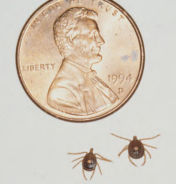
Fig. 85.11 Amblyomma americanum (lone star tick). Note the characteristic white dorsal spot on the female ticks. The female’s scutum often covers only a portion of the body to allow room for engorgement.
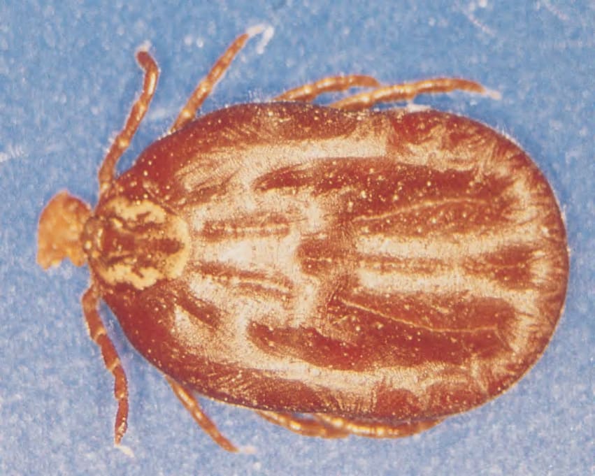
Fig. 85.12 Dermacentor variabilis tick (engorged female) with an ornate scutum.
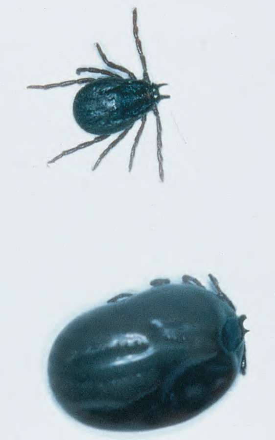
Fig. 85.13 Rhipicephalus ticks. Common brown dog ticks; the lower tick is engorged with blood.
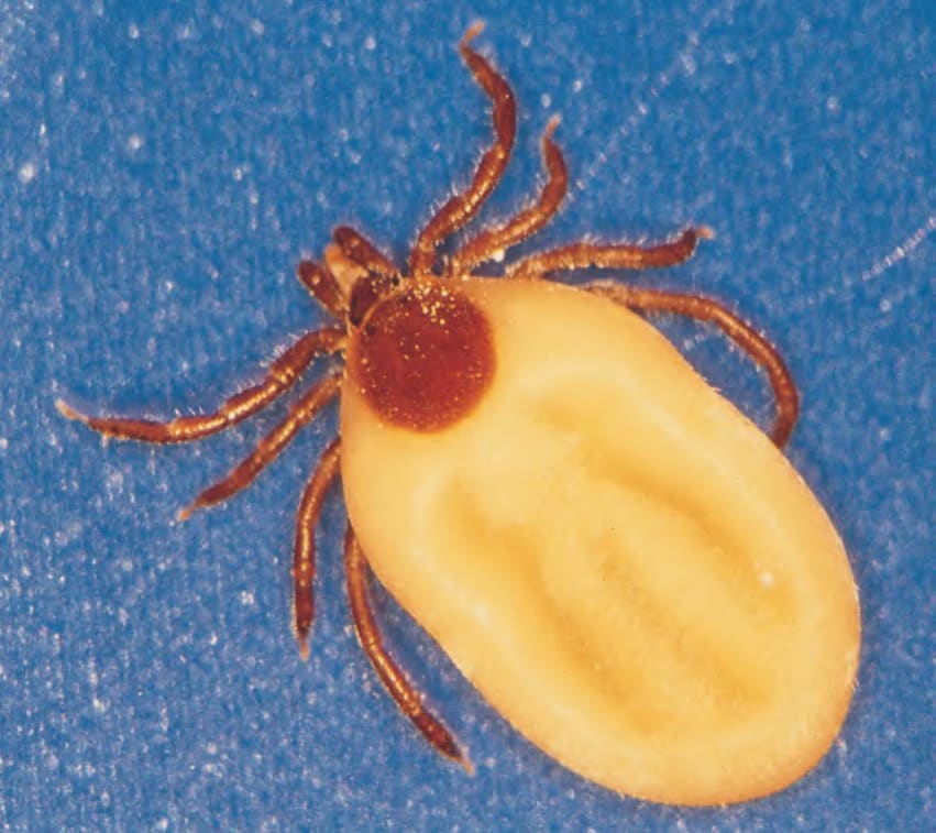
Fig. 85.14 Ixodes scapularis tick. The inornate brown scutum is overshadowed by the large, cream-colored, engorged abdomen.
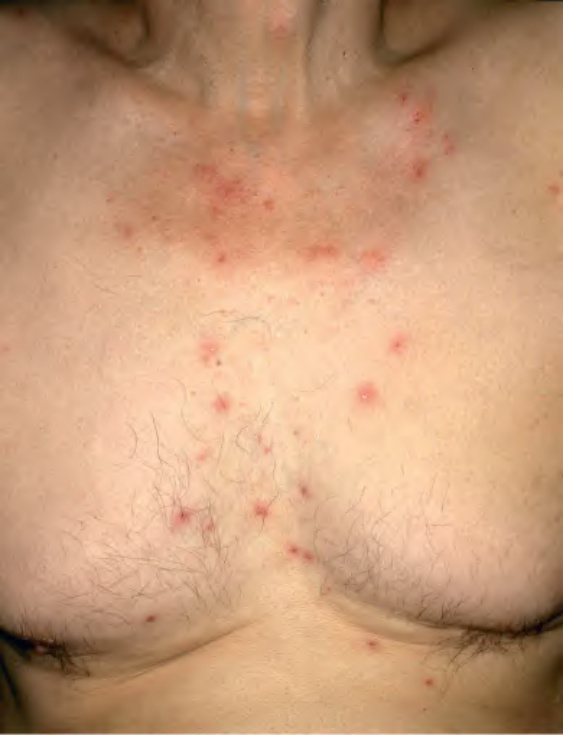
Fig. 85.15 Avian mite bites. Multiple erythematous pruritic papules, several of which have hemorrhagic crusts. The exposure was birds nesting on the patient’s house. Courtesy Jean L. Bolognia, MD.
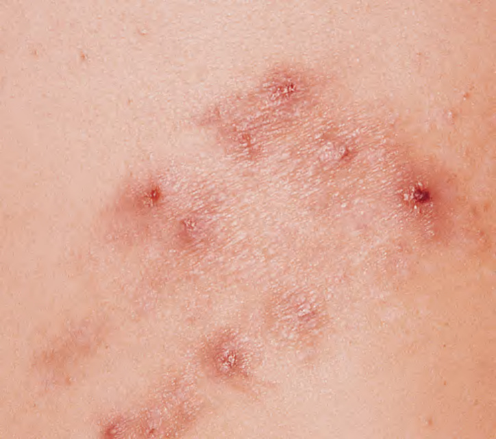
Fig. 85.16 Cheyletiella bites. These mites are non-burrowing and are found on cats, rabbits, and dogs.
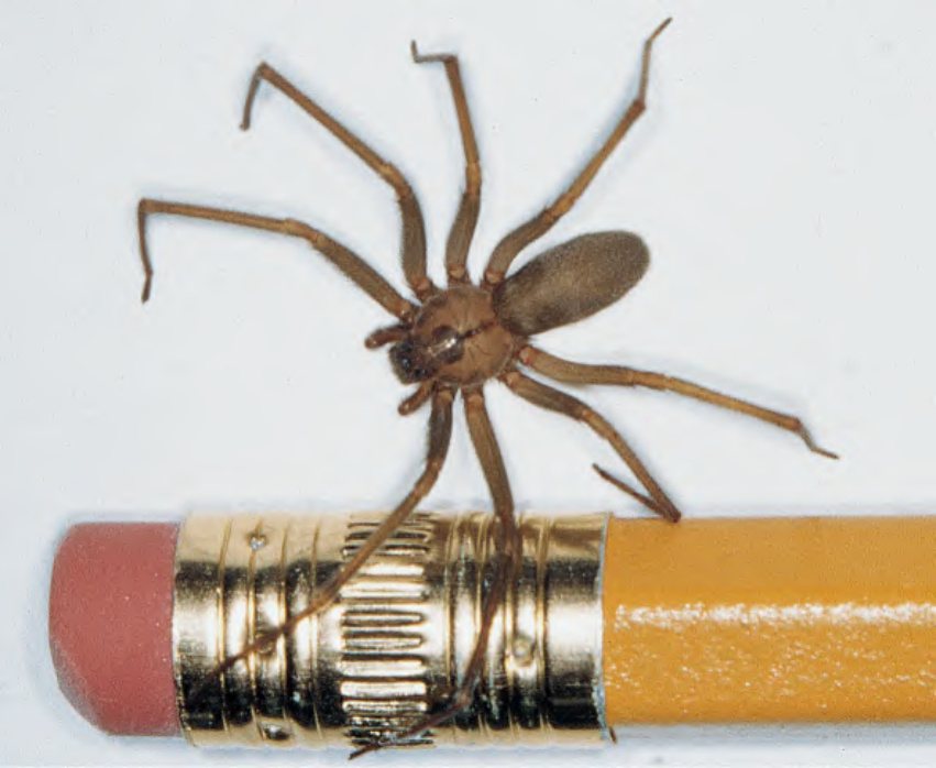
Fig. 85.17 Brown recluse spider (Loxosceles reclusa). This spider has a small body, long legs, three pairs of eyes, and a dorsal violin pattern on the cephalothorax.
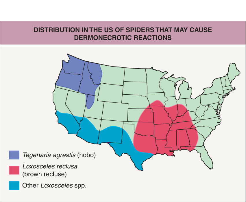
Fig. 85.18 Distribution in the US of spiders that may cause dermonecrotic reactions. The potential for hobo spider bites to result in dermonecrosis is debated in the literature. Adapted from: Sams HH, Dunnick CA, Smith ML, et al. Necrotic arachnidism. J Am Acad Dermatol. 2001;44:561–73.
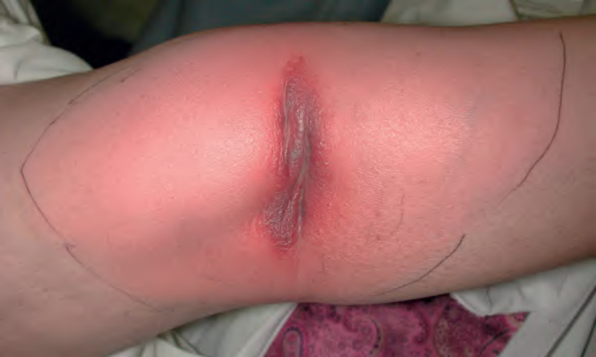
Fig. 85.19 Dermonecrotic spider bite. Note the central dusky necrosis. The patient was initially misdiagnosed with cellulitis and hospitalized after failure to respond to oral antibiotic therapy.
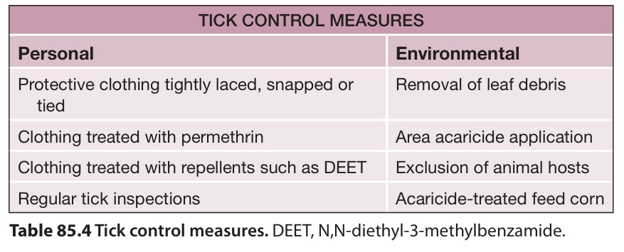
Table 85.4 Tick control measures. DEET, N,N-diethyl-3-methylbenzamide.
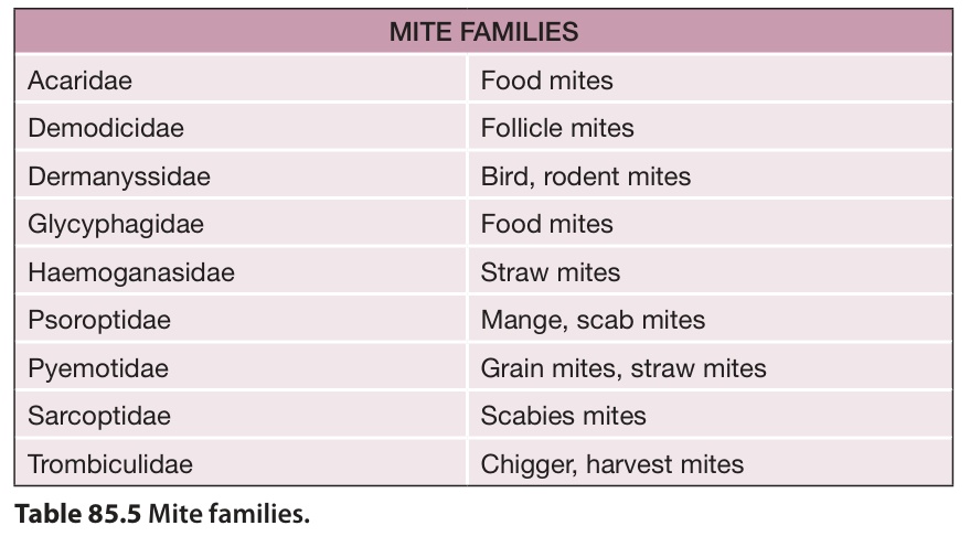
Table 85.5 Mite families.
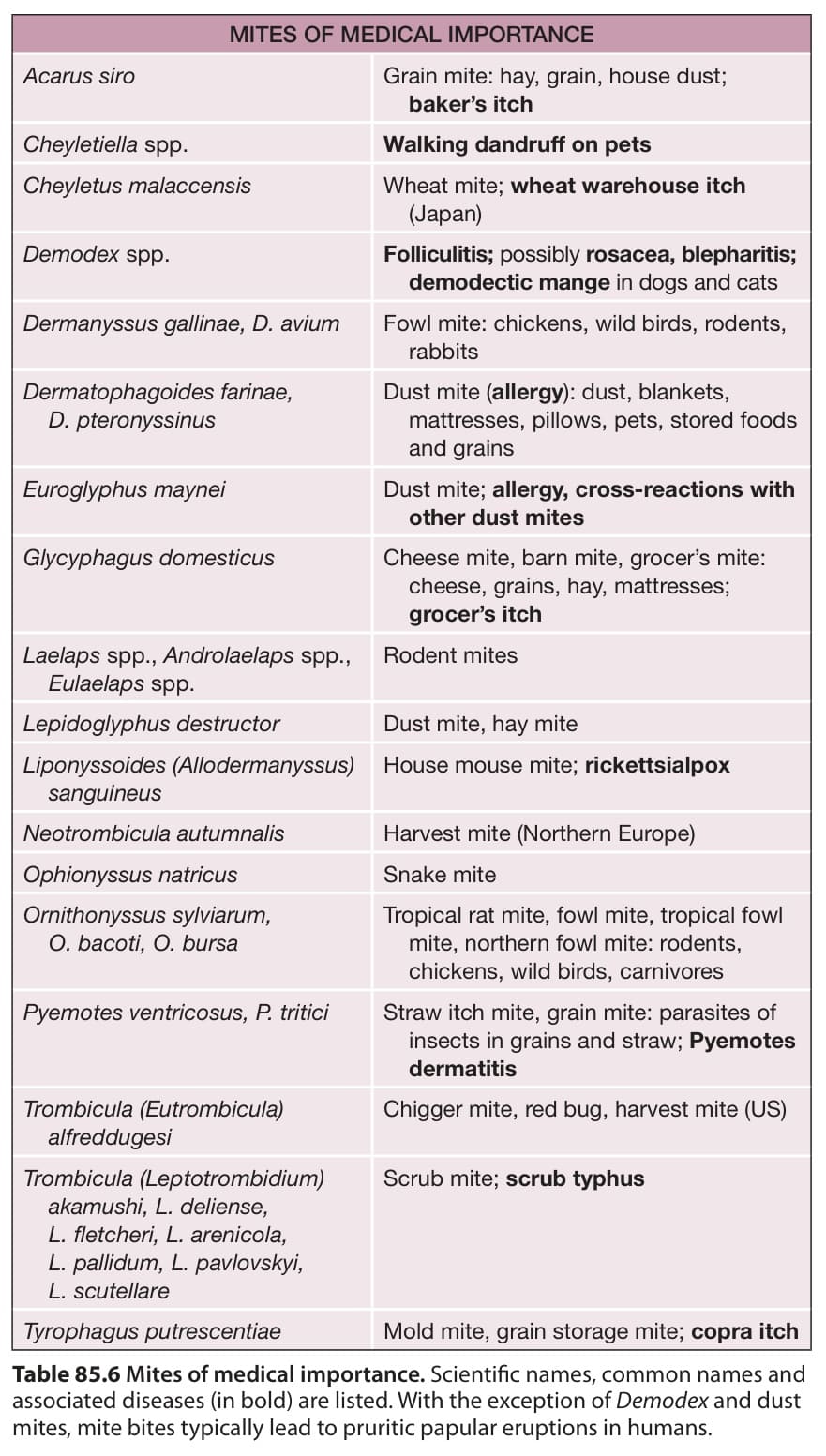
Table 85.6 Mites of medical importance. Scientific names, common names and associated diseases (in bold) are listed. With the exception of Demodex and dust mites, mite bites typically lead to pruritic papular eruptions in humans.
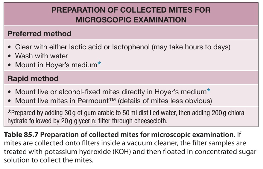
Table 85.7 Preparation of collected mites for microscopic examination. If mites are collected onto filters inside a vacuum cleaner, the filter samples are treated with potassium hydroxide (KOH) and then floated in concentrated sugar solution to collect the mites.
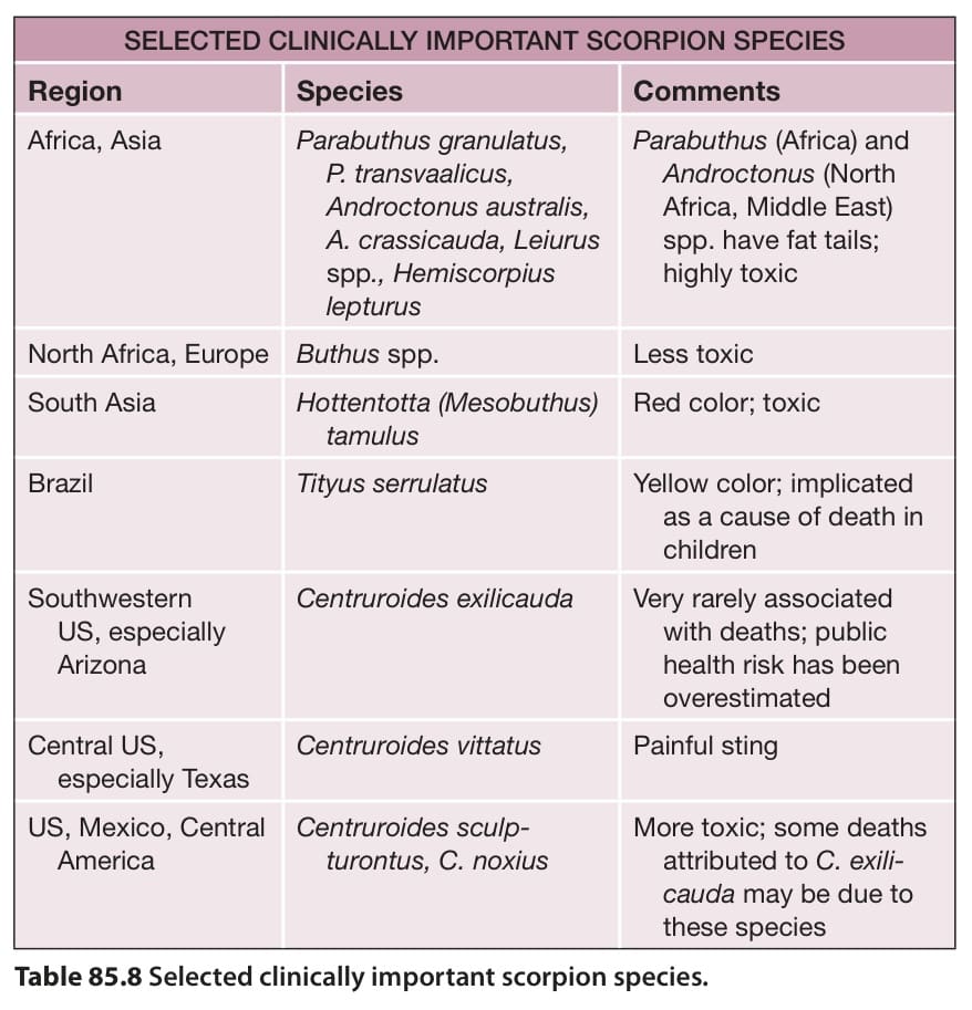
Table 85.8 Selected clinically important scorpion species.