MARINE INJURIES
Key features
Marine organisms contain some of the most potent toxins known
Drowning can occur after loss of consciousness following envenomation
Penetrating injuries are prone to secondary infection
Marine Vibrio infections are more common in patients with liver disease
First aid for most marine venoms includes immersion in hot, but not scalding, water
Introduction
Penetrating marine injuries may be complicated by secondary bacterial infection. S. aureus is the most common organism, but Vibrio infections also occur. Vibrio vulnificus sepsis is strongly associated with liver disease, especially cirrhosis (see Ch. 74). Although most infections are related to ingestion of oysters or injuries in brackish water, the organism can survive on the skin for more than 24 hours, and infection has been reported after a penetrating injury on dry land. With warming of the oceans, there has been a northward extension of the geographic range of Vibrio. Seal finger, resulting from seal bites to the hand, is caused by Mycoplasma infection. Cutaneous infections with Erysipelothrix rhusiopathiae (see Ch. 74), Aeromonas hydrophila, and Mycobacterium marinum (see Ch. 75) can also be acquired from trauma in an aquatic environment.
Pathogenesis
Cnidarians (including jellyfish, corals, and sea anemones) are aquatic animals that produce specialized epidermal cells called cnidocytes with organelles referred to as nematocysts or stinging capsules. A physical (e.g. contact with a foreign body) or osmotic trigger causes a coiled, inverted, hollow filament within the nematocyst to be ejected externally and discharge its toxic contents.
Clinical features
Sea urchins are echinoderms with fragile spines that break easily in the skin. Pigment from the spine often diffuses into the wound, making it difficult to find and remove the spine. Only about 80 of the roughly 600 species of sea urchin described are known to be hazardous to humans. Long-spined urchins such as the black sea urchin (Diadema setosum) are often a problem, and spines embedded near joints have been associated with synovitis and arthritis. Sea cucumbers, another type of echinoderm, can eject an irritating visceral liquid.
Seabather’s eruption (sea lice) is characterized by pruritic papules located under the bathing suit (Fig. 85.20) and in intertriginous areas. It may be caused by a variety of stinging larvae, including those of the thimble jellyfish (Linuche unguiculata, L. aquila), common in southern waters (e.g. the Florida coast and Caribbean). Seabather’s eruption acquired off the coast of Long Island, New York, is typically caused by larvae of a sea anemone (Edwardsiella lineata). Swimmer’s itch, in contrast, affects exposed skin after wading or swimming in bodies of fresh water; this condition is related to cercariae (free-swimming larvae) of avian schistosomes (see Ch. 83).
Jellyfish stings result in immediate pain as well as delayed and occasionally recurrent cutaneous reactions. Erythematous, urticarial, or hemorrhagic streaks may be observed (Fig. 85.21), and they tend to be longer and fewer in number following contact with larger jellyfish. A lichen planus-like eruption and inflammation distant from the site of envenomation have also been reported. Chironex fleckeri, the Pacific box jellyfish or sea wasp, is generally considered the most dangerous of the jellies, and stings often result in shock. Confirmation of enveno mation can be obtained by tape stripping of nematocysts from skin. Physalia physalis, the Portuguese man of war, is found in southern waters of the Atlantic while Physalia utriculus, the blue bottle jellyfish, is seen in the Pacific. These Physalia spp. contain nematocysts but are actually siphonophores, not true jellyfish. Cyanea and Chrysaora sea nettles are common causes of jellyfish dermatitis. Storms frequently drive jellies into shallow water in great numbers.
Haloclava producta, the “ghost anemone”, is a burrowing sea anemone found off the east coast of the US and in the Gulf of Mexico.
Painful red papules and papulovesicles in multiple linear streaks. Courtesy Joyce Rico, MD.
It is responsible for summer outbreaks of dermatitis off the coast of Long Island, New York. Corals are another group of stinging cnidarians in the same class (Anthozoa) as sea anemones. Coral cuts can be slow to heal and should be washed vigorously. Fire corals are false corals that produce intense stinging and a papular eruption that can last for several days. Cnidarian nematocysts found in seaweed and in the dorsal papillae of nudibranch sea slugs can also cause dermatitis. Exposure to algae alone can lead to dermatitis as well, referred to as seaweed dermatitis (see Ch. 17).
Red sponges and fire sponges (members of the phylum Porifera) both cause dermatitis. Sponge dermatitis is related to spicules embedded in the skin, but sponge diver’s disease is caused by the stings of sea anemones attached to the base of sponges. Local itching and burning progress to erythema and vesiculation. Systemic symptoms such as nausea, vomiting, and headache are common.
Two classes of the phylum Mollusca, cephalopods and gastropods, have been linked to human envenomation. The blue-ringed octopus contains a potent venom. This small cephalopod is often only 10 cm long and is found in waters off the coast of Australia. Envenomation from cone shells (marine gastropods) may be associated with a 20% mortality rate. The shells are about 10 cm in length and tend to be found in shallow water, mainly in tropical and subtropical climates.
Encounters with stingrays can result in soft tissue injury and embedded spines. Death has been reported after a penetrating chest injury from a stingray barb resulting in venom-induced myocardial necrosis. Injuries inflicted by stingrays occur in both fresh and salt water, and pain is the major manifestation.
Lionfish are common in subtropical and tropical waters. Since August 2000, lionfish have been reported along the eastern coast of the US, from Florida to New York. Lionfish belong to the family of scorpionfish (Scorpaenidae). The family includes the genera Pterois (lionfish), Scorpaena (“true scorpionfish”, bullrout and sculpin), and Synanceia (stonefish). Stonefish are the most toxic members of the family, although all produce local pain as well as systemic symptoms. Catfish spine injuries occur in both salt and fresh water. They may be associated with local pain, bleeding, and systemic symptoms.
Pathology
Marine Vibrio infections cause intense cellulitis affecting the subcutis and dermis. The superficial dermis is devitalized and lacks an inflammatory
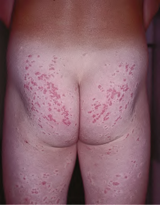
Fig. 85.20 Seabather’s eruption. Edematous pink papules in the same distribution as the bathing trunks. Courtesy Kalman Watsky, MD.
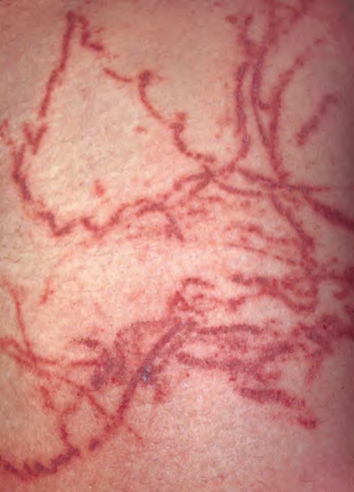
Fig. 85.21 Jellyfish sting.
infiltrate. Bullae are subepidermal in location and non-inflammatory. Many organisms are present, especially around vessels.
Sea urchin spines examined by polarized light microscopy exhibit an ornate symmetric structure produced by calcite crystals. Cutaneous granulomatous reactions may occur.
Histologic sections from sites of jellyfish stings may demonstrate nematocysts penetrating the epidermis and papillary dermis.
Avoidance is the best means of preventing injury, which can result from specimens in salt water fish tanks as well as in the ocean. When envenomation occurs in a swimmer, the individual should immediately be removed from the water to prevent drowning. Initial treatment for all marine envenomations includes soaking the site in hot, but not scalding, water to denature some of the venom proteins.
Antivenin is available for some of the more toxic species such as C. fleckeri, and it was shown to neutralize cardiovascular effects of C. fleckeri envenomation in an animal model. In contrast, verapamil did not improve cardiovascular status and increased mortality in the same animal model. In an animal model of envenomation by the Chiropsalmus species of box jellyfish, pretreatment with C. fleckeri antivenin or MgSO did not have any effect on the venom-induced hypertensive response or subsequent cardiovascular collapse. This highlights the importance of species identification following box jellyfish envenomation.
In a study of acute Carybdea alata box jellyfish stings, hot water immersion (40–41°C) was more effective than papain meat tenderizer or vinegar. Topical jellyfish sting inhibitors, including lotions in which they are combined with sunscreens, are available. Sea water can be used to remove jellyfish tentacles. Inactivation with acidic or alkaline solutions can be risky, as it may also lead to unexpected discharge of nematocysts. Treatment with topical corticosteroids and calcineurin inhibitors may be helpful for delayed reactions.
Most fish envenomations should be treated with hot water immersion. In a study of fresh water ray envenomation, hot water immersion controlled acute pain, but it did not prevent skin necrosis.
Additional figures and a table on Approach to the Evaluation and Management of Bedbug Infestations, available in our eBook (see inside front cover for access code).
collar formulations of imidacloprid/flumethrin and deltamethrin and of spot-on formulations of fipronil/(s)– methoprene, dinotefuran/pyriproxyfen/permethrin and dermatitis in a 2-year-old girl: case report and literature