MECHANISMS OF UV-BASED PHOTOTHERAPY
UV irradiation is an effective treatment for a variety of skin diseases, from psoriasis, atopic dermatitis, prurigo, and vitiligo to lichenoid dermatoses (e.g. lichen planus, pityriasis lichenoides, graft-versushost disease), morphea, and cutaneous T cell lymphoma (CTCL; see Ch. 134). The therapeutic mechanisms are complex and depend upon the particular disease and the administered wavelengths, e.g. NB-UVB versus UVA1. UV radiation has proapoptotic, immunomodulatory, antipruritic, antifibrotic, propigmentary, and proprebiotic properties, which alone or in combination lead to clinical improvement. Despite having diverse etiologies, most photoresponsive disorders have disturbances in the cutaneous immune microenvironment.
As depicted in Figs. 86.3 and 86.5, UV irradiation induces various mediators including cytokines, chemokines, AMPs, cis-urocanic acid, platelet activating factor (PAF), and PAF-like molecules that simultaneously influence the cutaneous micromilieu and its interplay with the microbiota of the skin. In particular, the induction of regulatory
T cells, along with downregulation of the Th17/IL-23 axis, may be crucial for months-long disease remission in disorders such as psoriasis even after treatment is discontinued. In mycosis fungoides, phototherapy can disrupt the cutaneous proinflammatory microenvironment, along with diminishing the growth of T cells via suppression of the production and secretion of IL-9. In both atopic dermatitis and CTCL, UV irradiation affects the cutaneous microbiota (e.g. inhibits the growth of Staphylococcus aureus) which suppresses the proinflammatory microenvironment of the skin. In fibrotic skin diseases such as morphea, phototherapy (UVA1) induces matrix metalloproteinase and Smad7 expression as well as downregulation of IL-6, leading to increased collagenase activity and effectively reducing tissue fibrosis.
UV irradiation can also lead to vitamin-dependent and -independent systemic effects that may affect gut microbiota, thereby playing a potential role in a variety of skin disorders. In patients with psoriasis, phototherapy decreases systemic inflammation as measured by serum levels of C-reactive protein (CRP) and IL-6; it can also increase levels of high-density lipoprotein (HDL)-P and restore HDL composition and function which may reduce aortic inflammation and cardiovascular risk, respectively. Lastly, exposure to UV light may benefit multiple sclerosis as well as alter mood, behavior and cognition, possibly due to increased synaptic release of glutamate and enhanced motor learning and memory.
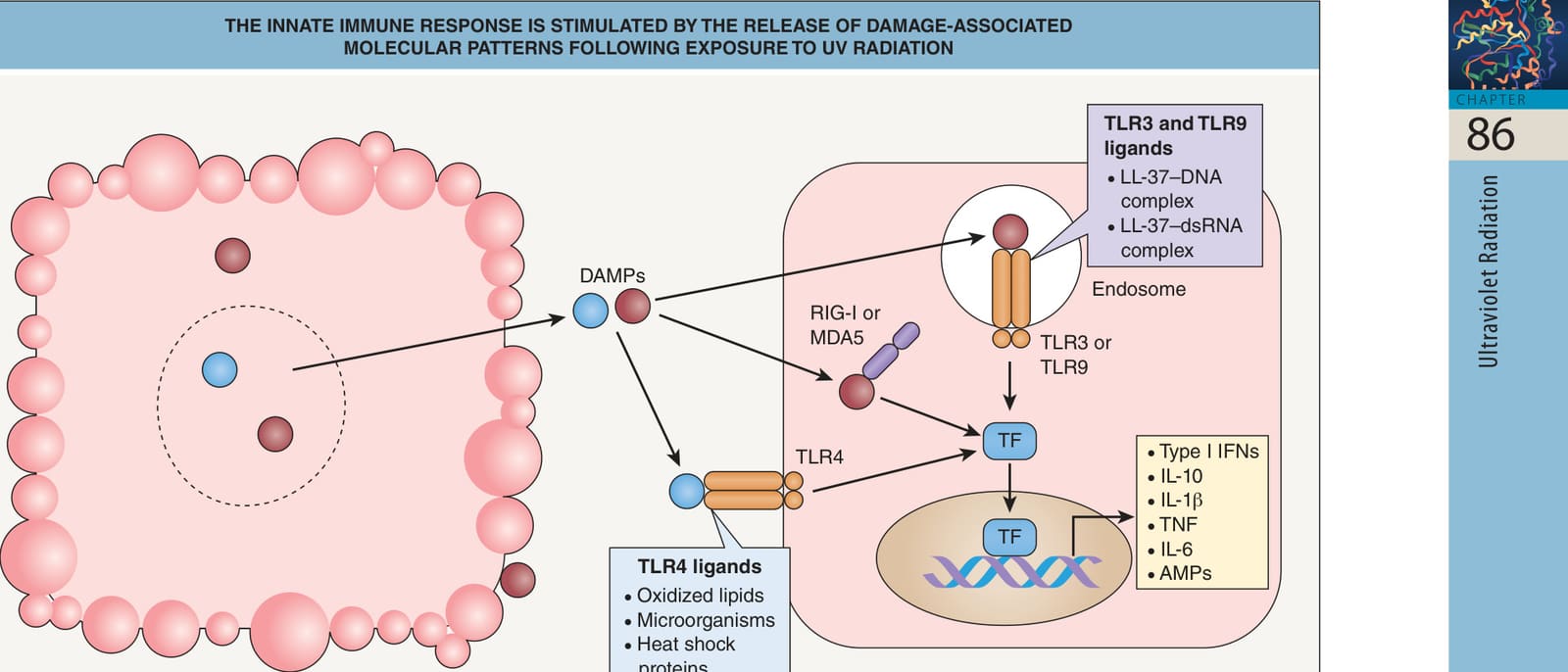
Fig. 86.3 The innate immune response is stimulated by the release of damage-associated molecular patterns following exposure to UV radiation (UVR). UVR induces the formation and release of damage-associated molecular patterns (DAMPs) from necrotic keratinocytes that activate Toll-like receptor (TLR) signaling. UVR can induce the formation and/or release of DAMPs that are recognized by the innate immune system as non-self, such as LL-37–DNA complex, LL-37–doublestranded RNA (dsRNA) complex, oxidized lipids, microorganisms, heat shock proteins, hyaluronic acid, and high-mobility group box 1 protein (HMGB1). TLR signaling in neighboring healthy keratinocytes leads to a type I interferon (IFN) response and the release of tumor necrosis factor (TNF), IL-10, IL-1β, IL-6, and antimicrobial peptides (AMPs). Cell responses to DNA and dsRNA are also mediated by the cytosolic receptors retinoic acid-inducible gene I (RIG-I) and melanoma differentiationassociated protein 5 (MDA5), which trigger the release of the above-mentioned cytokines and AMPs. TF, transcription factor. Reproduced with permission from Bernard JJ, Gallo RL, Krutmann J. Photoimmunology: how ultraviolet radiation affects the immune system. Nat Rev Immunol 2019;19:688–701.
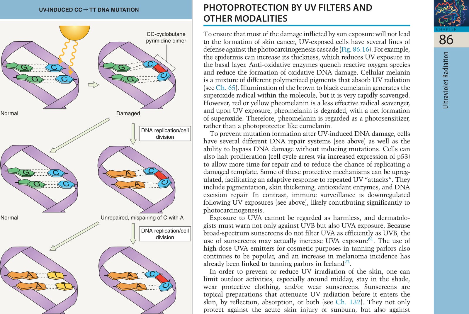
*Fig. 86.15 UV-induced CC→TT DNA mutation. Following formation of a pyrimidine dimer between two adjacent cytosines and subsequent cell division, a CC→TT mutation results. This represents a UV-signature mutation. May give rise to more C→T mutations.