OVERVIEW
The histiocytoses represent a group of proliferative disorders that share a common CD34+ progenitor cell in the bone marrow. Three “histiocytic cells” of cutaneous importance are: (1) the Langerhans cell, which initially migrates from the embryonic yolk sac to the epidermis and functions as a potent antigen-presenting cell (APC); (2) the mononuclear cell/macrophage, which migrates to and from the dermis and has both phagocytic and APC properties; and (3) the dermal dendrocyte/dendritic cell, of which there are two subtypes. Type 1 dermal dendrocytes are factor XIIIa-positive and generally reside within the papillary dermis, whereas type 2 dermal dendrocytes are CD34-positive and are generally found within the reticular dermis. Type 1 dermal dendrocytes may be involved in phagocytosis, antigen presentation, inflammation, collagen production, and wound healing, while the function of type 2 dermal dendrocytes is less certain. These two types represent myeloid dendritic cells and are distinct from plasmacytoid dendritic cells.
Dysfunction of these various histiocytes has led to a group of well-known, but poorly understood, disorders. For years, many of the histiocytoses were known by numerous names, reflecting the lack of understanding and agreement regarding their origin. Initially electron microscopy, then immunohistochemical stains, and most recently molecular pathology have provided insights into these conditions. Over the past decade, multiple types of histiocytoses were shown to be neoplasms of myeloid origin driven by somatic gain-of-function mutations that lead to activation of the mitogen-activated protein kinase (MAPK) signaling pathway. BRAF was found to be the most frequently altered gene. However, genes encoding membrane receptors (e.g. CSF-1R, NTRK1, RET, ALK) and components of the RAS-RAF-MEK-ERK (MAPK) pathway, both upstream (e.g. NRAS, KRAS) and downstream (e.g. MAP2K1/MEK1) of BRAF may also be involved (see Fig. 113.1). Less commonly, there is activation of the PI3K-AKT signaling pathway. In patients with disseminated disease, the mutation found in an affected organ can also be detected in blood cells and/or hematopoietic progenitors.
In the new classification scheme from the Histiocyte Society, histiocytoses are classified into five main groups based upon their clinical, histologic, and molecular characteristics: (1) Langerhans-related histiocytoses (L group); (2) cutaneous and mucocutaneous histiocytoses (C group); (3) malignant histiocytoses (M group); (4) Rosai–Dorfman disease (R group); and (5) hemophagocytic lymphohistiocytosis and macrophage activation syndrome (H group).
This chapter covers the most common histiocytic disorders as well as several rare forms (Table 91.1).
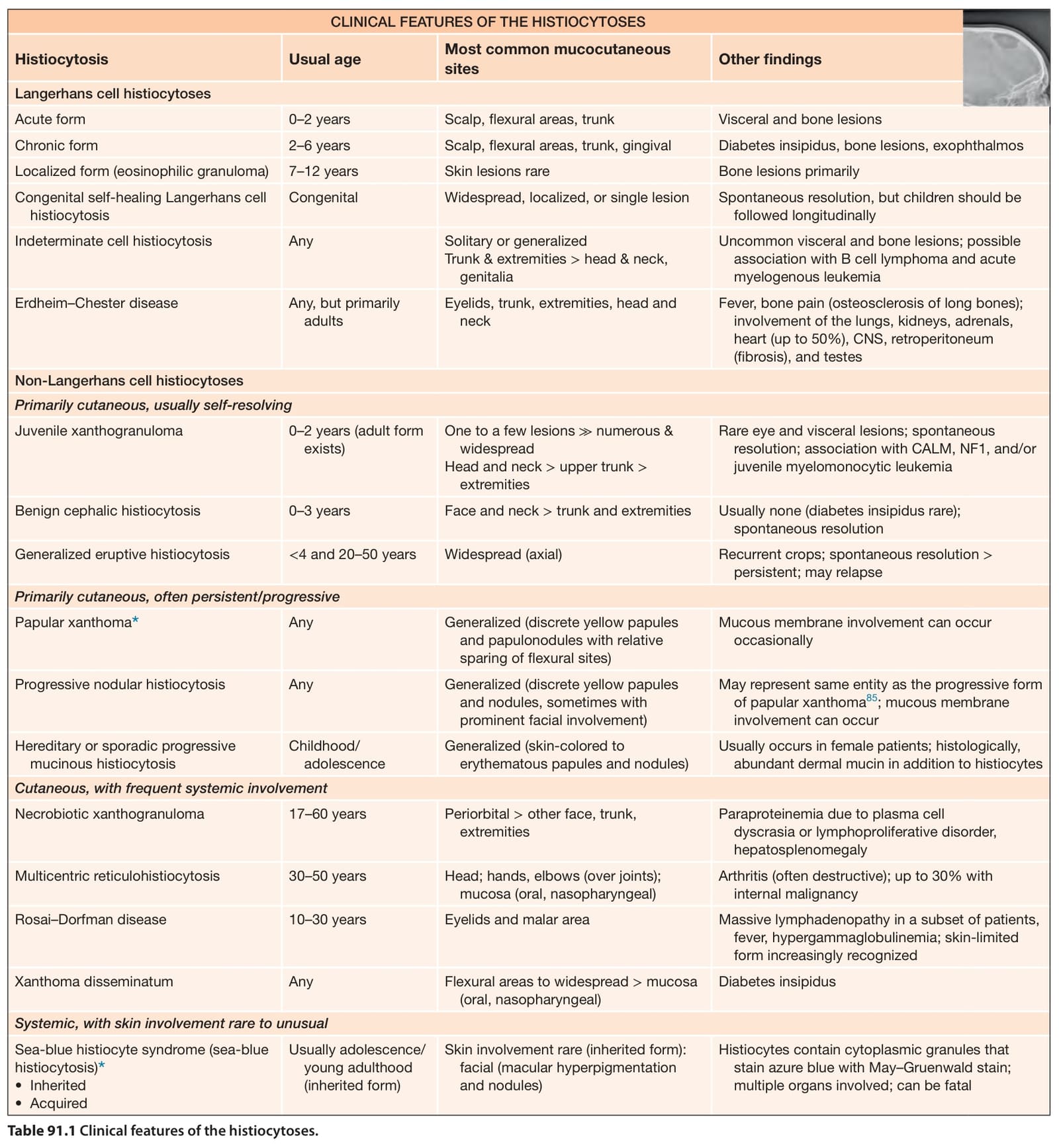
Table 91.1 Clinical features of the histiocytoses.