“L” GROUP: LANGERHANS-RELATED HISTIOCYTOSES
Langerhans Cell Histiocytosis
Langerhans cell histiocytosis (LCH): class I histiocytosis, histiocytosis X Subtypes (historical names):
Acute (Letterer–Siwe disease) Chronic (Hand–Schüller–Christian disease) Localized (eosinophilic granuloma) Congenital self-healing Langerhans cell histiocytosis (congenital self-healing reticulohistiocytosis; Hashimoto–Pritzker disease)
Histiocytoses 91
Sylvie Fraitag and Stéphane Barete
Key features
A clonal proliferation of Langerhans cells, CD1a+/CD207+ dendritic cells derived from the bone marrow
Somatic gain-of-function mutations, most commonly BRAF V600E, lead to activation of the mitogen-activated protein kinase (MAPK) signaling pathway; as a result, ERK phosphorylation is a consistent finding
Histopathologic diagnosis is aided by immunohistochemistry, e.g. anti-VEI antibody, and DNA sequencing to detect associated mutations
Systemic manifestations include osteolytic bone lesions and diabetes insipidus
Targeted therapy consists of selective BRAF inhibitors (e.g.
vemurafenib) and/or MEK inhibitors
Introduction
Langerhans cell histiocytosis (LCH) is a clonal proliferative disorder in which Langerhans cells, CD1a+/CD207+ dendritic cells, accumulate within a variety of organs. It is characterized by a broad spectrum of clinical presentations and outcomes, ranging from localized and self-healing to generalized and fatal.
History
Letterer–Siwe disease, Hand–Schüller–Christian disease, and eosinophilic granuloma were described in the early twentieth century. In 1953, Lichtenstein grouped these three disorders into a single entity which he called histiocytosis X. Twenty years later, Hashimoto and Pritzker described the entity congenital self-healing reticulohistiocytosis. Immunologic and ultrastructural studies confirmed the relationship of the pathologic cells in histiocytosis X and congenital self-healing reticulohistiocytosis to Langerhans cells, providing a basis for the Writing Group of the Histiocyte Society, in 1987, to reclassify them as “Langerhans cell histiocytoses”.
Epidemiology
LCH occurs worldwide and most commonly develops in children ages 1–3 years, although disease can occur at any age. The reported incidence of LCH varies widely. However, an annual incidence of at least five per million children is often quoted, with the adult incidence suspected to be less than one-third that of children. LCH is more common in boys, with a male : female ratio of nearly 2 : 1. In adults, there may be a slight female predominance.
Some cases of LCH appear to be familial. One study described simultaneous development of LCH in four of five monozygotic twin pairs and in one of three dizygotic pairs. Additionally, in two families with affected non-twin siblings, parental consanguinity was known in one and possible in the other.
Pathogenesis
Until recently, the pathogenesis of LCH was unknown. In 2010, a significant percentage (55%–60%) of LCH specimens were shown to harbor the BRAF V600E mutation. This strongly suggested that LCH was a clonal neoplasm, a finding supported by further studies in which clonality of CD1a+ histiocytes was uniformly demonstrated. In high-risk patients, BRAF V600E mutations were also detected in CD34+ hematopoietic progenitor cells within the bone marrow as well as circulating CD11c+/CD14+ monocytic cells. As a result, LCH is now regarded to be a myeloid neoplasm.
As in melanoma, somatic gain-of-function mutations in BRAF point to activation of the mitogen-activated protein kinase (MAPK) signaling pathway (see Fig. 113.1). Of note, BRAF V600E mutations have been more commonly observed in patients with multisystem LCH than in those with isolated disease. Mutations in genes that encode other kinases in the RAS-RAF-MEK-ERK (MAPK) pathway (e.g. MAP2K1 [also referred to as MEK1]) as well as membrane receptors were subsequently detected (see Overview). The end result is phosphorylation of ERK. BRAF and MEK1 mutations are mutually exclusive, and ~50% of LCH patients with wild-type BRAF have MEK1 mutations.
Clinical features
Clinical manifestations of LCH vary from unifocal or single-system (S-S) disease to multifocal involvement to multisystem (M-S) Letterer– Siwe-type disease. The latter nearly always develops prior to age 2 years, and commonly presents in children less than 1 year of age. Cutaneous involvement occurs in most patients as 1–2 mm pink to skin-colored or purpuric papules, pustules, and/or vesicles in the scalp, flexural areas of the neck, axilla and perineum, and on the trunk (Fig. 91.1A–C). Rarely, the lesions resemble mollusca contagiosa (Fig. 91.1D). Scale and crust with secondary impetiginization are common findings, as is the development of petechiae and purpura (Fig. 91.1E). The lesions tend to
coalesce and become tender, especially when fissures develop in inter-triginous zones (Fig. 91.1B,F). Palmoplantar and nail involvement can occur, as can soft tissue nodules (Fig. 91.1G). In patients with darkly pigmented skin, the lesions may appear relatively hypopigmented (Fig. 91.1H). The eruption is most often confused with seborrheic dermatitis, various forms of diaper dermatitis or intertrigo (see Figs. 13.4 and 13.12), and arthropod bites (including scabies) when pruritic. During the course of the disease, many organs can become infiltrated by clonal LCH cells. However, only if the key functions of the organ are affected is such involvement of prognostic significance. Lung, liver, lymph node, and bone involvement commonly occur at some point during the illness. Osteolytic bone lesions are painful, usually multiple, and most frequently involve the cranium. Occasionally, the hematopoietic system can be affected, with thrombocytopenia and anemia portending a poor prognosis.
Another early-onset form of cutaneous LCH is known as congenital self-healing Langerhans cell histiocytosis or Hashimoto–Pritzker disease. It is present at birth or develops during the first few days of life. Clinical features include vesicles and/or pustules that can be mistaken for varicella as well as red–brown, crusted papulonodular lesions that often have central necrosis (Fig. 91.2). Some newborns may have a “blueberry muffin baby” presentation (see Table 121.4). Rarely, there is a single neonatal nodule, referred to as a Langerhans cell histiocytoma. While congenital self-healing LCH is usually a benign, self-resolving disorder, its relationship to other LCH variants suggests a cautious approach with respect to prognosis, and longitudinal evaluation is recommended.
The triad of diabetes insipidus, bone lesions, and exophthalmos (formerly called Hand–Schüller–Christian disease) typically begins between the ages of 2 and 6 years. These patients tend to have a chronic, progressive course and often do not have the complete triad. Approximately 30% of patients develop skin or mucous membrane lesions (Fig. 91.1I). While early cutaneous lesions are similar to those seen in the M-S form, older lesions can become xanthomatous. Ulcerative nodules may develop in the oral and genital areas, with premature loss of teeth possible secondary to gingival lesions.
At least 80% of patients develop bone lesions, the cranium being preferentially involved. Chronic otitis media occurs commonly in these patients and in patients with all forms of LCH. Diabetes insipidus, secondary to infiltration of the posterior pituitary by LCH cells, develops in ~30% of patients and is more common in those individuals with cranial bone involvement.
A localized variant of LCH, formerly called eosinophilic granuloma generally affects older children, boys more than girls. The cranium is most frequently affected, though lesions can also develop within the ribs, vertebrae, pelvis, scapulae, and long bones. A spontaneous fracture or otitis media may be the first sign of disease. Skin and mucous membrane lesions are rare.
Adults rarely develop LCH, but when they do, the most commonly involved sites are the skin, lung, and bone (see Fig. 91.1F). Diabetes insipidus can also develop and, as in children, is more likely when bony involvement of the skull is present. Severe multisystem disease is rare in adults. However, LCH can be a progressive disease in adults, especially when both bone and extra-skeletal sites are involved. Pulmonary involvement can be isolated or be a component of multisystem disease and it favors men who smoke cigarettes.
There are two patterns of associations between LCH and malignancy. The first is the development of acute leukemias and solid tumors in patients with a history of LCH who have been treated with chemotherapy, radiotherapy, or both, and the malignancies are likely to be treatment-related. There are reports of a few patients who developed skin or solid tumors (e.g. basal cell carcinoma, osteosarcoma) within the fields of radiotherapy for their LCH.
A second more interesting association is an apparent increased incidence of LCH in patients with hematologic malignancies, including acute myelogenous leukemia, acute lymphocytic leukemia, chronic myelomonocytic leukemia, and lymphomas of both T and B cell origin. These malignancies can occur prior to, concurrently, or following the diagnosis of LCH. In contrast to treatment-related (often referred to as secondary) hematologic malignancies, it has been demonstrated in several patients that the LCH and the hematologic malignancy are clonally related, suggesting origin from a common neoplastic stem cell.
The prognosis of patients with LCH varies dramatically. Involvement of “risk organs” – hematopoietic system, liver, lungs, and/or spleen – substantially increases the risk of disease-related mortality. However, in individuals with single-system disease or multisystem disease that does not involve risk organs, mortality rates are very low (e.g. <5% in the latter group). As with hematologic malignancies, the response to initial systemic treatment is an important prognostic indicator in LCH. Of note, in one study patients with high-risk LCH had BRAF V600E mutations in their CD34+ hematopoietic progenitor cells, while the mutation was limited to lesional CD207+ (Langerin+) dendritic cells in low-risk LCH patients.
Pathology
In a typical papule of LCH, a proliferation of neoplastic cells is present in the papillary dermis (Fig. 91.3A). They are medium-sized cells, 10–15 microns in diameter, with a reniform (kidney-shaped) nucleus and a pink, non-xanthomatous (non-foamy) cytoplasm. The LCH cells are often admixed with eosinophils, lymphocytes, and red blood cells.
Epidermotropism is commonly observed, resulting into a spongiform pattern and focal parakeratosis (Fig. 91.3B). In nodular lesions of congenital self-healing LCH, including LC histiocytoma, epidermo tropism is not a feature and the dermal infiltrate can extend into the deep dermis. Some of the histiocytes within the infiltrate have abundant eosinophilic cytoplasm and are intermixed with LCH cells and giant cells.
Like Langerhans cells, LCH cells are characterized by a CD1a+, CD207+ (Langerin+) phenotype (Fig. 91.3C,D); CD207 is the major protein of Birbeck granules. Both CD207 and CD1a have important roles in the uptake, processing, and presentation of non-peptide (e.g. lipid) antigens by Langerhans cells (Table 91.2) In addition, LCH cells commonly stain positive for CD68, while they are negative for CD163 (macrophage/monocyte marker) and FXIIIa (Fig. 91.4). Electron microscopy demonstrates Birbeck granules, which are rod- or racquetshaped cytoplasmic structures pathognomonic for Langerhans cells and LCH cells (Fig. 91.5). However, electron microscopy is no longer routinely performed.
Molecular analysis and immunohistochemistry are increasingly being utilized as diagnostic tools in that up to 60% of cases of LCH harbor a clonal BRAF V600E mutation. The VE1 mutant-specific antibody is useful in detecting this mutation. In addition, pERK expression as well as cyclin D1 nuclear positivity are always observed in LCH, irrespective of the BRAF V600E mutational status, since mutations other than BRAF V600E can lead to activation of the MAPK pathway (see Fig. 113.1).
Differential diagnosis
The clinical differential diagnosis is vast and includes seborrheic dermatitis, various forms of diaper dermatitis or intertrigo (see Figs. 13.4 and 13.12), and arthropod bites (including scabies), as well as leukemia, B and T cell lymphomas, multiple myeloma (bony lesions), maculopapular cutaneous mastocytosis, and the non-Langerhans cell histiocytoses (see Table 91.1). However, the characteristic histologic features of LCH will lead one to suspect the diagnosis, which is confirmed by the combination of positive immunostaining for CD1a and CD207 (Fig. 91.6). Occasionally, dense dermal infiltrates of CD1a+ dendritic cells can be seen in insect bite reactions, including scabies, and lead to misdiagnosis (see Fig. 85.3). However, these cells do not express CD207.
Treatment
All patients diagnosed with LCH should undergo evaluation of the hematologic, pulmonary, hepatosplenic, renal, and skeletal systems to determine the extent of disease. Depending on age and extent of involvement, some patients may not require specific treatment, while others will require systemic therapy. The latter include corticosteroids, vinblastine, cladribine, and/or interferon alpha-2a as well as targeted therapies such as selective BRAF and MEK inhibitors.
For mild, single-system skin disease (if treatment is required), topical agents, including corticosteroids, antimicrobials, mechlorethamine (nitrogen mustard) and imiquimod, as well as phototherapy (narrowband UVB, PUVA) have been reported to be effective in case series. For more extensive cutaneous disease, based upon case reports, thalidomide, azathioprine, or methotrexate may be effective. Other chemotherapeutic treatments such as cladribine and cytarabine have been used for refractory disease as have autologous hematopoietic stem cell transplants.
Targeted therapy in the form of selective BRAF and MEK inhibitors has revolutionized treatment strategies for patients with aggressive, resistant disease; however, aspects of their use such as duration are still under debate. Updated information regarding ongoing clinical trials is available at www.clinicaltrials.gov and www.histiocytesociety.org.
A A somewhat lichenoid infiltrate of large LCH cells with some involvement of the epidermis; the infiltrate also contains lymphocytes and numerous eosinophils. Inset: note the reniform-shaped nuclei. B Another example with larger collections of Langerhans cells within the epidermis. C CD1a immunostain highlighting large LCH cells within the epidermis. D Positive CD207 (Langerin) immunostaining. B, D, Courtesy Lorenzo Cerroni, MD
Indeterminate Cell Histiocytosis
Key features
Currently classified within the L group (Langerhans-related histiocy- toses) due to mutations in BRAF V600E in nearly all patients
Occurs in adults and children with both generalized and solitary variants
Cells display the phenotype of dendritic cells: CD1a+, S100+, CD207−
Primarily a cutaneous disorder, but occasionally there is visceral involvement and associated hematologic malignancies
Introduction
Indeterminate cell histiocytosis (ICH) is a very rare proliferative disorder comprised of histiocytes that have both LCH and non-LCH immunophenotypic features. There are two clinical variants – solitary and generalized.
History
Indeterminate cells were described in 1963, and a patient with features first recognized as ICH was reported by Wood et al. in 1985. To date, ~60 cases have been reported in the literature. ICH is thought to be closely related to LCH because the pathologic cells are positive for CD1a, although they do not express CD207.
Epidemiology
ICH is quite rare and has no apparent sex predominance. It can occur at any age, from infancy to adulthood, and a congenital form has been reported.
Pathogenesis
Most cases of ICH harbor a gain-of-function mutation in BRAF V600E. Translocations involving NCOA2 (nuclear receptor co-activator 2) and ETV3 (Ets variant 3) have also been reported.
Clinical features
While the majority of patients with ICH have involvement of the trunk and extremities, lesions have also been observed on the genitalia and in the head and neck region, including the eyelids and ears. Both a generalized form and a solitary form have been described. In the generalized form, cutaneous lesions usually begin as firm red to brown papules or nodules (Fig. 91.7). The solitary form presents as a single, often soft, erythematous nodule that is usually ~1 cm in diameter. Ulceration can occur and as lesions age, they become brown to yellow. The clinical course is variable, with some patients experiencing a partial or complete regression of lesions (which may wax and wane), while others have a progressive course.
Although mucous membrane involvement is rare, both corneal and conjunctival lesions have been reported. Patients are usually in good health, but because visceral involvement and death can occasionally occur, periodic evaluation is warranted. There is a report of an infant who developed an aggressive form of ICH with a fatal outcome; the
histiocytosis.Courtesy Lorenzo Cerroni, MD.
disease initially presented in the bone and then eventually spread to visceral organs and the skin. Several cases have been associated with hematologic malignancies including mast cell leukemia, acute myelogenous leukemia, and low-grade B cell lymphoma.
Pathology
There is usually a predominantly monomorphous infiltrate of histiocytes, either well-circumscribed or more diffusely distributed within the dermis. Epidermotropism is less common than in LCH. Histiocytes often have an oncocytic appearance, with occasional spindled, scalloped, or vacuolated cell variants. Touton giant cells can be present. Lymphocytes are commonly present and eosinophils are occasionally observed.
The immunophenotype of ICH consists of positivity for CD68 and CD1a, but negativity for CD207. S100 may be either positive or negative, making this marker less helpful in establishing the diagnosis (see Fig. 91.4 & Table 91.2).
Differential diagnosis
The clinical appearance of the cutaneous lesions of ICH is not unique. Similar-appearing papules in the same distribution can be found in generalized eruptive histiocytoma, juvenile xanthogranuloma, and congenital self-healing LCH (see Table 91.1). When lesions are limited to the head and neck region, benign cephalic histiocytosis should be considered as well. The clinical differential diagnosis also includes eruptive syringomas, maculopapular cutaneous mastocytosis, and lymphomatoid papulosis. While the diagnosis of ICH can be suspected based upon histopathology, confirmation requires demonstration of the characteristic CD1a+, CD207− immunophenotype.
Treatment
Treatment of cutaneous lesions is usually not required as the lesions are asymptomatic and ICH is often either self-limited or non-progressive. A
single lesion can be removed surgically. Patients with extensive, disfiguring cutaneous disease have been treated with systemic chemotherapy (e.g. vinblastine, 2-chlorodeoxyadenosine, cyclophosphamide, and/or etoposide). There are also reports of the use of thalidomide, isotretinoin, methotrexate, NB-UVB, PUVA, or total skin electron beam therapy. As with LCH, selective BRAF and MEK inhibitors can be considered. Because visceral involvement and leukemia can occur, longitudinal evaluation of all patients with indeterminate cell histiocytosis is recommended.
Erdheim–Chester Disease
Key features
Erdheim–Chester disease (ECD) is a severe form of histiocytosis which usually affects adults
As in LCH, gain-of-function mutations in BRAF V600E are observed in >50% of patients; mutations in other genes that lead to activation of the MAPK pathway (e.g. MAP2K1/MEK1) also occur
Cutaneous lesions occur in ~25% of patients and are accompanied by symmetric diaphyseal and metaphyseal osteosclerosis of the long bones as well as periaortic, retroperitoneal, and perirenal fibrosis
Histologically, there are dermal infiltrates of histiocytes
Treatment primarily consists of a selective BRAF inhibitor (e.g.
vemurafenib) and/or a MEK inhibitor (e.g. cobimetinib)
Introduction
Erdheim–Chester disease (ECD) is a severe form of histiocytosis with visceral involvement characterized by xanthomatous or xanthogranulomatous infiltrates within tissues. It is a rare histiocytosis classified in the “L” group of histiocytoses.
History
ECD was first described in 1930 by the American physician Dr. William Chester while he was doing a postdoctoral fellowship with the Viennese pathologist Dr. Jakob Erdheim. Although originally considered an aggressive histiocytosis of unclear origin, ECD is now regarded as a clonal hematopoietic disorder characterized in nearly all patients by an underlying BRAF V600E mutation or mutations in other genes that lead to activation of the RAS-RAF-MEK-ERK (MAPK) signaling pathway (see Fig. 113.1). More rarely, there is activation of the PI3K-AKT signaling pathway.
Epidemiology
The exact incidence of ECD is not known. Although it is considered an extremely rare disease, the incidence appears to be increasing. It occurs during adulthood (mean age = 55 years), with exceptional cases in children.
Pathogenesis
Originally classified as a non-Langerhans cell histiocytosis, it is now included with LCH in the “L” group in light of the detection of BRAF V600E mutations in ~55%–60% of patients. Mutations in other genes that lead to activation of the MAPK pathway (e.g. NRAS, KRAS, MAP2K1/MEK1, ALK) have also been described. Nearly 20% of patients with ECD have concomitant features of LCH, sometimes with cellular infiltrates of both entities within the same biopsy, further supporting placing ECD and LCH within the same group of histiocytoses.
Clinical features
Facial sinus osteosclerosis is suggestive of ECD, although it is often asymptomatic and discovered incidentally by CT or MRI. Bilateral hypermetabolic and sclerotic metaphyseal–diaphyseal involvement of long bones is highly suggestive of ECD, and this is usually associated with bone pain. Plain radiography (skeletal survey) may not detect these lesions, but whole body low-dose CT, MRI, skeletal scintigraphy (bone scan), or F-FDG PET scan represent more sensitive imaging tests. Diabetes insipidus may be an initial clinical manifestation. Patients can also develop respiratory symptoms, with pleural and pericardial (as well as peritoneal) thickening seen by CT or MRI. Approximately half of patients have periaortic fibrosis, described radiographically as a “coated aorta”. Retroperitoneal fibrosis is a hallmark sign of ECD, with ~60% of patients having perirenal fibrosis (“hairy kidney”) on CT scans.
Cutaneous involvement is present in 25% of patients, typically presenting as bilateral, symmetric, periocular xanthelasma-like lesions. Multiple yellow papules or plaques favor the medial upper eyelids and may precede other symptoms by several years. Yellow to brown plaques and papulonodules can also develop on the trunk, extremities, and head and neck region (Fig. 91.8), but mucosal involvement is rare. Recently described cutaneous manifestations include panniculitis-like nodules and granuloma annulare-like lesions.
Pathology
Within the dermis, there is a granulomatous infiltrate with foamy histiocytes, a few multinucleated histiocytes or Touton giant cells, lymphocytes, and occasional plasma cells and eosinophils. Fibrosis may be prominent. The histiocytes express CD68, CD163, and focally FXIIIa, but are CD1a- and CD207-negative. However, pERK is consistently positive due to activation of the MAPK pathway, and VE1 mutantspecific antibody staining is positive in ~55%–60% of cases, reflecting the presence of the BRAF V600E mutation.
Differential diagnosis
The primary differential diagnosis includes xanthelasma and the yellow periocular plaques seen in patients with necrobiotic xanthogranuloma. When there is involvement of the trunk and extremities, other histiocytoses or granulomatous disorders need to be considered. An associated
myeloid malignancy may develop in 20%–30% of patients and clonal hematopoiesis has been detected in almost half of patients with ECD; the most frequently mutated genes in the bone marrow are TET2, ASXL1, DNMT3A, and NRAS.
Treatment
Selective BRAF inhibitors such as vemurafenib are an effective treatment for patients who harbor the BRAF V600E mutation. MEK inhibitors (e.g. cobimetinib) are very effective in patients with other mutations that activate the MAPK pathway. For this reason, it is important to determine the mutational status of patients.
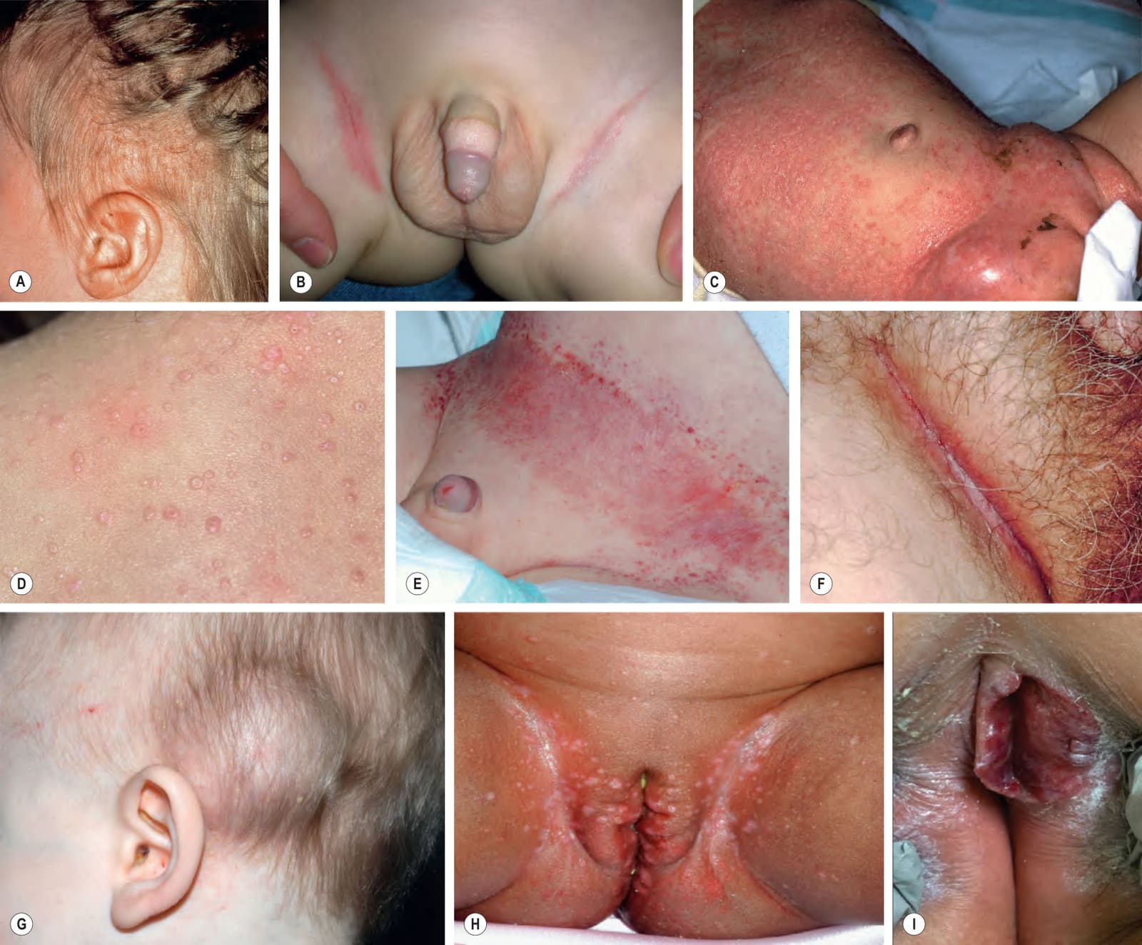
Fig. 91.1 Langerhans cell histiocytosis – clinical spectrum.A Scalp involvement may initially be diagnosed as seborrheic dermatitis; however, there are usually more discrete papules and crusting. B Pink, thin plaques with fissuring along the inguinal creases can also resemble seborrheic dermatitis. C Advanced disease with coalescence of papules into large plaques and prominent inguinal lymphadenopathy. D Pale pink to skin-colored papules, several of which have central umbilication and resemble mollusca contagiosa. E The presence of petechiae and purpuric papules is a clue to the diagnosis. F In an adult, the clinical presentation of inguinal involvement is similar to that of infants. G This subcutaneous nodule on the scalp involved the skull and represented the initial manifestation of multisystem Langerhans cell histiocytosis in an infant. H In patients with highly pigmented skin, the papules can be hypopigmented. I Eroded erythematous plaque of the labial mucosa. B, Courtesy Richard Antaya, MD; D, Courtesy Kristen Hook, MD; E, G, H, Courtesy Julie V. Schaffer, MD.
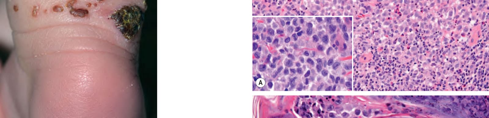
Fig. 91.2 Congenital self-healing Langerhans cell histiocytosis (Hashimoto–Pritzker disease). Cluster of crusted red–brown papules as well as a large hemorrhagic crust on the posterior ankle of a neonate. Courtesy Kristen Hook, MD.
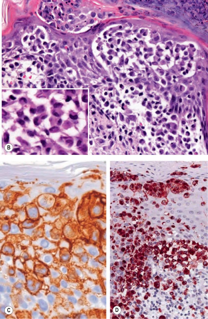
Fig. 91.3 Langerhans cell histiocytosis (LCH) – histopathologic features.
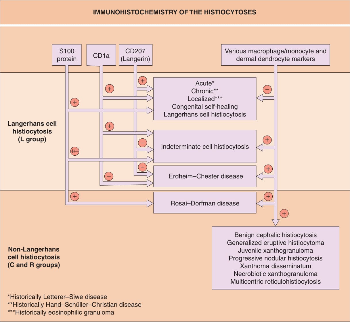
Fig. 91.4 Immunohistochemistry of the histio- cytoses. Examples of classic macrophage/ monocyte markers are CD68 and CD163; an example of a classic dermal dendrocyte marker is factor XIIIa.
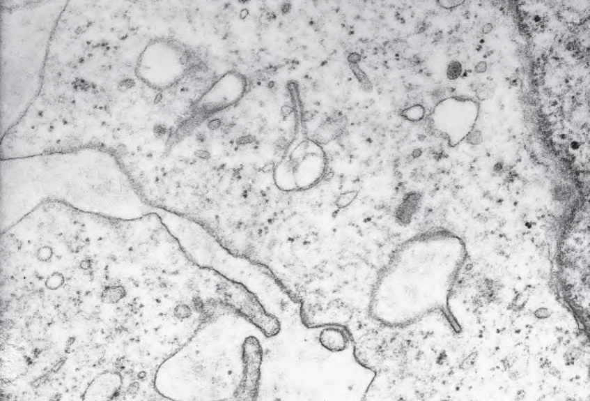
Fig. 91.5 Langerhans cell histiocytosis (Birbeck granules). Electron micrograph demonstrates classic racquet-shaped Birbeck granules in the cytoplasm of an LCH cell.
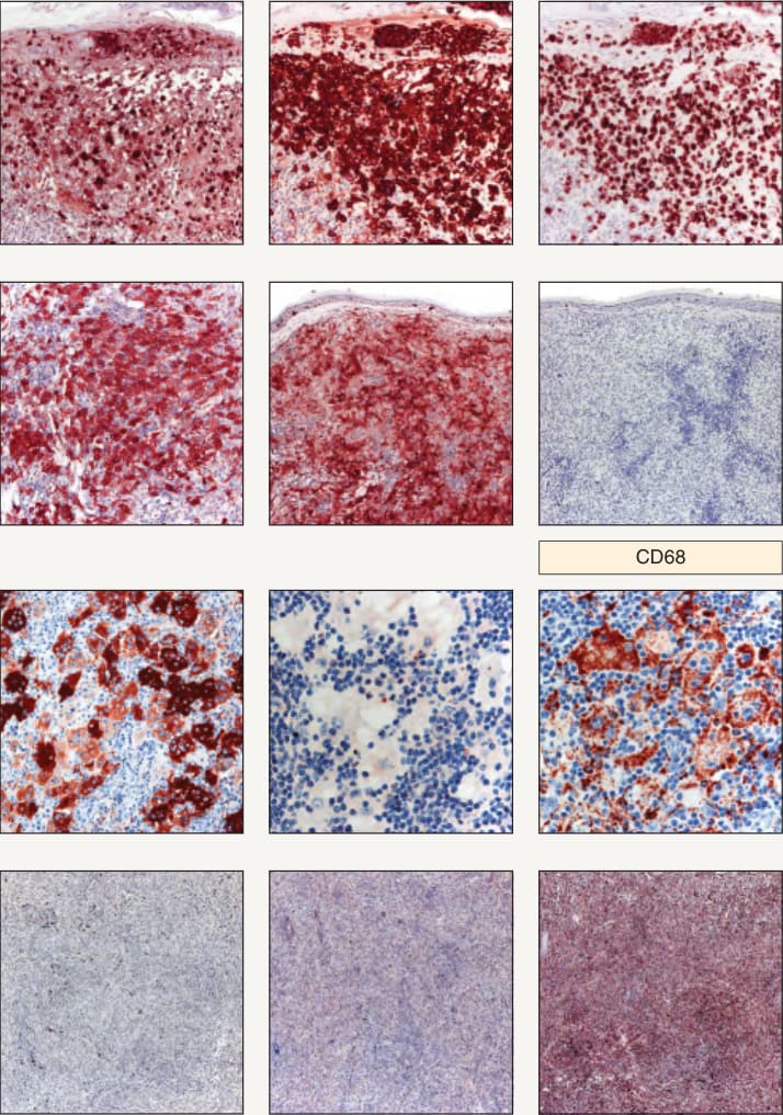
Fig. 91.6 Use of a panel of four immunostains to distinguish between Langerhans cell histiocytosis and several types of non-Langerhans cell
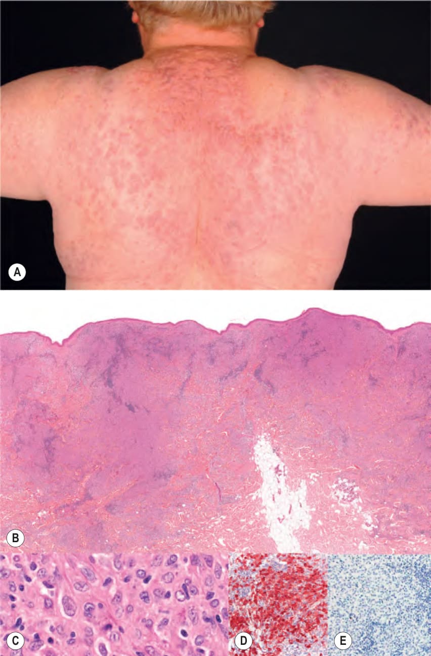
Fig. 91.7 Indeterminate cell histiocytosis.A Multiple pink-violet papules, plaques, and small nodules on the back. B Histologically, dense infiltrates were seen throughout the entire dermis and superficial subcutaneous fat. These infiltrates were composed of medium-to-large histiocytic cells (C) that were positive for S100 (D) and negative for CD1a (E). Courtesy Lorenzo Cerroni, MD.
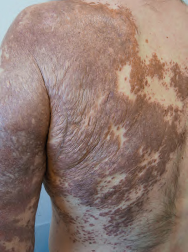
Fig. 91.8 Erdheim–Chester disease. Red–brown papules and large confluent yellow–brown and brown plaques. The patient had concomitant features of LCH (mixed histiocytosis).
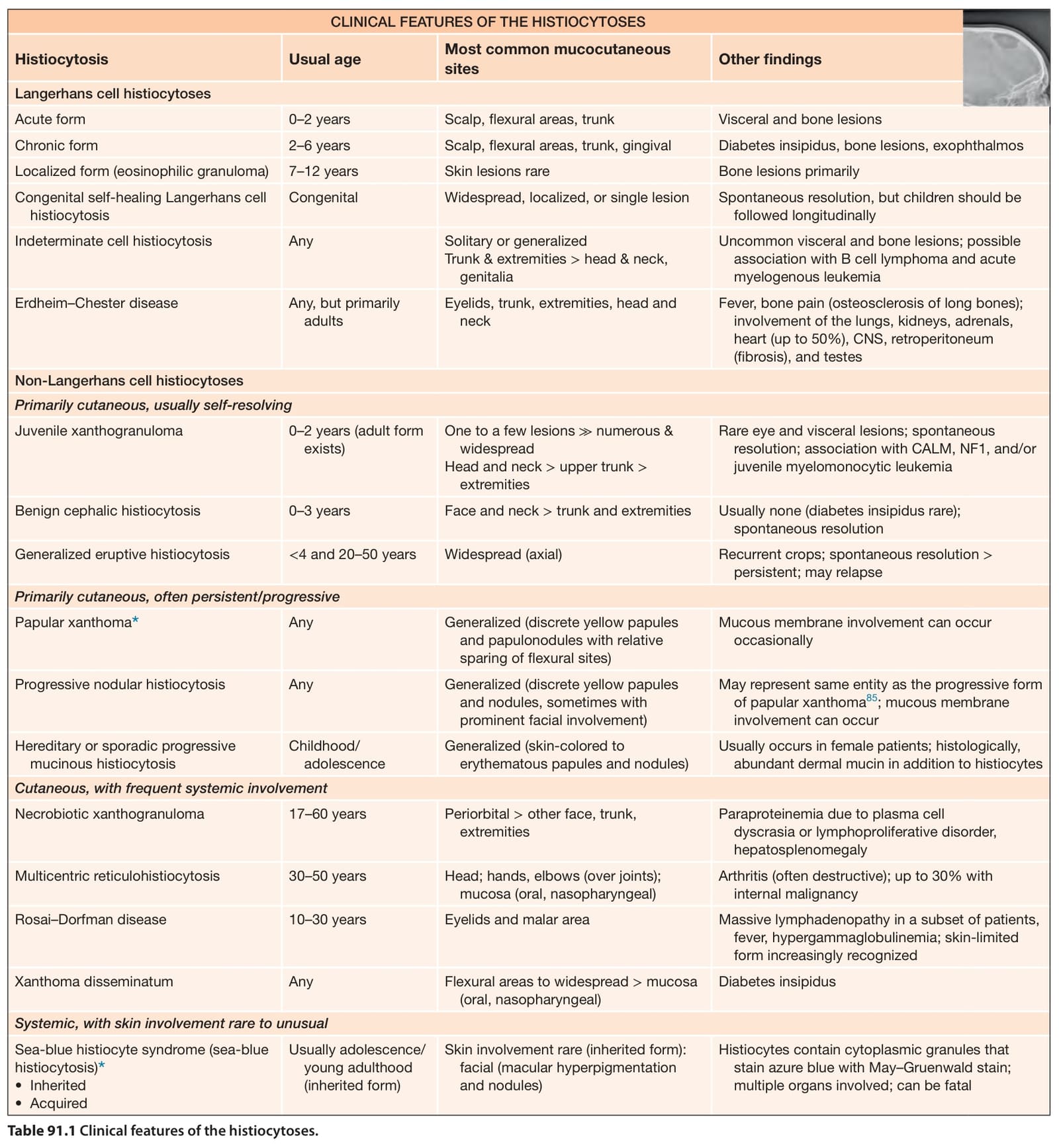
Table 91.1 Clinical features of the histiocytoses.
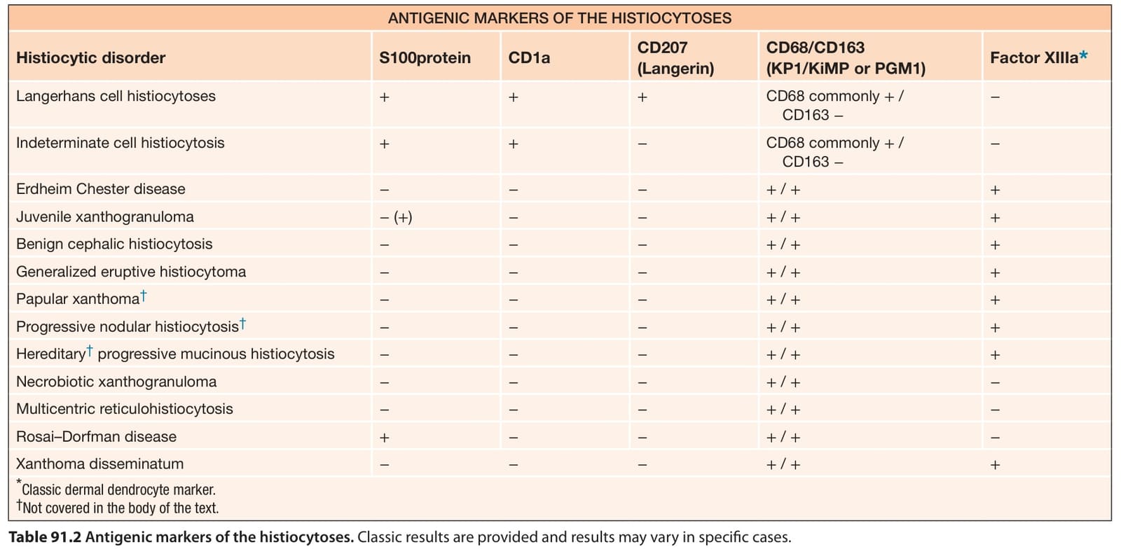
Table 91.2 Antigenic markers of the histiocytoses. Classic results are provided and results may vary in specific cases.