Benign Cephalic Histiocytosis
Synonym: Infantile histiocytosis with intracytoplasmic worm-like bodies
Key features
Infants <1 year of age are most commonly affected
Red to brown macules and papules on the face and neck
Self-limiting disease
Introduction
Benign cephalic histiocytosis (BCH) is a rare, self-limited histiocytic proliferative disorder of young children, primarily affecting the face.
History
Gianotti et al. in 1971 described the condition as “infantile histiocytosis with intracytoplasmic worm-like bodies”, based upon findings of comma-shaped structures by ultrastructural studies. When it became clear that several histiocytic disorders (e.g. LCH, juvenile xanthogranuloma) had similar ultrastructural findings, the disorder was renamed “benign cephalic histiocytosis”, based upon typical clinical findings.
Epidemiology
BCH is a rare disorder but is probably under-reported. It typically begins by the age of 1 year and nearly always within the first 3 years of life. There may be a male predominance.
Pathogenesis
The pathogenesis of BCH is not known. However, due to the histopathologic and immunohistochemical features it shares with juvenile xanthogranuloma and generalized eruptive histiocytoma, several investigators have suggested that these three non-Langerhans cell histiocytoses should be viewed as a single disease with a spectrum of clinical presentations.
Clinical features
The eruption of BCH is characterized by the evolution of 2–5 mm, pink–red to red–brown macules and papules, initially on the face (Fig. 91.9A,B), with subsequent appearance on the ears and neck. Occasionally, lesions may develop on the trunk and arms, with infrequent involvement of the buttocks and thighs. The papules eventually flatten, often leaving residual hyperpigmentation that fades over time. Spontaneous resolution occurs over a period of months to years. Most children are otherwise healthy without involvement of the mucous membranes or internal organs. However, diabetes insipidus has been reported in a young girl with BCH. The clinical course can be marked by exacerbations or evolution into generalized eruptive histiocytosis.
Pathology
The most typical finding is a well-circumscribed infiltrate of histiocytes that is confined to the upper dermis. Occasionally, a diffuse dermal infiltrate has been described. The histiocytes have regularly shaped nuclei and slightly vacuolated eosinophilic cytoplasm (Fig. 91.9C). Foamy cells and eosinophils are rarer than in juvenile xanthogranuloma. While multinucleated histiocytes and Touton giant cells were previously said to be absent, their presence has been described.
The histiocytes in BCH express typical (non-Langerhans) histiocytic markers including CD11b, CD11c, CD14b, CD68, CD163, HAM56, and factor XIIIa (see Fig. 91.4, Table 91.2). However, usually only immunostains for CD68 or CD163 and occasionally factor XIIIa are routinely performed.
Differential diagnosis
The differential diagnosis includes LCH, urticaria pigmentosa, and other non-LCH disorders (see Table 91.1). BCH overlaps with generalized eruptive histiocytoma and multiple juvenile xanthogranulomas in addition to sharing some features with progressive nodular histiocytosis and papular xanthoma. However, the clinical appearance and number of lesions as well as the distribution pattern usually allows for distinction of BCH from other non-LCH disorders while the immunophenotype of the histiocytic cells assists in excluding LCH (see Fig. 91.4).
Treatment
BCH is a self-limiting disorder and no treatment is usually needed. However, longitudinal evaluation of all patients is recommended, as both exacerbations of the disease and diabetes insipidus can occur.
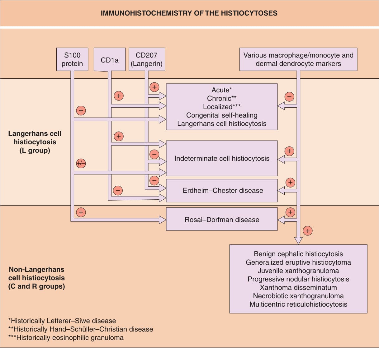
Fig. 91.4 Immunohistochemistry of the histio- cytoses. Examples of classic macrophage/ monocyte markers are CD68 and CD163; an example of a classic dermal dendrocyte marker is factor XIIIa.
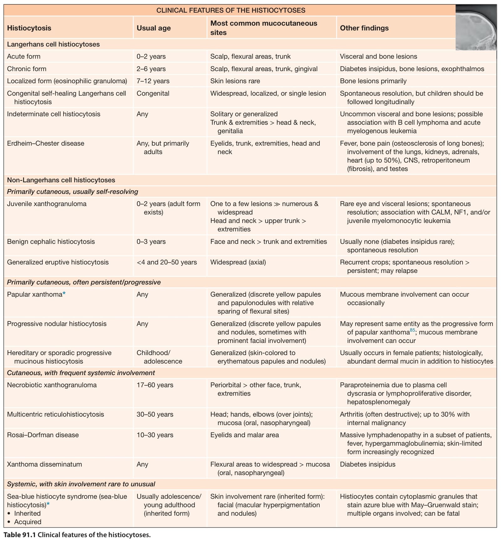
Table 91.1 Clinical features of the histiocytoses.
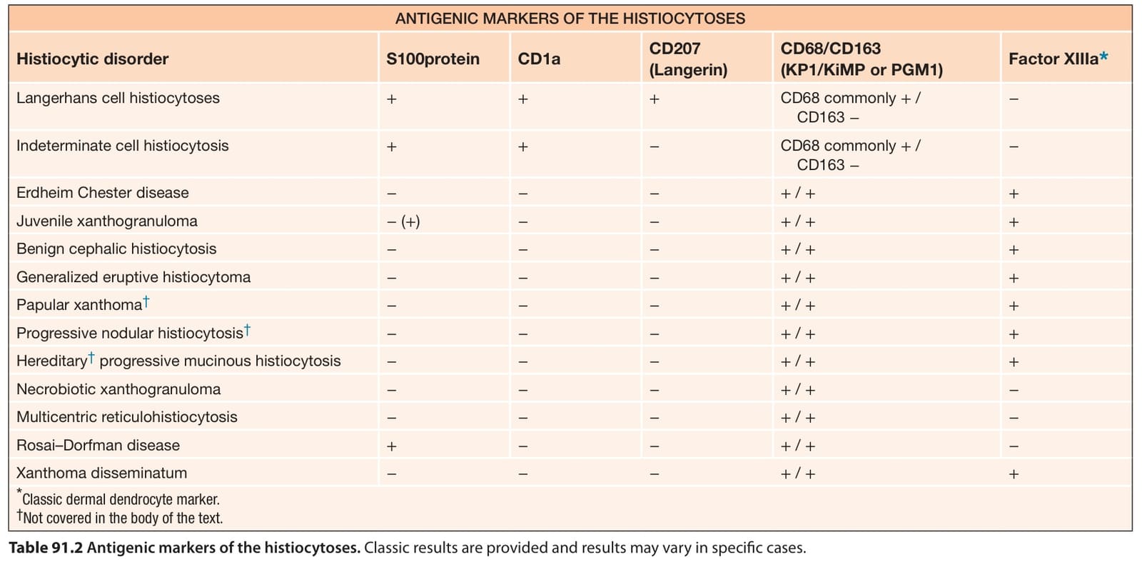
Table 91.2 Antigenic markers of the histiocytoses. Classic results are provided and results may vary in specific cases.