Generalized Eruptive Histiocytoma
Synonyms: Eruptive histiocytoma Generalized eruptive histiocytosis
Key features
Adults affected more commonly than children
Recurrent crops of numerous, small, red to brown papules
Widespread axial distribution
Self-limiting disease
Rarely associated with leukemia (primarily acute or chronic myelomonocytic)
Introduction
Generalized eruptive histiocytoma is a rare disorder characterized by recurrent crops of small papules that are axially distributed and heal with hyperpigmented macules.
History
In 1963, Winkelmann and Muller first described three adult patients with generalized eruptive histiocytoma. The disorder has since been reported in children.
Epidemiology
Generalized eruptive histiocytoma is a rare disease that is probably under-reported. Approximately one-quarter of the cases in the literature have been in children. There may be a male predilection. Onset in adults is from the third to sixth decade while in children, it is usually before the age of 4 years.
Pathogenesis
The cause of generalized eruptive histiocytoma is unknown, but there was a case report of a novel LMNA-NTRK1 fusion. As discussed in the section on BCH, several authors have suggested that generalized eruptive histiocytoma, BCH, and juvenile xanthogranuloma represent different expressions of the same disorder. Generalized eruptive histiocytoma-like presentations have been seen early in the course of other non-LCH disorders, leading some investigators to suggest that generalized eruptive histiocytoma may be an early, indeterminate stage of non-LCHs (e.g. xanthoma disseminatum, progressive nodular histiocytosis). There are a few case reports of generalized eruptive histiocytoma in patients with leukemia, in particular acute or chronic myelomonocytic leukemia. In one patient, interphase FISH analysis showed that the cutaneous lesions of generalized eruptive histiocytoma were clonally linked to the hematologic malignancy.
Clinical features
The eruption is characterized by recurrent crops of red to brown papules. At each occurrence, hundreds of papules, less than 1 cm in diameter, are distributed on the trunk, proximal extremities, and occasionally the face (Fig. 91.10). The papules are usually arranged symmetrically, and mucosal surfaces are occasionally involved. Complete resolution may occur within several months or lesions may persist; recurrences have been observed. In some children, there may be a less symmetric distribution pattern and papules can become xanthomatous. Internal involvement has not been described and the affected individuals are otherwise healthy.
Pathology
The superficial and mid dermis typically contain a nearly uniform infiltrate of slightly vacuolated histiocytes with a few lymphocytes. Xanthomatous cells are rare as are multinucleated cells. The histiocytes stain for mature histiocytic markers such as CD68, CD163, and factor XIIIa (see Fig. 91.4, Table 91.2).
Differential diagnosis
The differential diagnosis includes LCH, urticaria pigmentosa, eruptive syringomas, widespread papular granuloma annulare, and other non-LCH disorders. Of the latter, juvenile xanthogranuloma, xanthoma disseminatum, BCH, indeterminate cell histiocytosis, and papular xanthoma (see Table 91.1) can be the most difficult to differentiate from generalized eruptive histiocytoma. However, the clinical features plus the histologic and immunohistochemical findings usually allow for distinction. Occasionally
Multiple firm red to red– brown papules on the trunk.
infections that lead to granulomatous inflammation (e.g. leishmaniasis, leprosy, atypical mycobacterial infections) need to be excluded.
Treatment
No treatment is usually required as the disorder lacks systemic involvement and is often self-limited. There are, however, scattered reports of the use of isotretinoin, cryotherapy, and PUVA. Longitudinal evaluation is recommended, as more serious forms of non-LCH (e.g. xanthoma disseminatum) and rare cases of leukemia have been reported to develop in patients with generalized eruptive histiocytoma.
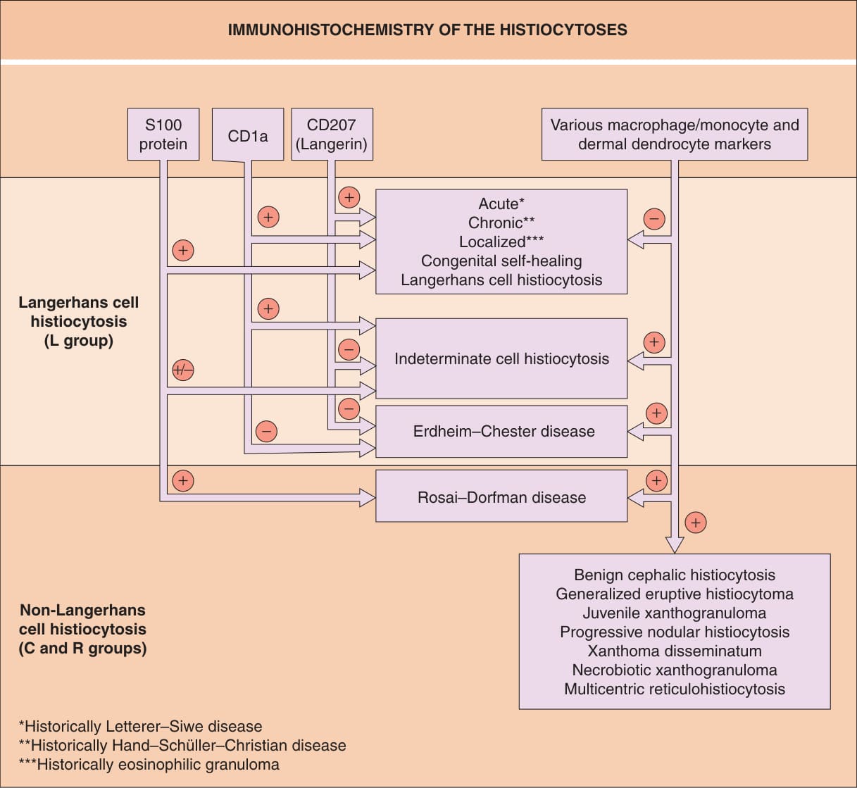
Fig. 91.4 Immunohistochemistry of the histio- cytoses. Examples of classic macrophage/ monocyte markers are CD68 and CD163; an example of a classic dermal dendrocyte marker is factor XIIIa.
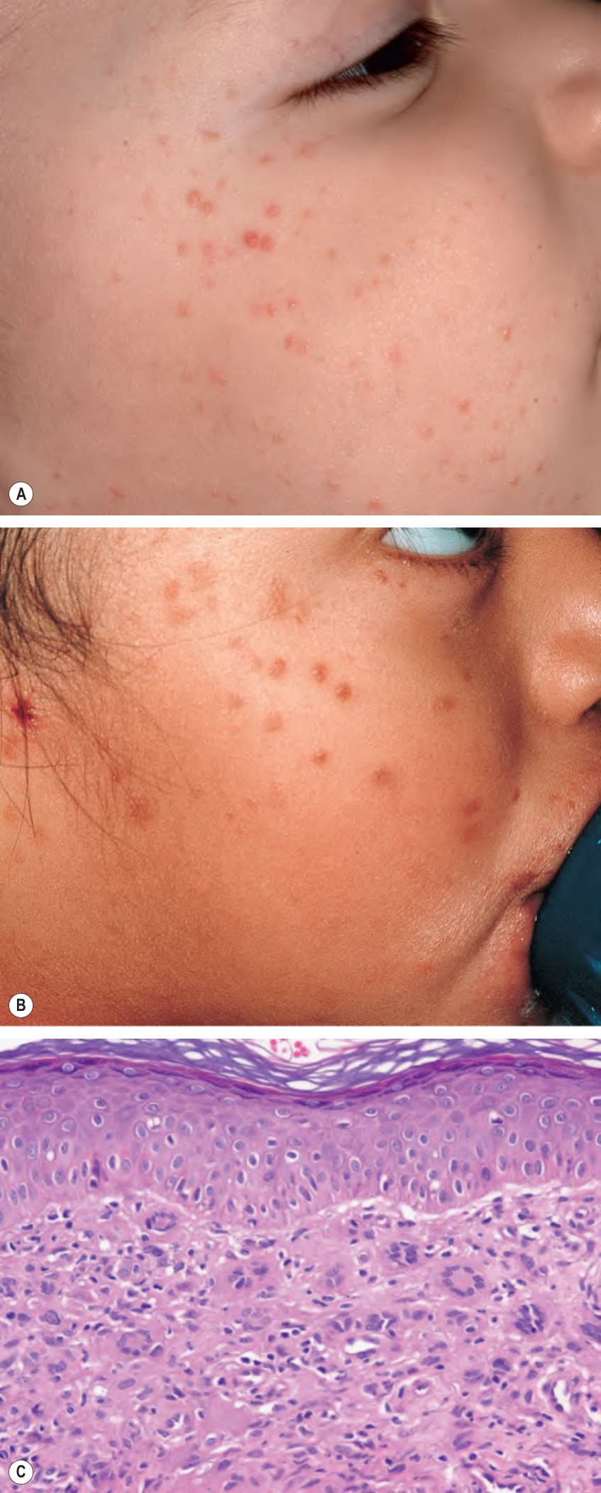
Fig. 91.9 Benign cephalic histiocytosis. Multiple papules that vary in color from light pink–brown (A) to brown (B) on the face of two young children with different skin phototypes. C Photomicrograph of the upper dermal infiltrate of histiocytes admixed with occasional Touton-like giant cells. A, Courtesy Julie V. Schaffer, MD.
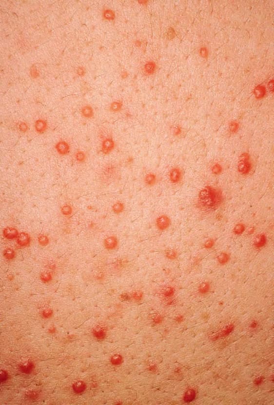
Fig. 91.10 Generalized eruptive histiocytoma.
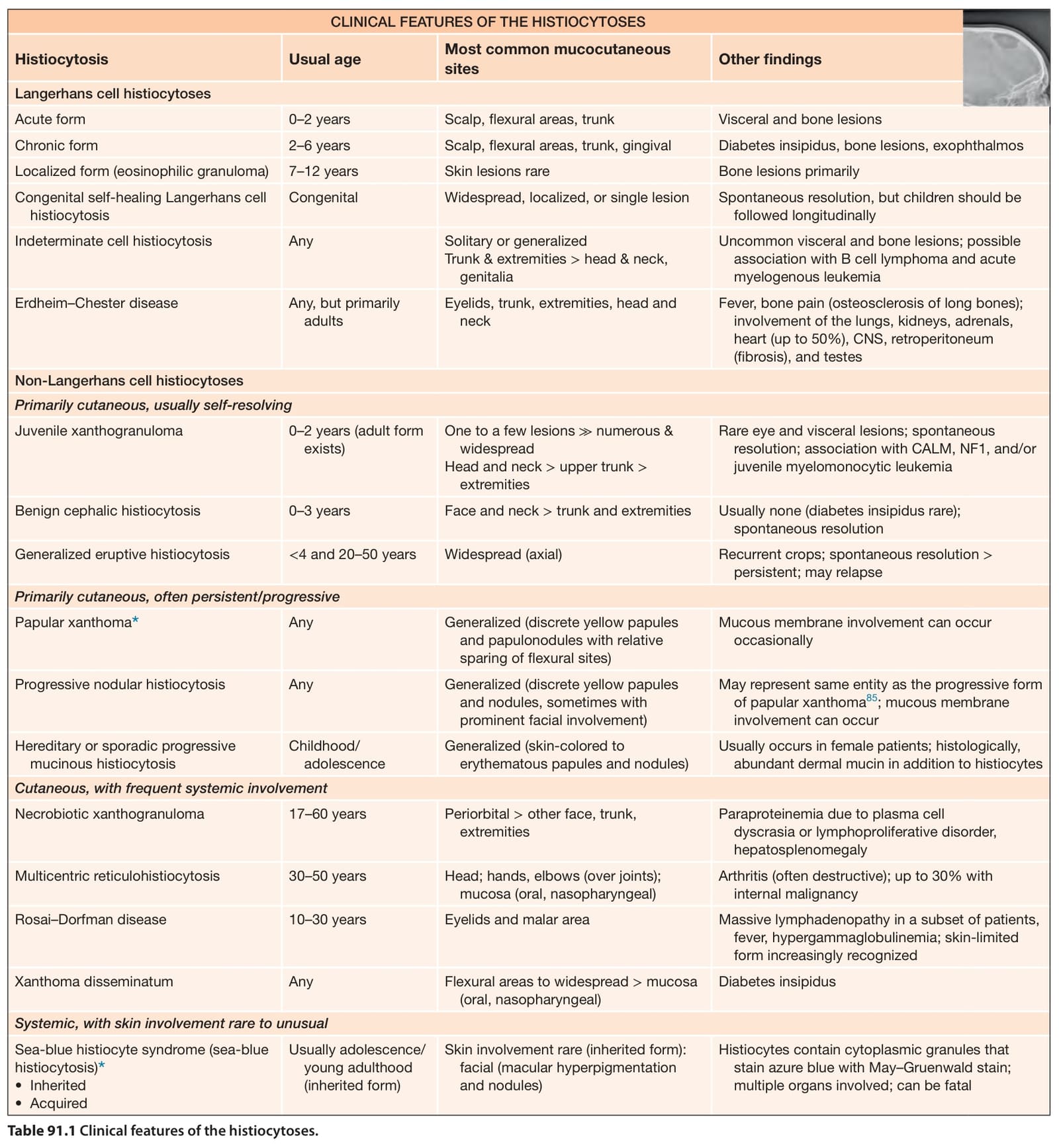
Table 91.1 Clinical features of the histiocytoses.
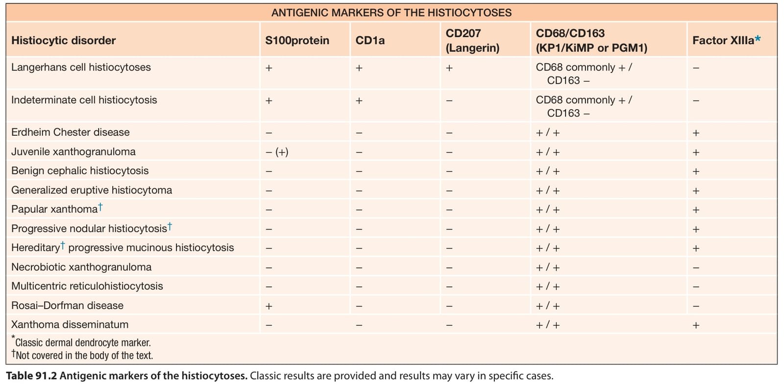
Table 91.2 Antigenic markers of the histiocytoses. Classic results are provided and results may vary in specific cases.