Juvenile Xanthogranuloma
Key features
Most common histiocytosis
Usually occurs in infants and young children, with involvement of the head and neck region and upper body
There are both small and large nodular forms, and lesions acquire a yellow color as they mature
In patients with multiple lesions, a very small minority also have ocular involvement
Rare association with both neurofibromatosis type 1 and juvenile myelomonocytic leukemia
Introduction
Juvenile xanthogranuloma (JXG) is a fairly common non-LCH, most often affecting infants and young children. Cutaneous lesions usually resolve and most patients have an otherwise unremarkable course. Although ocular JXG can lead to blindness and there is an association of JXG with neurofibromatosis type 1 (NF1) and juvenile myelomonocytic leukemia, these are unusual occurrences.
History
The term “juvenile xanthogranuloma” was suggested by Helwig and Hackney in 1954 based upon histologic findings of xanthomatous histiocytes and giant cells. However, multiple cases of JXG, under different names, had previously been reported during the first half of the twentieth century. The first case of JXG was actually reported in 1905 by Adamson, who named the disorder “congenital xanthoma multiplex”.
Epidemiology
JXG is a fairly common disorder and the most common histiocytic disease of childhood. However, the true incidence may be underestimated, as many lesions, especially those which are solitary and small, may go unrecognized. In children, there is a male predominance of approximately 1.5 : 1; no sex predilection is noted in adults. A significant majority of those reported are White individuals. In almost 75% of cases lesions appear during the first year of life, with >15% of patients having them at birth. JXG is rare in adults, with a peak incidence during the late twenties to early thirties, although lesions have also been reported in the elderly. Most adult patients have solitary lesions.
Pathogenesis
The cause of JXG is not known; it is often suggested that the condition is reactive, with histiocytes possibly responding to a traumatic or infectious stimulus. However, in a recent series of 55 patients with both cutaneous and extracutaneous JXG, ~75% had a genetic mutation that would lead to activation of the MAPK pathway. The reason for the progressive lipidation of histiocytes in the absence of hyperlipidemia is not clear. However, it has been shown that in adult patients with xanthogranulomas, the uptake of low-density lipoprotein and the synthesis of cholesterol within macrophages is increased. As discussed in the section on BCH, several authors have suggested that generalized eruptive histiocytoma, BCH, and JXG may represent different expressions of the same disorder.
Clinical features
Gianotti and Caputo described two common clinical variants of JXG: a small nodular form and a large nodular form. Patients with the small nodular form, also known as the micronodular form, present with multiple pink to red–brown, dome-shaped papules, 2–5 mm in diameter. The lesions are widely scattered on the upper part of the body and rapidly become yellow. In contrast, the more common large nodular form is characterized by one or a few nodules 1–2 cm in diameter. The two forms may coexist.
The most common location for JXG is the head and neck (Fig. 91.11), followed by the upper torso, the upper extremities, and then the lower extremities. Oral JXG is rare and usually presents as a solitary yellow nodule on the lateral aspect of the tongue or the midline of the hard palate. Unusual morphologic presentations include keratotic, pedunculated, subcutaneous, clustered, plaque-like, and giant lesions.
Extracutaneous lesions have been reported in many organs, with the eye being most commonly affected. The lung represents the second most frequent extracutaneous site of disease. Other visceral, bone, and CNS involvement is unusual and disease-related deaths are rare. Ocular JXG is almost always unilateral and develops in <0.5% of patients with cutaneous lesions. However, ~40% of patients with ocular JXG have cutaneous lesions, always multiple, at the time of diagnosis. Ocular involvement usually occurs before 2 years of age and often affects the iris. Hyphema (hemorrhage into the anterior chamber) and glaucoma are serious complications which can result in blindness. Early referral to an ophthalmologist for evaluation and possible treatment is important.
A well-recognized association exists between JXG and café-au-lait macules. Some of these patients have a family history of NF1, while others fulfill the clinical criteria for NF1 (see Table 61.3). Another well-known association is that of JXG and juvenile myelomonocytic leukemia; patients with NF1 also have an increased risk of developing juvenile myelomonocytic leukemia. A so-called “triple association” consisting of JXG, NF1, and juvenile myelomonocytic leukemia has been observed in several patients, and patients with JXG and NF1 have at least a 20× increased risk of developing juvenile myelomonocytic leukemia. Other hematologic malignancies have been reported in association with JXG, e.g. chronic lymphocytic leukemia or B cell lymphoma in adults with multiple lesions.
In the majority of patients with disease limited to the skin, the course is self-limited and benign. These patients are otherwise in good health and lesions usually regress within 3–6 years. Hyperpigmentation, mild atrophy, or anetoderma may remain. However, in adults, the xanthogranulomas are usually solitary and persistent.
Pathology
In small lesions, there is a well-demarcated, dense infiltrate of histiocytes within the superficial dermis, which extends into the subcutis in larger lesions. A loss of the rete ridges is often seen, and ulceration occurs in some cases. Early lesions usually have monomorphous histiocytes with abundant eosinophilic cytoplasm (Fig. 91.12A). In mature lesions, the histiocytes develop lipid in their cytoplasm, creating a foamy “xanthomatous” appearance. Touton giant cells are a characteristic finding, but they may be absent in early lesions (Fig. 91.12B). Lymphocytes, eosinophils, plasma cells, and even neutrophils are also scattered throughout the infiltrate. Variants with spindle cells have been reported, which may be misinterpreted as another spindle cell tumor. Other morphologic variants include mononuclear cells with an oncocytic appearance, vacuolated cells, and cells with a polygonal or spiculated cytoplasm.
The cells in JXG stain positively for mature histiocyte markers such as CD68, CD163, and factor XIIIa (see Table 91.2). In some cases there is expression of S100 protein on the large-sized cells, whereas CD1a and CD207 (Langerin) are always negative.
Differential diagnosis
The major entities to consider when there are multiple lesions are other histiocytic disorders including LCH. In a child with a single JXG, the initial clinical differential diagnosis can be broad (e.g. molluscum contagiosum, Spitz nevus, mastocytoma). A solitary lesion in an adult may be diagnosed clinically as a more common melanocytic nevus or dermatofibroma. The so-called “solitary reticulohistiocytoma” is simply a xanthogranuloma in which oncocytic macrophages and giant cells with ground-glass cytoplasm dominate.
In general, the constellation of clinical and histopathologic features allows for distinction between JXG and either BCH or generalized eruptive histiocytoma, although some cases of BCH can have giant cells (see Fig. 91.9C), and these three disorders may indeed exist within a spectrum. LCH can be differentiated from JXG via immunophenotyping (see Fig. 91.6).
Treatment
Due to the self-limiting nature of JXG, no treatment is usually required. Occasionally, lesions are removed for cosmetic concerns, despite the
anticipated spontaneous resolution. While ocular lesions often require intervention (e.g. topical corticosteroids for iris lesions, excision for limbal lesions), sites of systemic involvement can be followed without treatment unless their location interferes with normal function. For these patients, various chemotherapeutic regimens, radiotherapy, high-dose systemic corticosteroids, and cyclosporine have been administered. Given the possibility of spontaneous involution, however, it is sometimes difficult to evaluate the response to treatment in a given patient.
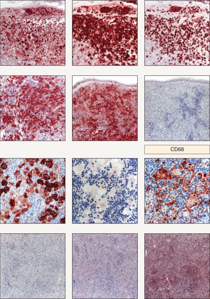
Fig. 91.6 Use of a panel of four immunostains to distinguish between Langerhans cell histiocytosis and several types of non-Langerhans cell
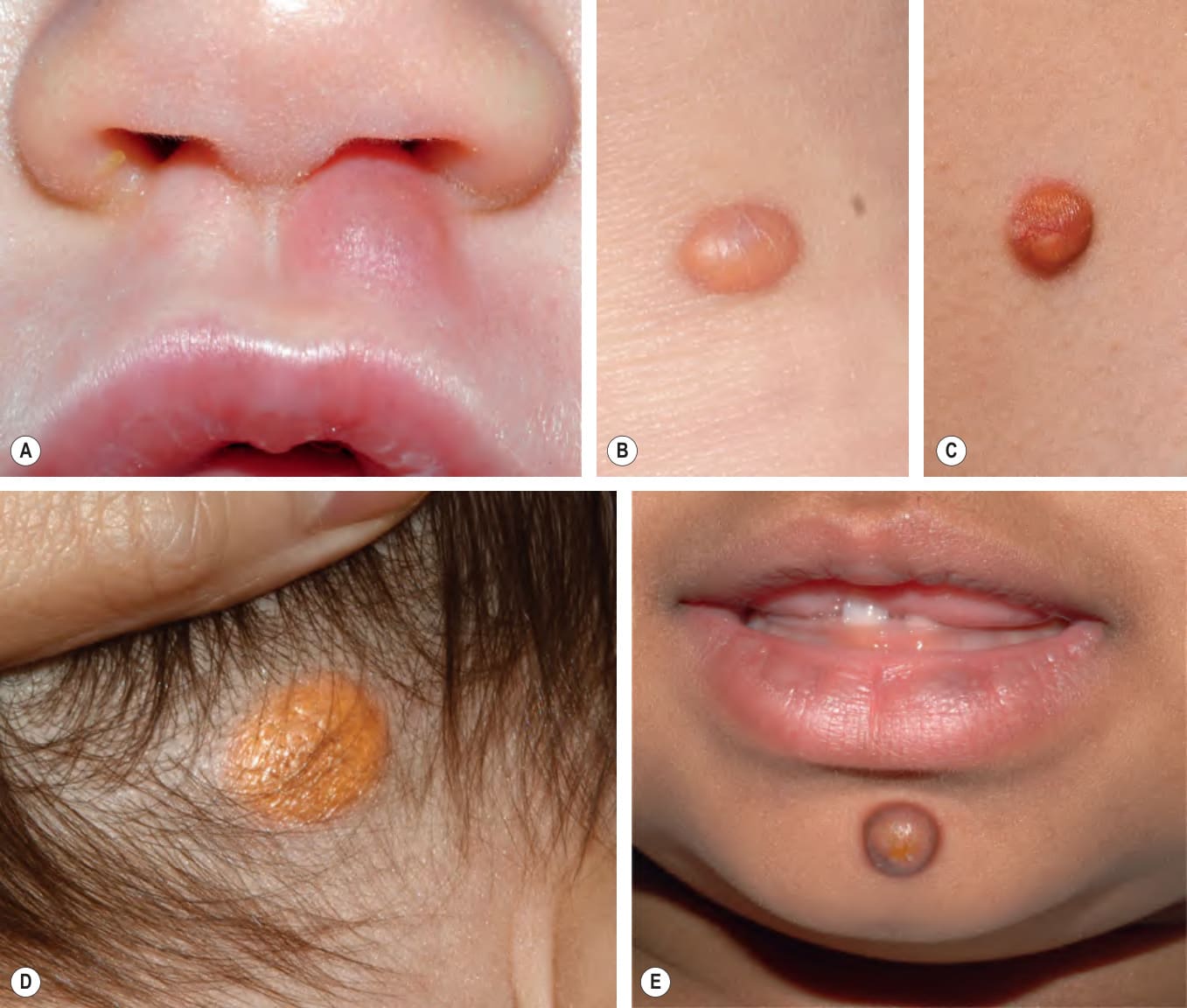
Fig. 91.11 Juvenile xantho- granuloma.A Pink nodule with a few telangiectasias representing an early lesion. Well-developed lesions that vary in color from pale yellow (B) to red–yellow (C) to yellow–orange (D) to yellow– brown (E), with the yellow hue reflecting the accumulation of lipid within histiocytes. Courtesy Julie V. Schaffer, MD.
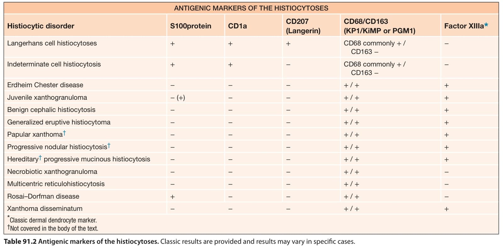
Table 91.2 Antigenic markers of the histiocytoses. Classic results are provided and results may vary in specific cases.