Progressive Nodular Histiocytosis
Key features
Affects primarily the elderly of either sex
Hundreds of lesions of two types: small superficial papules and nodules or deep nodules and tumors
Mucosal lesions may be seen
Chronic without spontaneous regression
Introduction, epidemiology, and pathogenesis
Progressive nodular histiocytosis (PNH) is an extremely rare, clinically distinct disorder affecting primarily the elderly. It has a chronic course and an unknown etiology and pathogenesis. However, an IRF2BP2-NTRK1 fusion was detected in a patient with PNH/ xanthogranulomatosis.
Clinical features
Patients typically develop multiple lesions that progressively become widespread, but usually spare the flexural areas. Two distinct types of lesions have been described: (1) superficial xanthomatous papules and nodules that are 2–10 mm in diameter; and (2) deep fibrous nodules and tumors, 1–3 cm in diameter, that are primarily on the trunk. The face is
often involved and confluent nodules can lead to a “leonine facies” (see Table 46.5). A key to the diagnosis is the presence of these two distinct types of lesions. There may be conjunctival, oral, and laryngeal involvement.
In patients followed over a course of many years, the lesions have rarely regressed. Visceral involvement has not been observed nor have consistent associations with other diseases. The significance of a report of a patient with PNH and chronic myeloid leukemia, hepatosplenomegaly, hyperuricemia, and hypocholesterolemia is unclear.
Pathology
The smaller papules contain foamy xanthomatous cells and occasional Touton giant cells, mimicking papular xanthomas. The larger nodules are distinctive and show features of spindle-cell xanthogranulomas. The cells express markers of mature dendritic histiocytes such as CD68, CD163, and FXIIIa.
Differential diagnosis
Other non-LCHs, especially those that do not regress spontaneously such as progressive mucinous histiocytosis, as well as sarcoidosis and leprosy represent the primary entities in the clinical differential diagnosis.
Treatment
The natural history is chronic and progressive, with an increase in the number of lesions. Treatment consists of removal of nodules via surgical excision or CO laser.
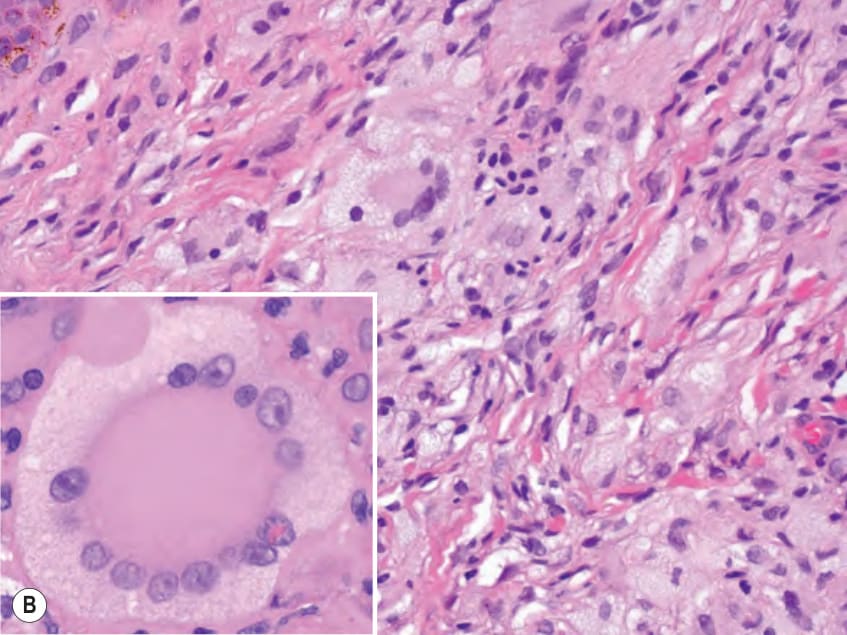
Fig. 91.12 Juvenile xanthogranuloma.A Earlier lesion – the dermal histiocytic infiltrate contains relatively few Touton giant cells. Lymphocytes and eosinophils are also present (inset). B Older lesion – multiple foamy (xanthomatous) histiocytes and giant cells as well a classic Touton giant cell with a “wreath-like” arrangement of nuclei (inset).
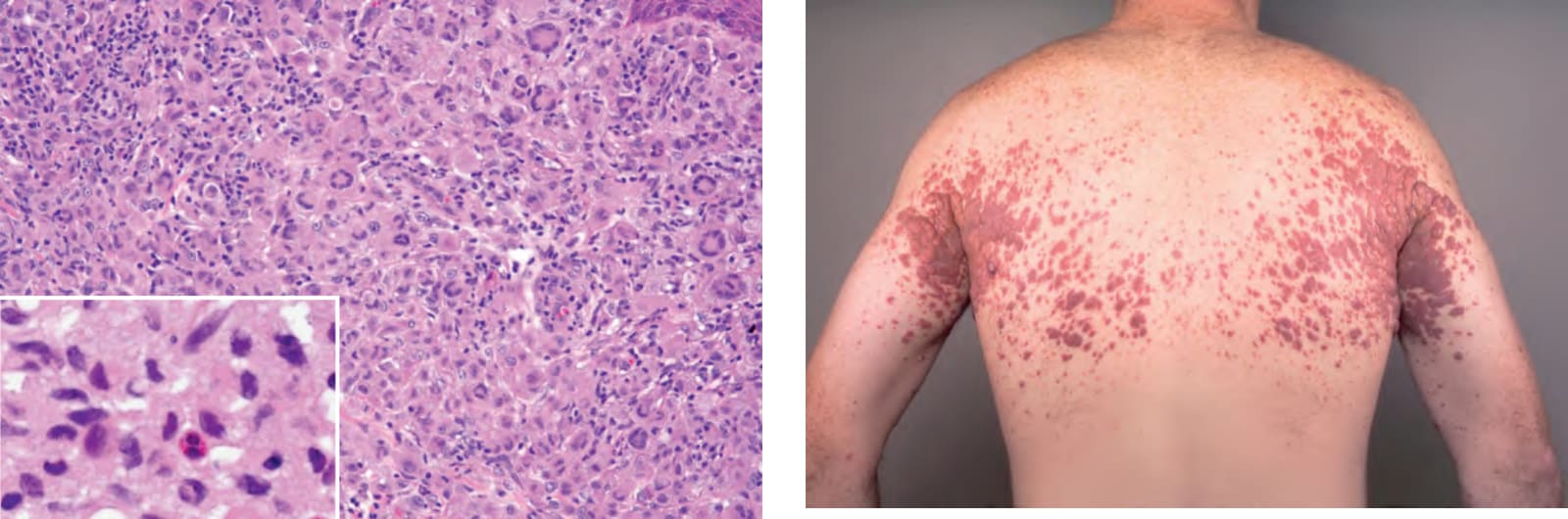
Fig. 91.13 Xanthoma disseminatum. Symmetric involvement of the major flexures is a characteristic finding. Note the yellow discoloration of some of the coalescing papulonodules. Courtesy David Wetter, MD.