Xanthoma Disseminatum
Synonym: Xanthoma disseminatum of Montgomery
Key features
Classic triad of cutaneous xanthomas, mucous membrane xanthomas, and diabetes insipidus
Symmetric distribution of lesions
Favors flexural areas and intertriginous zones
Corneal and conjunctival lesions can threaten vision
Introduction
Xanthoma disseminatum is a rare, normolipemic, histiocytic proliferative disorder that affects the skin, mucous membranes, and frequently, the hypothalamus and pituitary gland, leading to transient diabetes insipidus.
History
Four cases of “xanthomatosis disseminata” were described by Montgomery and Osterberg in 1938. A review and characterization of the disorder was provided by Altman and Winkelmann in 1962.
Epidemiology
Xanthoma disseminatum is a rare condition, with ~100 cases reported to date. Men are more commonly affected than women. Age of onset ranges from 8 months to 85 years, with >60% of patients developing the disease before the age of 25.
Pathogenesis
The etiology is unknown. As most patients have normal lipid levels, it has been suggested that xanthoma disseminatum represents a reactive proliferative disorder of histiocytes with secondary accumulation of lipid.
Clinical features
Patients may have the classic triad of cutaneous xanthomas, xanthomas of mucous membranes, and diabetes insipidus. The primary xanthomatous lesion is a yellow, red, or brown papule. Disease onset is marked by an eruption of hundreds of papules, symmetrically arranged on the face and in the major body folds and flexural areas of the trunk and proximal extremities (Fig. 91.13). The lesions tend to cluster into well-formed, potentially disfiguring plaques (Fig. 91.14). Older lesions may become atrophic.
Mucous membrane lesions are found in 40%–60% of patients, with the upper airway and oral mucosa being commonly involved. Corneal
and conjunctival lesions can threaten vision. CNS involvement of the hypothalamus and pituitary stalk results in diabetes insipidus in ~40% of patients. The latter is usually mild, transient, and sensitive to vasopressin. Rarer associations include monoclonal gammopathies due to plasma cell dyscrasias and thyroid disorders.
Caputo et al. suggested that patients follow one of three clinical courses: (1) a rare, self-healing form with spontaneous resolution of lesions; (2) the common persistent form in which lesions may never resolve; and (3) the very rare progressive form with organ dysfunction and CNS involvement. A few disease-related deaths have been reported, including one patient with CNS involvement outside the pituitary gland and hypothalamus.
Pathology
In the early stages, there is a dense dermal infiltrate of scalloped macrophages in addition to a few foamy cells or other inflammatory cells. Well-developed lesions contain a mixture of scalloped cells, foamy cells and other inflammatory cells, as well as Touton and foreign body giant cells. The histiocytes express typical non-LCH markers such as CD68, CD163, CD11b, CD14, CD11c, and factor XIIIa (see Fig. 91.4, Table 91.2).
Differential diagnosis
The disorders most likely to be confused with xanthoma disseminatum include generalized eruptive histiocytoma, papular xanthomas, eruptive and plane xanthomas (see Ch. 92), ECD, multiple JXGs, progressive nodular histiocytosis, and multicentric reticulohistiocytosis. In general, these can be fairly easily differentiated based on clinical and histopathologic features.
Treatment
As xanthoma disseminatum is a rare disease, there are no controlled trials of systemic therapies. Radiotherapy has been used to control obstructive airway disease. No significant improvement in cutaneous and mucosal lesions has been observed with oral corticosteroids. Physical modalities including surgery, radiotherapy, cryosurgery, and laser therapy have all been tried, as have systemic medications such as cyclophosphamide, cyclosporine, and 2-chlorodeoxyadenosine (2-CdA). In a case series, all five patients who received 2-CdA experienced a remission and long-term control of cutaneous lesions.
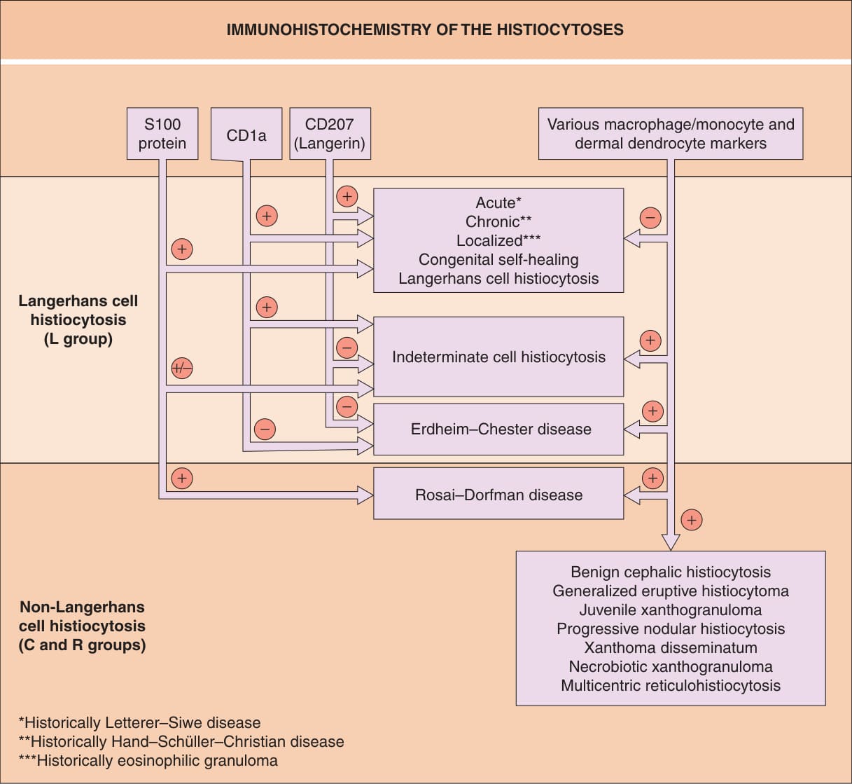
Fig. 91.4 Immunohistochemistry of the histio- cytoses. Examples of classic macrophage/ monocyte markers are CD68 and CD163; an example of a classic dermal dendrocyte marker is factor XIIIa.
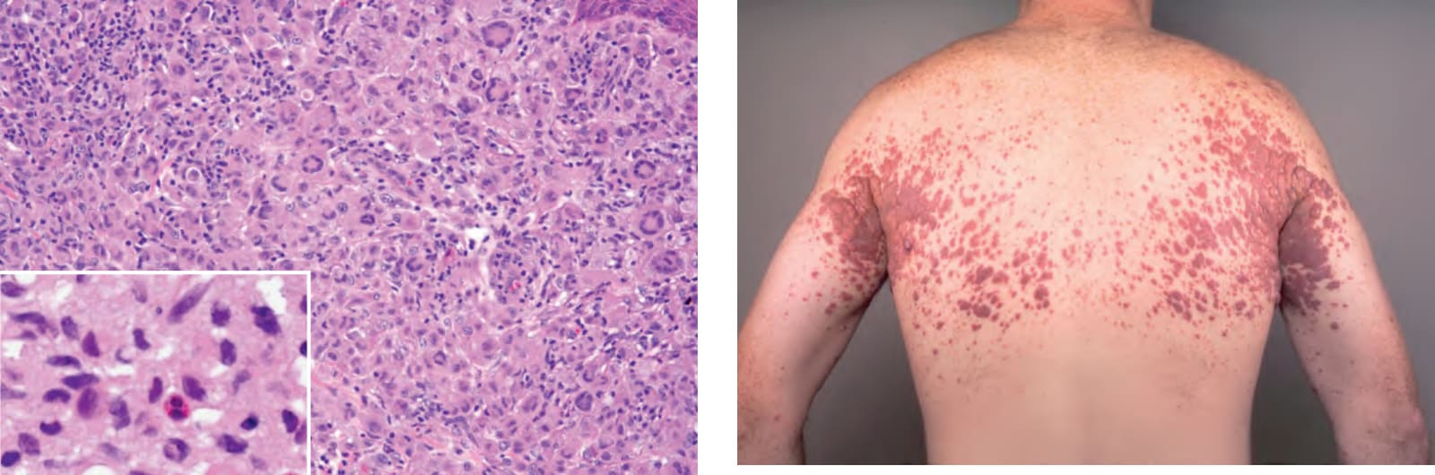
Fig. 91.13 Xanthoma disseminatum. Symmetric involvement of the major flexures is a characteristic finding. Note the yellow discoloration of some of the coalescing papulonodules. Courtesy David Wetter, MD.
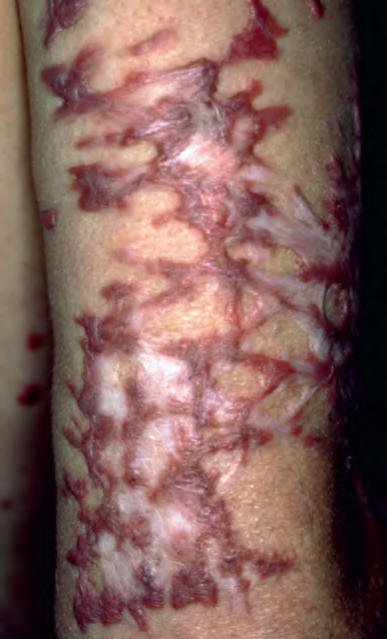
Fig. 91.14 Xanthoma disseminatum. Sclerotic form of xanthoma disseminatum in a patient who developed multiple myeloma. Note the associated scarring.
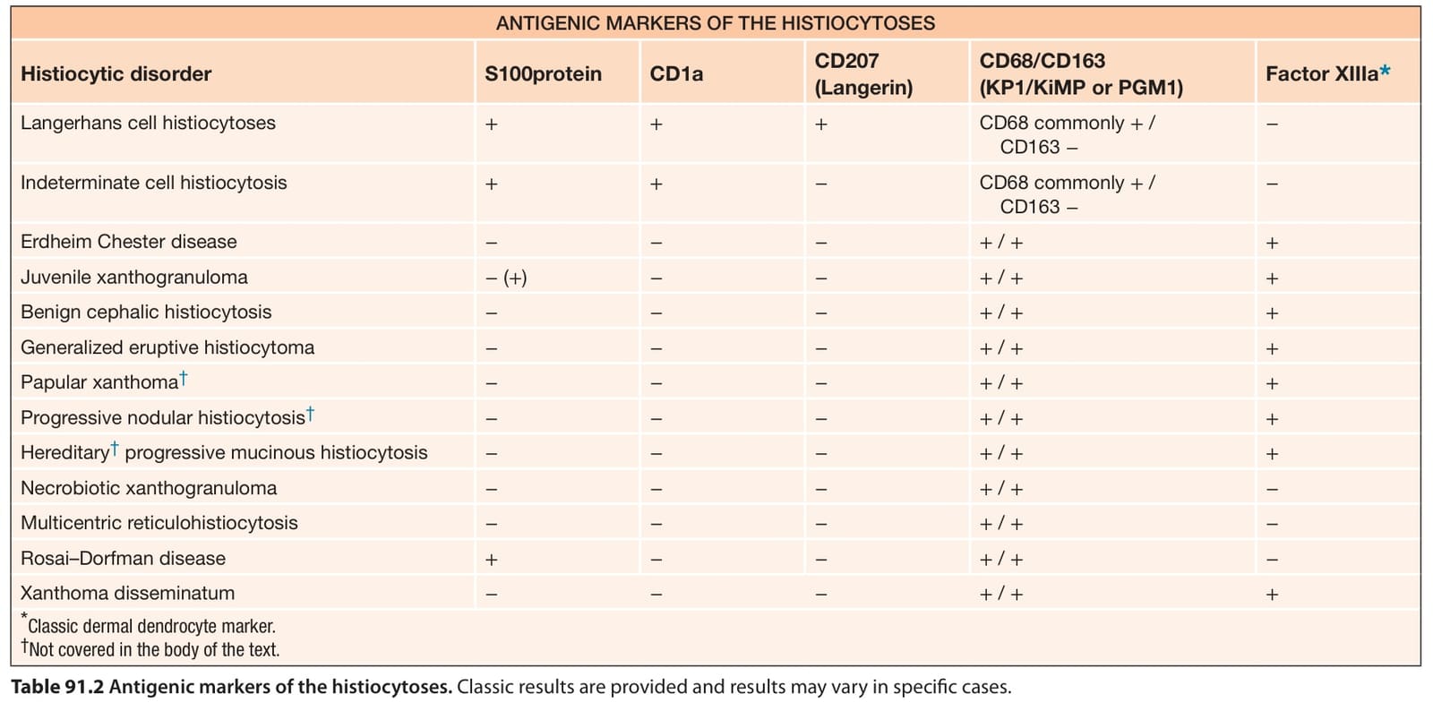
Table 91.2 Antigenic markers of the histiocytoses. Classic results are provided and results may vary in specific cases.