Necrobiotic Xanthogranuloma
Synonym: Necrobiotic xanthogranuloma with paraproteinemia
Key features
Multisystem histiocytic disease that can present with hepato- splenomegaly as well as ocular and skin involvement
In at least 70% of patients, there is an IgG monoclonal gammopathy due to a plasma cell dyscrasia or lymphoproliferative disorder (e.g. B cell chronic lymphocytic leukemia)
Average age of onset is the sixth decade
Firm nodules and plaques with a yellow hue are seen and they may develop ulceration
Most common site of involvement is the periorbital region
Introduction
Necrobiotic xanthogranuloma (NXG) is a rare, progressive multisystem histiocytic disease in which characteristic cutaneous lesions point to the diagnosis. The majority (~80%) of patients have a monoclonal gammopathy due to either a plasma cell dyscrasia or a B cell lympho proliferative disorder (see Ch. 119).
History
Necrobiotic xanthogranuloma was codified in 1980 by Kossard and Winkelmann. Their original eight patients demonstrated yellowish plaques, subcutaneous nodules, and an associated dysproteinemia. These authors also noted several reports published prior to 1980 representing unrecognized cases of NXG.
Epidemiology
Necrobiotic xanthogranuloma is a rare disorder with over 200 cases reported to date. In 1992, Mehregan and Winkelmann described 32 patients with NXG and reviewed the 16 reported cases from the world literature. Men and women are generally equally affected. The average age of onset is the sixth decade.
Pathogenesis
While the pathogenesis of NXG is as yet unknown, its strong association with monoclonal gammopathies has led to several hypotheses. The M protein may act either as a primary inciting agent or as a cofactor in eliciting a giant cell granulomatous reaction. In a review of 17 cases, there was no evidence of a monoclonal proliferation of plasma cells within the inflammatory infiltrate of skin lesions. It has been suggested that NXG and normolipemic plane xanthoma exist within a spectrum, given their shared relationship to monoclonal gammopathies.
Clinical features
Cutaneous lesions, typically multiple, develop in all patients with NXG. The classic skin lesion is an asymptomatic indurated papule, nodule, or plaque with a yellow, “xanthomatous” hue (Fig. 91.15). Other reported features include telangiectasias, atrophy, ulceration and scarring, with scars being common sites for the development of new lesions.
The periorbital region is the most common site of involvement. The trunk, remainder of the face, and proximal extremities are also frequently involved. From 15% to 50% of patients have ophthalmic manifestations, which include orbital masses, ectropion, ptosis, conjunctival lesions, keratitis and scleritis, episcleritis, anterior uveitis, and proptosis.
A hallmark feature of NXG is the associated IgG monoclonal gammopathy, which is found in ~80% of patients. The M protein can be detected by serum protein electrophoresis and/or the more sensitive immunofixation electrophoresis. Other common findings include hepatomegaly, splenomegaly, an increased ESR/CRP, leukopenia, hypocomplementemia, and an underlying plasma cell dyscrasia (including multiple myeloma). Less commonly, cryoglobulinemia and/ or an underlying B cell lymphoproliferative disorder are observed. Of the 48 patients described by Mehregan and Winkelmann, a plasma cell dyscrasia was found in 17 patients (with 8 fulfilling the criteria for multiple myeloma) and a lymphoproliferative disorder in 2 patients. In a more recent retrospective study from the same institution, a higher percentage of patients (12 of 17 [71%]) had a monoclonal gammopathy,
Yellow–brown papules and plaques in a periorbital distribution in a patient with chronic lymphocytic leukemia. Such lesions may initially be mistaken for xanthelasma. Courtesy Kalman Watsky, MD.
with 3 patients fulfilling the criteria for multiple myeloma. Postmortem examination has demonstrated involvement of multiple organ systems, with endocardial NXG found in most cases.
In one series, when patients with NXG developed a plasma cell dyscrasia or a lymphoproliferative disorder (10 of 26 patients), these disorders often arose after the onset of cutaneous manifestations and did not tend to be aggressive. Overall survival was 100% at 10 years and 90% at 15 years.
Pathology
Classic, non-ulcerated lesions have a normal epidermis and superficial dermis. A palisading xanthogranuloma is found in the mid dermis extending into the panniculus (Fig. 91.16). The granulomas consist of histiocytes, foam cells, lymphoid follicles, plasma cells, and giant cells with zones of collagen degeneration (“necrobiosis”). Cholesterol clefts are found in areas of necrobiosis. A prominent feature is the presence of both Touton giant cells and large, bizarre foreign body giant cells. These cells are found scattered within the granuloma and often at the margins of areas of necrobiosis. Lymphoid or lymphoplasmacytic nodules are also common, and clonal infiltrates can be seen in patients with B cell chronic lymphocytic leukemia (CLL). Histiocytic cells stain positively for non-LCH markers such as CD68 and CD163, but are negative for FXIIIa (see Fig. 91.4, Table 91.2).
Differential diagnosis
The clinical differential diagnosis includes necrobiosis lipoidica, normolipemic plane xanthomas, xanthelasma, non-LCH disorders (xanthoma disseminatum, ECD, multicentric reticulohistiocytosis, JXG), adultonset asthma and periocular xanthogranuloma (AAPOX), foreign body granulomas, and sarcoidosis. However, most of these disorders can be excluded on the basis of morphology, distribution pattern, histologic features, and associated systemic manifestations (if any).
Treatment
No controlled clinical studies are available regarding the treatment of NXG. In the past, alkylating agents (e.g. chlorambucil, melphalan) were often tried but due to the availability of other therapeutic options and the risk of myelodysplastic syndrome and acute myelogenous leukemia, their use has declined. In a multicenter cohort of 34 patients, IVIg was found to be the most effective therapy (9/9 patients), followed by antimalarial drugs (4/5) and intralesional triamcinolone (6/8). When the patient meets the criteria for multiple myeloma, combination therapy that can include an immunomodulatory drug (e.g. lenalidomide), proteosome inhibitor (e.g. bortezomib, carfilzomib), anti-CD38 antibody (e.g. daratumumab), and dexamethasone consistently leads to disappearance of the M protein. These medications can also be considered for patients with severe recalcitrant disease, even if they do meet the criteria for symptomatic
myeloma. For B cell CLL, current treatments include BTK inhibitors (e.g. ibrutinib, acalabrutinib, zanabrutinib), bcl-2 inhibitors (e.g. venetoclax), and anti-CD20 antibodies (e.g. obinutuzumab, rituximab).
Topical JAK inhibitors (e.g. tofacitinib 2% cream) may lead to improvement of cutaneous lesions. A recurrence rate of ~40% was observed in one series following surgical excision of lesions.
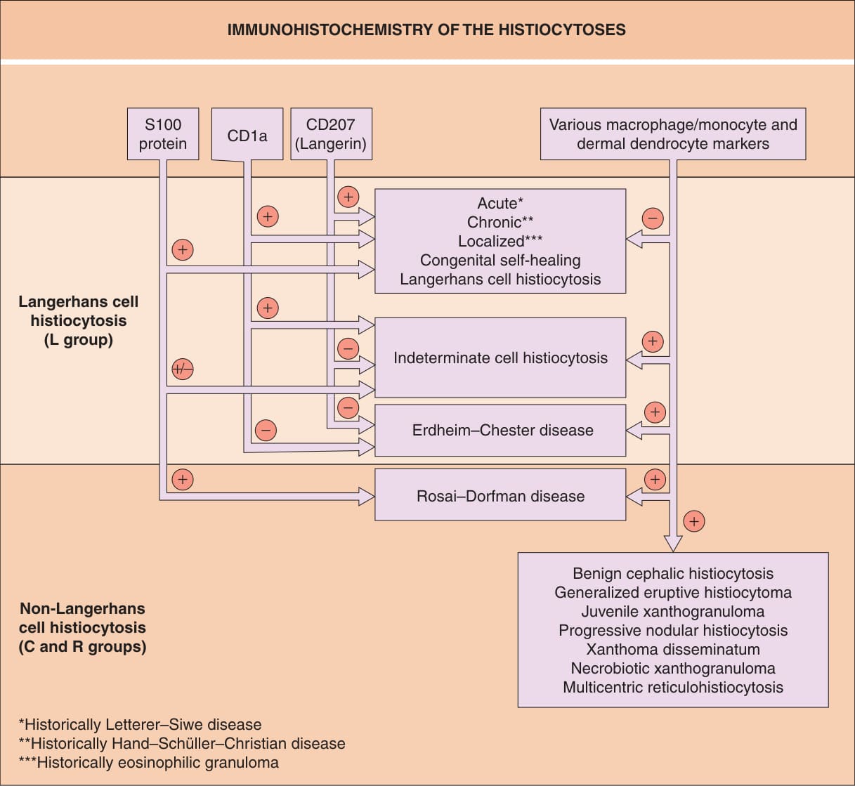
Fig. 91.4 Immunohistochemistry of the histio- cytoses. Examples of classic macrophage/ monocyte markers are CD68 and CD163; an example of a classic dermal dendrocyte marker is factor XIIIa.
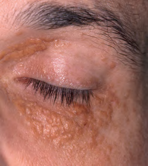
Fig. 91.15 Necrobiotic xanthogranuloma.
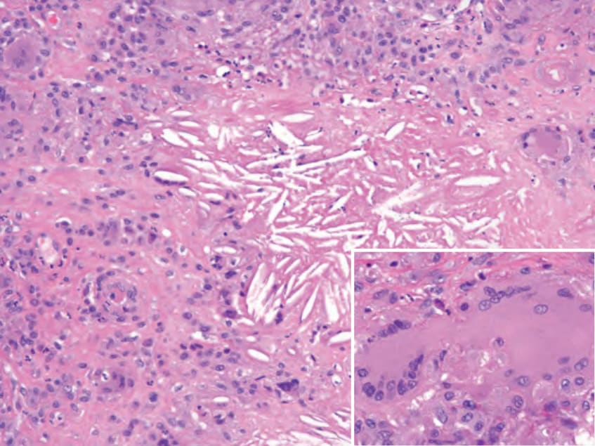
Fig. 91.16 Necrobiotic xanthogranuloma. A necrobiotic area contains cholesterol clefts. The presence of bizarre, large multinucleated giant cells is a typical feature (inset).
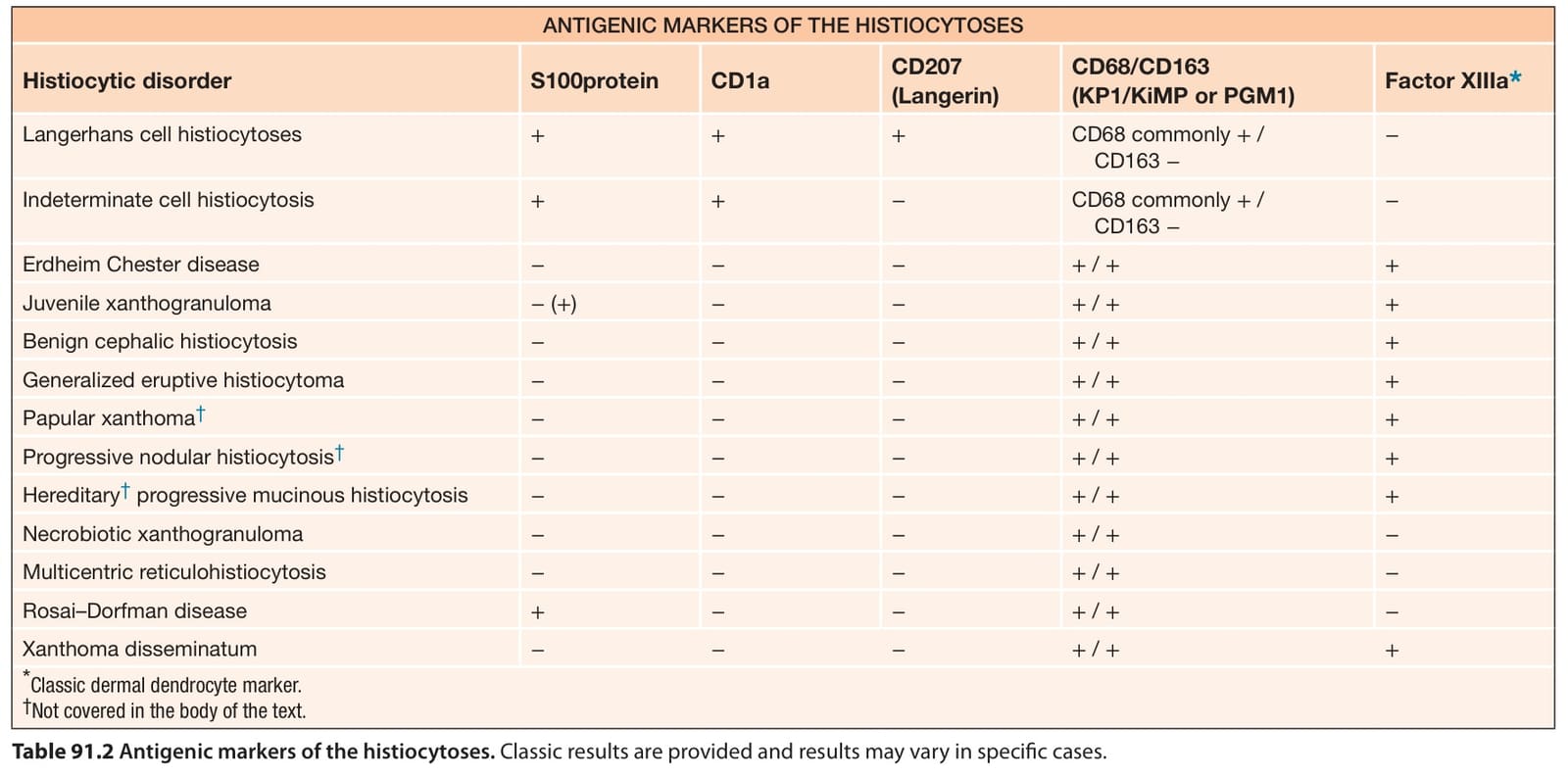
Table 91.2 Antigenic markers of the histiocytoses. Classic results are provided and results may vary in specific cases.