Multicentric Reticulohistiocytosis
Key features
Occurs primarily in adults
Papules and nodules favor the head, hands, and elbows; periungual papules have been likened to “coral beads”
50% of patients develop mucous membrane lesions (oral and nasopharyngeal)
Multicentric reticulohistiocytosis is associated with a destructive arthritis and, in some patients, solid organ malignancies
Histiocytes have a characteristic “ground glass” appearance
Introduction
Multicentric reticulohistiocytosis (MRH) is a rare form of non-LCH which most commonly affects adults. This disorder is characterized by both cutaneous and systemic features. Well-developed lesions demonstrate a classic histopathology consisting of mononucleated and multi-nucleated giant cells with a “ground glass” appearance.
History
The term multicentric reticulohistiocytosis was introduced in 1954 by Goltz and Laymon to define cases with both cutaneous and systemic manifestations. Familial histiocytic dermatoarthritis, described in
1973, is a very rare variant of MRH, characterized by a familial occurrence and associated glaucoma, uveitis, and cataracts.
Epidemiology
MRH is uncommon, occurring most frequently in women during their fifth or sixth decade of life.
Pathogenesis
The pathogenesis of MRH is unknown. In one study from the 1960s, 56% of patients had a positive tuberculin skin test, leading to the suggestion that mycobacteria could be a possible trigger. In addition, MRH has been reported to respond to antituberculosis treatment. However, confirmation of mycobacteria within skin nodules is lacking. Others have proposed that the histiocytic response in MRH is an immunologic process related to an underlying neoplastic or autoimmune disorder. There is evidence that TNF, which can promote osteoclast formation, may play a role in its pathogenesis.
Clinical features
Patients with MRH usually have cutaneous and mucous membrane involvement in addition to a severe arthropathy, although histiocytic infiltrates can be found in multiple organs (see below). There is an association with hyperlipidemia, a positive tuberculin skin test, systemic vasculitis, and autoimmune disease (e.g. systemic lupus erythematosus, Sjögren syndrome). Up to 25%–30% of patients have reportedly had an associated malignancy, with a range of solid and hematologic malignancies. An elevated ESR and anemia have been noted in approximately half of patients, and one-third demonstrate hypercholesterolemia. Rarely, monoclonal gammopathies or cryoglobulinemia have been observed. Fever and weight loss can also occur.
Cutaneous lesions range from a few millimeters to 2 cm and are skin-colored to pink, red–brown, or yellow. Lesions tend to have an acral distribution. Favored sites include the head, hands, fingers, ears, and articular regions of the limbs (Fig. 91.17). Small papules aligned
Grouped, firm, red–brown papules on the dorsal surface of the fingers, hand, and wrist in this 73-year-old African-American woman. B Grouped pink papulonodules on the elbow in a second patient. C A more subtle presentation in a third patient, with small pink papules and thin plaques that favor the skin overlying the small joints of the hands; this is the form that can initially be confused with dermatomyositis. D Telescoping of fingers due to arthritis mutilans. A, Courtesy Susan D. Laman, MD; B, Courtesy Jean L. Bolognia, MD; C,D, Courtesy Kalman Watsky, MD.
along the proximal and lateral nail folds result in a characteristic “coral bead” appearance. Approximately one-half of patients develop papules and nodules of the oral, pharyngeal, and/or nasal mucosae. Less common findings include a “leonine facies” secondary to severe facial involvement, periarticular rheumatoid-like nodules, an initial photodistribution, and secondary nail changes.
A 6- to 8-year course of symmetric, erosive arthritis of multiple joints is common, and there is progression to arthritis mutilans in ~45% of patients. The joints of the fingers and hands, as well as the knees and wrists, are most commonly involved, although any joint may be affected. Cartilaginous destruction of the nose and ears can lead to facial disfigurement. Rarely, there is histiocytic infiltration of the heart, eye, lungs, thyroid, liver, kidney, muscle, salivary gland, and/or bone marrow. In most patients the disease resolves spontaneously within 5–10 years, although patients are often left with significant disability. Fatal disease has been rarely reported in patients with widespread systemic involvement.
Pathology
In well-developed lesions there are numerous multinucleated giant cells and oncocytic macrophages that have eosinophilic, finely granular cytoplasm, often with a “ground glass” appearance (Fig. 91.18). The abundant eosinophilic granular cytoplasm (oncocytic change) is a reflection of accumulated mitochondria and lysosomes, which is evident by electron microscopy. The multinucleated cells have nuclei arranged haphazardly, aligned at the periphery, or clustered in the center.
Histiocytes in MRH express CD68, CD163, CD11b, CD14, and HAM56 (see Fig. 91.4, Table 91.2); S100 protein is usually negative as is FXIIIa.
Differential diagnosis
MRH must be differentiated from rheumatoid arthritis, dermatomyositis (especially when lesions are photodistributed), paraneoplastic reactive granulomatous dermatitis, and other non-LCH histiocytoses such as JXG, generalized eruptive histiocytosis, and cutaneous Rosai– Dorfman disease. The entity that may prove the most challenging to exclude is fibroblastic rheumatism; some authors consider fibroblastic rheumatism to be a type of histiocytosis while others view it as a fibromatosis.
Treatment
Systemic therapies for MRH often focus on treating the symptoms, and frequently they are not effective in achieving remission or in altering the course of the disease. Based upon case reports or case series, various combinations of NSAIDs, oral corticosteroids, JAK inhibitors, methotrexate, tocilizumab, cyclophosphamide, TNF inhibitors, and anakinra (an interleukin-1 antagonist) have been employed. Other
immunosuppressive drugs (e.g. azathioprine, leflunomide, cyclosporine) have also been tried, as well as hydroxychloroquine and isoniazid.
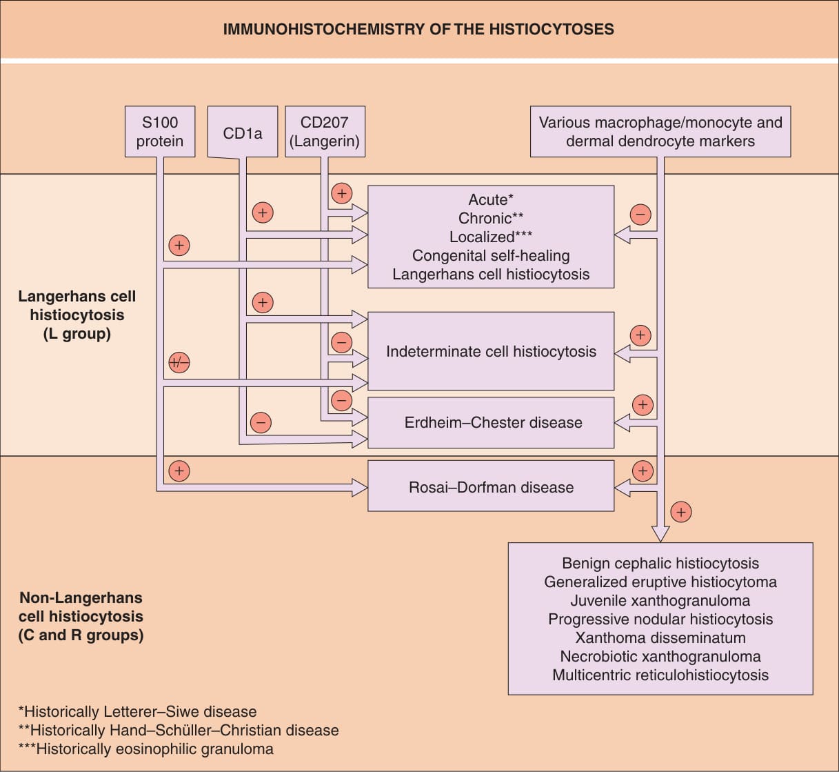
Fig. 91.4 Immunohistochemistry of the histio- cytoses. Examples of classic macrophage/ monocyte markers are CD68 and CD163; an example of a classic dermal dendrocyte marker is factor XIIIa.
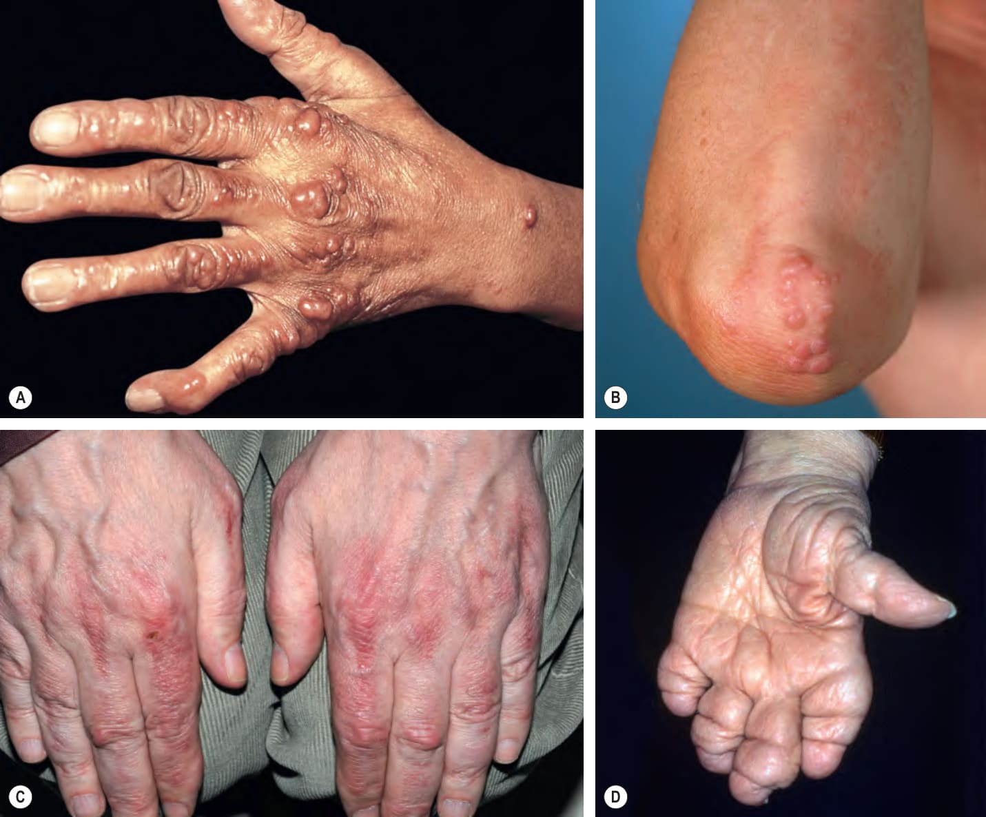
Fig. 91.17 Multicentric reticulohistiocytosis.A
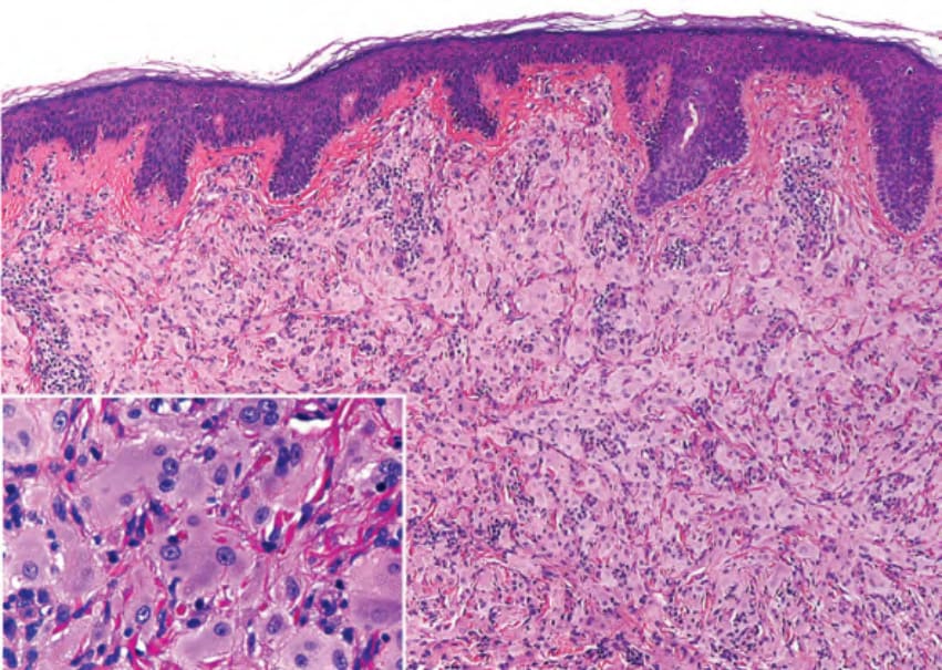
Fig. 91.18 Multicentric reticulohistiocytosis. Numerous mononucleated and multinucleated histiocytes within the dermis. The histiocytes have abundant eosinophilic, finely granular cytoplasm, creating a “ground glass” appearance (inset). Courtesy Luis Requena, MD.
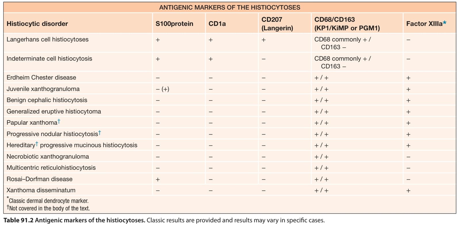
Table 91.2 Antigenic markers of the histiocytoses. Classic results are provided and results may vary in specific cases.