“R” GROUP: ROSAI–DORFMAN DISEASE
Synonyms: Sinus histiocytosis with massive lymphadenopathy Destombes–Rosai–Dorfman syndrome
Key features
Children and young adults are most commonly affected
Massive, painless, bilateral cervical lymphadenopathy is characteristic
Involvement of extranodal sites can be the sole manifestation of the disease, with a skin-limited form increasingly being recognized
In the systemic form, cutaneous lesions occur in a minority of patients (~10%), are usually multiple, and are clinically nonspecific
Fever and polyclonal hypergammaglobulinemia are often seen
In the lymph nodes and the skin, emperipolesis is a common histo- logic feature
A rare familial form due to mutations in SLC29A3 falls within the histiocytosis–lymphadenopathy plus syndrome
Introduction
Rosai–Dorfman disease (RDD) is an uncommon, often protracted, histiocytic proliferative disorder. It is characterized by massive, but painless, bilateral cervical lymphadenopathy, fever, anemia, elevated ESR/CRP, neutrophilia, and polyclonal hypergammaglobulinemia. Cutaneous lesions occur in a minority of patients with systemic disease, are usually multiple, and are clinically nonspecific. However, there is a form of RDD with only cutaneous involvement, and this skin-only form is currently classified within histiocytosis group C not group R.
History
Sinus histiocytosis with massive lymphadenopathy was described as a distinct entity by Rosai and Dorfman in 1969. However, the eponymous designation “Rosai–Dorfman disease” is more appropriate as some patients have extranodal lesions as the sole manifestation of the condition.
Epidemiology
A repository for cases of RDD, rather than a true registry, was available during 1980s–1990s. In 1990, the repository contained 423 patients, and by 1995, almost 600 cases had been reported. RDD has a widespread geographic distribution, although there is a higher incidence in the West Indies. The systemic form occurs most commonly in children and young adults and is more frequently seen in male patients. In 1990, an equal percentage of White and Black patients, 44% of each, were in the repository. On the other hand, the skin-limited form occurs primarily in adults and favors women.
Pathogenesis
Somatic genetic mutations that lead to activation of the MAPK pathway have been detected in patients with RDD (see Fig. 113.1). These genes include MAP2K1/MEK1 and NRAS, as well as MET, KRAS, and ARAF but rarely BRAF. Heterozygous germline mutations in FAS, which lead to defects in Fas-mediated apoptosis, lead to type IA autoimmune lymphoproliferative syndrome and may be associated with RDD (see Table 60.6). There is also a familial form of RDD due to germline mutations in SLC29A3, which encodes an equilibrative nucleoside transporter hENT3. These latter patients fall within the histiocytosis– lymphadenopathy plus syndrome which includes the H syndrome (see Table 70.2), pigmented hypertrichosis with insulin-dependent diabetes mellitus syndrome, and Faisalabad histiocytosis.
In addition, a viral pathogenesis has been postulated, including an association with HHV-6, cytomegalovirus, and Epstein–Barr virus. While HHV-6 sequences have been detected in involved lymph nodes, this finding is nonspecific given the presence of HHV-6 in many reactive and infectious disorders of lymphoid tissue. To date, no clear link has been proven.
Clinical features
While RDD is often an indolent, self-limited disease, a protracted course marked by remissions and exacerbations occurs in a number of patients. The characteristic clinical feature is massive, painless, bilateral cervical lymphadenopathy. However, any nodal site may be involved, unilateral involvement may occur, and an absence of lymph node involvement has also been reported. Fever is a frequent occurrence and most individuals have an elevated ESR/CRP and polyclonal hypergammaglobulinemia. A common finding is mild anemia, although severe anemia has been observed. Neutrophilia is occasionally seen. There are multiple reports of Hodgkin and non-Hodgkin lymphomas occurring in association with, or prior to, development of RDD. Of note, there was a report of a histiocytic sarcoma arising in a patient with autoimmune lymphoproliferative syndrome and associated RDD.
Immune disorders occur in ~15% of patients with RDD. Anti-red blood cell autoantibodies and joint disease are the most common findings. Patients with immunologic abnormalities have an unfavorable prognosis. In one analysis, 10 of 14 fatal cases were noted to have some form of immune dysfunction. Other unfavorable prognostic signs include disseminated nodal disease or involvement of the liver, kidney, or lower respiratory tract.
Over 40% of patients with RDD have at least one extranodal site of involvement. The most common extranodal sites are the skin and soft tissue, the eyelid and orbit, the upper respiratory tract, major salivary glands, CNS, and bone. However, virtually any organ can be involved. In patients with hearing loss, there is overlap with H syndrome (see above).
Cutaneous involvement occurs in ~10% of patients with the systemic form. The skin may be the only organ involved, with skin-limited disease increasingly being recognized. Cutaneous lesions are often multiple and appear as nonspecific red to red–brown or xanthomatous macules, papules, nodules, or plaques (Fig. 91.19); in the skin-limited form, there may be a single lesion. Panniculitis has also been reported. The eyelids and malar regions are frequent sites of involvement.
Pathology
The diagnosis requires the presence of histiocytic cells with abundant emperipolesis, i.e. engulfment of erythrocytes, plasma cells, and lymphocytes. The histiocytic cells stain positively for S100, CD68, CD14, and CD163; they are negative for CD1a and CD207 (Fig. 91.20; see Table 91.2). In addition, involved tissues usually contain abundant polyclonal plasma cells. Most cases demonstrate cyclin D1 and pERK expression, indicating activation of the MAPK pathway. Recently, OCT2-positive nuclear staining was observed in all cases of RDD (51/51), but this finding is nonspecific as a variable number of neoplastic cells show positive staining in other histiocytoses102a. Although IgG4-positive plasma cells are found within the infiltrate of RDD, the relationship between RDD and IgG4-related disease, if any, is yet unclear.
Differential diagnosis
Given the clinically nonspecific cutaneous lesions found in RDD, the differential diagnosis includes the other histiocytoses, sarcoidosis, granulomatous infectious diseases, and adnexal tumors. Of the histiocytoses, JXG in particular may resemble RDD as the former can also have large S100+ macrophages and, rarely, emperipolesis. The diagnosis of cutaneous RDD is based upon the combination of characteristic histologic and immunohistochemical findings.
The differential diagnosis of massive lymphadenopathy includes Hodgkin and non-Hodgkin lymphoma, chronic lymphocytic leukemia, metastases, infectious lymphadenopathies, and Kikuchi disease (a histiocytic necrotizing lymphadenitis that often affects young Asian women). RDD is differentiated by a typical combination of clinical and laboratory findings plus confirmatory histology and immunohistochemistry.
Treatment
Many lesions are asymptomatic and heal spontaneously, thus not requiring treatment. When treatment is indicated due to destructive lesions, disseminated disease or lesions causing physical compromise, radiotherapy, surgical excision, and/or systemic corticosteroids are usually employed, but there are no controlled trials to date. Based
upon case reports, improvement has been observed with thalidomide, imatinib, 2-chlorodeoxyadenosine, and clofarabine.
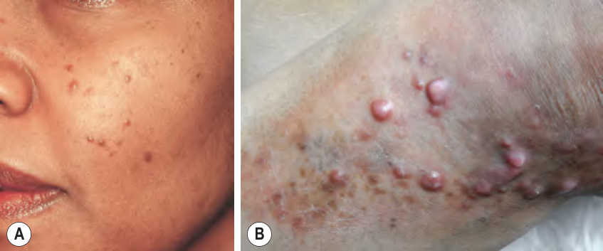
Fig. 91.19 Cutaneous Rosai–Dorfman disease.A Discrete, dome-shaped, brown papules on the cheek. B Multiple firm pink papulonodules arising within a yellow–brown plaque. B, Courtesy Boni Elewski, MD.
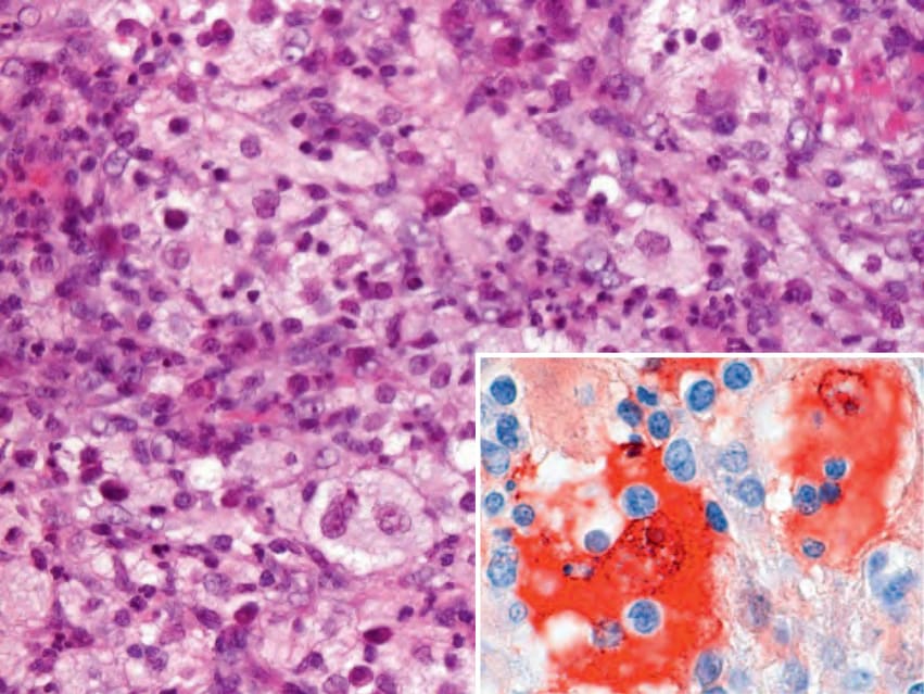
Fig. 91.20 Rosai–Dorfman disease. Large histiocytes with clear cytoplasm admixed with small lymphocytes and plasma cells. The large cells stain positively for S100 (inset), and the immunostain highlights lymphoid cells within the cytoplasm of large histiocytes (emperipolesis). Courtesy Lorenzo Cerroni, MD.
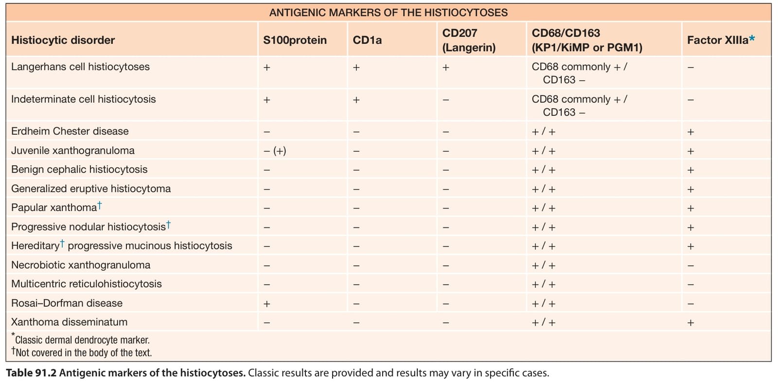
Table 91.2 Antigenic markers of the histiocytoses. Classic results are provided and results may vary in specific cases.