PATHOGENESIS
There is strong evidence to support the theory that the lipids found in the various xanthomas are the same as those in the circulation. The majority of plasma lipids are transported in complex structures known as lipoproteins. The basic structure of the lipoprotein allows the delivery of triglycerides and cholesterol to peripheral cells for their metabolic needs. This structure consists of a hydrophilic outer shell
William Trent Massengale Xanthomas 92
and a hydrophobic core. The outer shell consists of phospholipids, free cholesterol, and non-covalently linked specialized proteins known as apolipoproteins or apoproteins (apo). The inner core contains triglycerides and cholesterol esters.
Lipoproteins differ in their core lipid content. Triglycerides are the major core lipids in chylomicrons and very-low-density lipoproteins (VLDLs), while cholesterol esters dominate the core of low-density lipoproteins (LDLs), high-density lipoproteins (HDLs), and remnants of chylomicrons and VLDLs. The apoproteins found in the outer shell can also differ amongst the various lipoproteins (Table 92.1). These apoproteins serve several important functions, such as mediating the binding of lipoproteins to their respective receptors in target organs and activating enzymes involved in their metabolism.
There are two major pathways of lipoprotein synthesis (Fig. 92.1A). The exogenous pathway begins with dietary fat intake. Through the action of pancreatic lipase and bile acids, dietary triglycerides are degraded to fatty acids and monoglycerides. After absorption by the intestinal epithelium, the triglycerides are reformed and packaged with a small amount of cholesterol esters into the central core of a chylomicron. The outer shell of the chylomicron consists of phospholipids, free cholesterol, and several apoproteins, including B-48, E, A-I, A-II, and C-II.
Chylomicrons then enter the lymphatics and eventually the systemic circulation via the thoracic duct. Once in the circulation, hydrolysis of the core triglycerides occurs, releasing free fatty acids to the peripheral tissues. This is mediated through the action of the enzyme lipoprotein lipase that is bound to capillary endothelium. The activation of the lipoprotein lipase system is complex and involves not only hormones such as insulin, but also apoproteins such as C-II, located on the lipoprotein outer surface, and GPIHBP1, a protein expressed on endothelial cells that binds lipoprotein lipase and shuttles it to its site of action in the capillary lumen.
After hydrolysis of ~70% of the original triglyceride content, a chylomicron “remnant” exists. The central core now contains predominantly cholesterol ester that has been acquired from circulating HDL molecules. The chylomicron remnant is taken up by the liver via specialized high-affinity apo B-100/E receptors that recognize the apoproteins E or E on the remnant’s outer shell. Once in the liver, the remaining lipids enter hepatic storage and apoproteins such as B-48 are degraded.
Lipoprotein lipase (LPL) is activated by the C-II apoprotein.
The endogenous pathway begins with the hepatic formation of VLDL particles. The central core of the VLDL consists primarily of triglycerides, which are derived from circulating free fatty acids and hepatic triglyceride stores. Important apoproteins found on the outer shell include B-100, E, and C-II. In a fashion similar to the chylomicron, lipoprotein lipase mediates hydrolysis of the VLDL molecule, removing the majority of its triglyceride content, and its cholesterol esters are acquired from HDL molecules. Lipoprotein lipase activation requires the presence of apo C-II on the VLDL outer shell. After removal of the majority of the triglyceride content, the VLDL “remnant”, also known as an intermediate-density lipoprotein (IDL), can then be taken up by the liver via apo B-100/E receptors and degraded. IDLs that escape uptake by the hepatocyte are stripped of their remaining core triglycerides by extracellular hepatic lipases and enter the circulation as LDLs.
The LDL contains predominantly cholesterol ester in its central core and expresses B-100 on its surface. LDL delivers cholesterol ester to peripheral tissues, where it can be converted to free cholesterol. Cholesterol has several important functions within the body, including being an essential component of cell membrane bilayers. It is also important in the production of the myelin sheath of nerves, adrenal and gonadal steroidogenesis, and the production of bile acids. Hepatocytes play the major role in the catabolism of LDLs. Their uptake is mediated through the high-affinity apo B-100/E receptor found on the cell surface of the hepatocytes. Of note, the LDL receptors on hepatocytes are catabolized by PCSK9 (proprotein convertase subtilisin/kexin type 9). Free cholesterol in excess of metabolic needs is re-esterified for storage.
HDLs serve several important functions in cholesterol metabolism (see Fig. 92.1A). One of the primary functions of HDLs is the removal of cholesterol from the peripheral tissues. During this process, free cholesterol and phospholipids are transferred from the cell membranes of peripheral cells to the HDL molecules. The free cholesterol is then esterified by the enzyme lecithin:cholesterol acyltransferase, or LCAT. This enzyme requires the presence of the HDL apoprotein A-I. HDL molecules then transfer the cholesterol esters to other lipoproteins such as LDLs and remnants of chylomicrons or VLDLs for transportation back to the liver.
The liver plays the central role in the overall cholesterol economy. Hepatic intracellular cholesterol levels have a direct impact on the activity of HMG-CoA reductase, the rate-limiting enzyme of cholesterol synthesis, and on the expression of the high-affinity apo B-100/E receptor. When intracellular cholesterol levels are low, HMG-CoA reductase becomes activated and high-affinity apo B-100/E receptor expression increases. The increase in high-affinity receptors leads to increased uptake of cholesterol-containing lipoproteins such as chylomicron remnants, IDLs, and LDLs. This is followed by the lowering of plasma cholesterol levels. As discussed later, this mechanism will be the basis for many of the pharmacologic interventions aimed at lowering cholesterol levels.
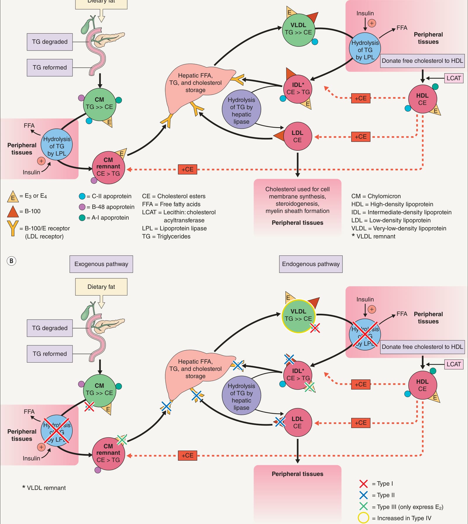
Fig. 92.1 Exogenous and endogenous pathways of lipoprotein synthesis (A) and sites of dysfunction that lead to the major forms of hyperlipidemia (B).
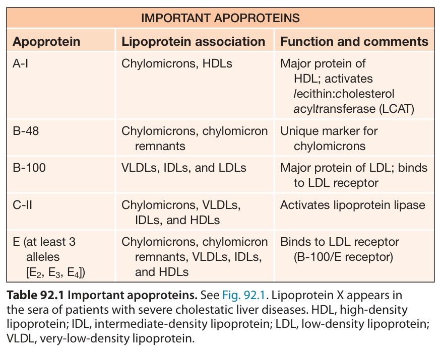
Table 92.1 Important apoproteins. See Fig. 92.1. Lipoprotein X appears in the sera of patients with severe cholestatic liver diseases. HDL, high-density lipoprotein; IDL, intermediate-density lipoprotein; LDL, low-density lipoprotein; VLDL, very-low-density lipoprotein.