CLINICAL FEATURES
Due to the complexity of cholesterol homeostasis, there are several possible ways in which hyperlipidemia may occur, from inherited disorders to metabolic diseases such as diabetes mellitus. Genetic mutations can affect important enzymes, receptors, or receptor ligands, with such defects leading to the overproduction of lipoproteins or the inhibition of their clearance (Fig. 92.1B). Each possible defect would lead to a different abnormal lipid profile.
In 1965, Lees and Frederickson published a system for classifying various disorders of lipid metabolism based upon the electrophoretic migration of the serum lipoproteins present. This system for phenotyping hyperlipoproteinemias is used today in a modified form (Table 92.2). In the next section, descriptions of underlying lipid disorders will reference not only the Frederickson classification system, but also the specific molecular defects when possible.
With the exception of the homozygous form of familial hypercholesterolemia (type II) and rare disorders such as sitosterolemia and biliary atresia, most cutaneous xanthomas do not appear until adulthood. Once the diagnosis is established, treatment of associated disorders (e.g. metabolic syndrome [see Table 53.5]) should reduce the incidence of potential systemic sequelae such as myocardial infarctions, cerebrovascular accidents, and hepatic steatosis.
Eruptive Xanthomas
Eruptive xanthomas appear as erythematous to yellow papules, approximately 1 to 5 mm in diameter (Fig. 92.2 & 92.3). They are usually distributed on the extensor surfaces of the extremities, buttocks, and hands. Early in their development, lesions may have an inflammatory halo (likely due to their triglyceride component), which may be accompanied by tenderness and pruritus. The Koebner phenomenon has been reported to occur with eruptive xanthomas.
Eruptive xanthomas can be seen in the setting of primary or secondary hypertriglyceridemia. Triglyceride levels in patients with eruptive xanthomas often exceed 3000 to 4000 mg/dl. In the Frederickson classification of hyperlipidemias, hypertriglyceridemia can be seen in type I (elevated chylomicrons), type IV (elevated VLDLs), and type V (elevated chylomicrons and VLDLs).
One reason for elevated triglyceride levels is failure to remove such lipids from the circulation (Fig. 92.4). Deficient activity of lipoprotein lipase will lead to accumulation of triglyceride-rich chylomicrons and VLDLs (see Fig. 92.1B). This can be related to either abnormalities in the enzyme itself, as in lipoprotein lipase deficiency (chylomicronemia syndrome), or in other controlling factors such as dysfunctional apoprotein C-II or impaired insulin activity. In addition to deficiency of GPIHBP1, autoantibodies against GPIHBP1 can also lead to hypertriglyceridemia.
Another reason for increased triglyceride levels is hepatic overproduction of triglyceride-rich lipoproteins via the endogenous pathway. In endogenous familial hypertriglyceridemia, a genetic defect exists that causes the liver to respond abnormally to dietary carbohydrates and insulin, with overproduction of hepatic VLDLs. The result is a Frederickson type IV pattern of hypertriglyceridemia. Secondary acquired defects in lipoprotein lipase activity, such as those due to diabetes mellitus, are not uncommon in these patients. With this second insult, the lipoprotein lipase system can become saturated and, as a result, no longer handles dietary lipids, leading to chylomicron elevations as well. This pattern is classified as a Frederickson type V phenotype.
Environmental factors and underlying diseases commonly exacerbate genetic defects of triglyceride metabolism, leading to worsening of the hypertriglyceridemia with eruptive xanthoma formation (see Fig. 92.4). Such factors include obesity, high caloric intake, diabetes mellitus, alcohol abuse, oral estrogen replacement, and systemic medications that can lead to hypertriglyceridemia (e.g. retinoids, protease inhibitors, olanzapine). The resultant pattern usually leads to a Frederickson type IV phenotype. Oral retinoid therapy, especially bexarotene, can elevate triglyceride levels through an elevation in hepatic VLDL secretion. With isotretinoin, this elevation seems to be more prevalent in genetically predisposed individuals and may signal an increased risk for future metabolic syndrome. Two of the five criteria for the clinical diagnosis of metabolic syndrome are lipid abnormalities – elevated triglycerides and reduced HDLs (see Table 53.5).
The treatment of eruptive xanthomas involves the identification and treatment of the underlying causes of the hypertriglyceridemia (see Fig. 92.4). Failure to recognize and treat the patient with hypertriglyceridemia could lead to complications such as acute pancreatitis. Pharmacologic and dietary lowering of the circulating triglycerides to reasonable levels will result in the prompt resolution of the eruptive lesions.
Tuberous/Tuberoeruptive Xanthomas
Tuberoeruptive and tuberous xanthomas are clinically and pathologically related and often described as being on a continuum. Tuberoeruptive xanthomas present as pink–yellow papules or nodules on extensor surfaces, especially the elbows and knees (Fig. 92.5). Tuberous lesions are noted to be larger than tuberoeruptive lesions and may exceed 3 cm in diameter (Fig. 92.6). Together, these lesions can be seen in hypercholesterolemic states such as dysbetalipoproteinemia (Frederickson type III) and familial hypercholesterolemia (Frederickson type II; see below). In contrast to eruptive xanthomas, tuberous xanthomas are usually slow to regress following institution of appropriate therapy.
Dysbetalipoproteinemia, or broad beta disease, is a genetic disorder of lipid metabolism that is usually inherited in an autosomal recessive fashion. It is caused by the presence of an isoform of apo E, primarily
apo E, that is a poor ligand for the high-affinity apo B-100/E receptor (see Fig. 92.1B). This results in the poor hepatic uptake of chylomicron and VLDL remnants. As a result, the serum levels of both triglycerides and cholesterol are elevated. The cutaneous lesions that are most characteristic of this disease are tuberous or tuberoeruptive xanthomas
(present in 80% of patients) and plane xanthomas of the palmar creases (xanthoma striatum palmare), which are present in two-thirds of patients (see below).
Tendinous Xanthomas
Tendinous xanthomas are firm, smooth, nodular lipid deposits that can affect the Achilles tendons (Fig. 92.7) or the extensor tendons of the hands (Fig. 92.8), knees, or elbows. The overlying skin is normal in appearance. Ultrasound can aid in the diagnosis of subtle lesions of the Achilles tendon by demonstrating hypoechoic nodules or an increase in the anteroposterior diameter of the tendon. The presence of tendinous xanthomas is almost always a clue to an underlying disorder of lipid metabolism. Lipid disorders that have been associated with this type of xanthoma include familial hypercholesterolemia, dysbetalipoproteinemia, and hyperlipidemia secondary to hypothyroidism (Table 92.3).
Tendinous xanthomas are most frequently seen in the setting of familial hypercholesterolemia. This disorder results from a deficiency of normal LDL receptors on cell membranes, which leads to the poor hepatic clearance of circulating LDLs and, therefore, elevated LDL cholesterol levels (Frederickson type II). This condition is inherited in an autosomal dominant fashion with a high degree of penetrance. Homozygotes can have LDL cholesterol levels of 800 to 1000 mg/dl with widespread atherosclerosis and the appearance of xanthomas during the first decade of life. Heterozygosity for the disorder is more common and is estimated to occur in 1 in 500 individuals in the US. The types of xanthomas seen in this disorder include tendinous, tuberous, tuberoeruptive, and plane (including xanthelasma). Because of its relatively
The yellowish hue is influenced by the degree of pigmentation of the skin. Note the clustering of some of the lesions.
common occurrence, patients with tendinous xanthomas are more likely to have the heterozygous form of the disease than the rare homozygous state. Plane xanthomas of the intertriginous web spaces of the fingers are thought to be pathognomonic for the homozygous condition (see Fig. 92.8).
There is a closely related disorder, known as familial defective apolipoprotein B-100. In this dominantly inherited genetic disorder, the LDL receptor is normal. However, there is decreased affinity of LDL for the LDL receptor because the mutation affects its ligand, apo B-100 (see
Fig. 92.1B). Patients may present with identical clinical findings as in familial hypercholesterolemia, although usually not as severe. From a therapeutic standpoint, distinguishing this disorder from dysfunction or accelerated degradation of the LDL receptor (see Table 92.2) will become more important in the future as treatments aimed at correcting LDL receptor function become commercially available (e.g. evinacumab)13a.
Rarely, tendinous xanthomas can develop in the absence of a lipoprotein disorder. Two examples are cerebrotendinous xanthomatosis and sitosterolemia. In cerebrotendinous xanthomatosis, an enzymatic
Note intertriginous plane xanthomas of the web spaces.
defect exists in the bile acid synthetic pathway, leading to the abnormal accumulation of an intermediate known as cholestanol. This intermediate is deposited in most tissues, including the brain, and can also form tendinous xanthomas. In sitosterolemia-1 and -2, an abnormal accumulation of plant sterols occurs, leading to tendinous and tuberous xanthoma formation.
Plane Xanthomas and Xanthelasma
Plane xanthomas appear as yellow to orange, non-inflammatory macules, papules, patches, and plaques. They can be circumscribed or diffuse. While anatomic locations vary, the site often serves as a clue to the particular underlying disease state. For example, intertriginous plane xanthomas may occur in body folds including the antecubital fossae (Fig. 92.9 & 92.10) or the web spaces of the fingers (see Fig. 92.8), where they are almost pathognomonic for homozygous familial hypercholesterolemia. Plane xanthomas of the palmar creases, or xanthoma striatum palmare, are nearly diagnostic for dysbetalipoproteinemia, especially when accompanied by tuberous xanthomas.
Xanthelasma, or xanthelasma palpebrarum, are commonly observed plane xanthomas of the eyelids (Fig. 92.11). Although the presence of xanthelasma warrants investigation for hyperlipidemia, the latter is present in only about one-half of the patients with these lesions. Younger patients or those with a strong family history of hyperlipidemia are more likely to have an underlying lipid disorder and should be appropriately screened.
Plane xanthomas of cholestasis may occur as a complication of diseases such as biliary atresia or primary biliary cholangitis (previously primary biliary cirrhosis). In these conditions, unesterified cholesterol begins to accumulate in the blood, leading to plane xanthoma formation. The lesions often begin as localized plaques on the hands and feet, but can become generalized.
Plane xanthomas can also occur in a normolipemic patient, where they may signal the presence of an underlying monoclonal gammopathy (Fig. 92.12), usually due to a plasma cell dyscrasia, but occasionally secondary to a lymphoproliferative disorder such as B cell lymphoma or Castleman disease (see Ch. 119). This type of xanthoma can also be seen in patients with chronic myelomonocytic leukemia. In gammopathy-associated plane xanthomas, monoclonal IgG is thought to bind to circulating LDL, rendering the antibody–LDL complex more susceptible to phagocytosis by macrophages. Favored locations include the neck, upper trunk, flexural folds, and periorbital region. In the setting of a monoclonal gammopathy, the coexistence of plane xanthomas and necrobiotic xanthogranuloma has been observed, suggesting a disease overlap. Of note, there is an entity known as hyperlipidemic myeloma in which xanthomas appear on the elbows and knees as well as within the palmar creases.
Verruciform Xanthomas
Verruciform xanthomas are asymptomatic, planar or verrucous, solitary plaques that are usually 1 to 2 cm in diameter. They occur primarily in the mouth (Fig. 92.13), but sometimes in periorificial, anogenital (including the scrotum), and acral sites. The latter are sometimes referred to as Vegas (verruciform genital-associated) and Vacas (verruciform acral-associated) xanthomas. These lesions may persist for years and there is usually no associated hyperlipidemia.
Single or multiple lesions are also seen in the setting of lymphedema, epidermolysis bullosa, pemphigus, discoid lupus erythematosus, lichen planus, and GVHD as well as within dermatofibromas and cutaneous lesions of the X-linked dominant disorder CHILD (congenital hemidysplasia with ichthyosiform erythroderma and limb defects) syndrome. The latter is due to mutations in NSDHL, which encodes
3β-hydroxysteroid dehydrogenase, an enzyme involved in cholesterol biosynthesis (see Ch. 57). In a molecular study of sporadic verruciform xanthomas, two of nine lesions were found to have a missense somatic mutation in exon 6 of NSDHL (only exons 4 and 6 were examined) which differed from the mutations seen in CHILD syndrome.
One proposal is that enzymatic dysfunction leads to an excess formation and accumulation of lipid storage droplets, followed by formation of lipid-laden dermal macrophages. Another hypothesis suggests that the foam cells result from damage to the epithelium
The large thin plaques have a yellow–orange color. The patient was found to have a monoclonal gammopathy. Courtesy Whitney High, MD, JD.
(e.g. from lichenoid dermatoses) and subsequent engulfing of lipids by macrophages within the dermal papillae, analogous to the amyloid deposits within the dermal papillae in lichen amyloidosis. The few foam cells beneath the epithelium may be subtle and easy to miss, in which case verruciform xanthomas may be confused histologically with warts and other papillomatous disorders. Surgery is generally curative.

Fig. 92.2 Eruptive xanthomas due to hypertriglyceridemia. In this patient the lesions favored the extensor surface of the lower extremities, in particular the knees.
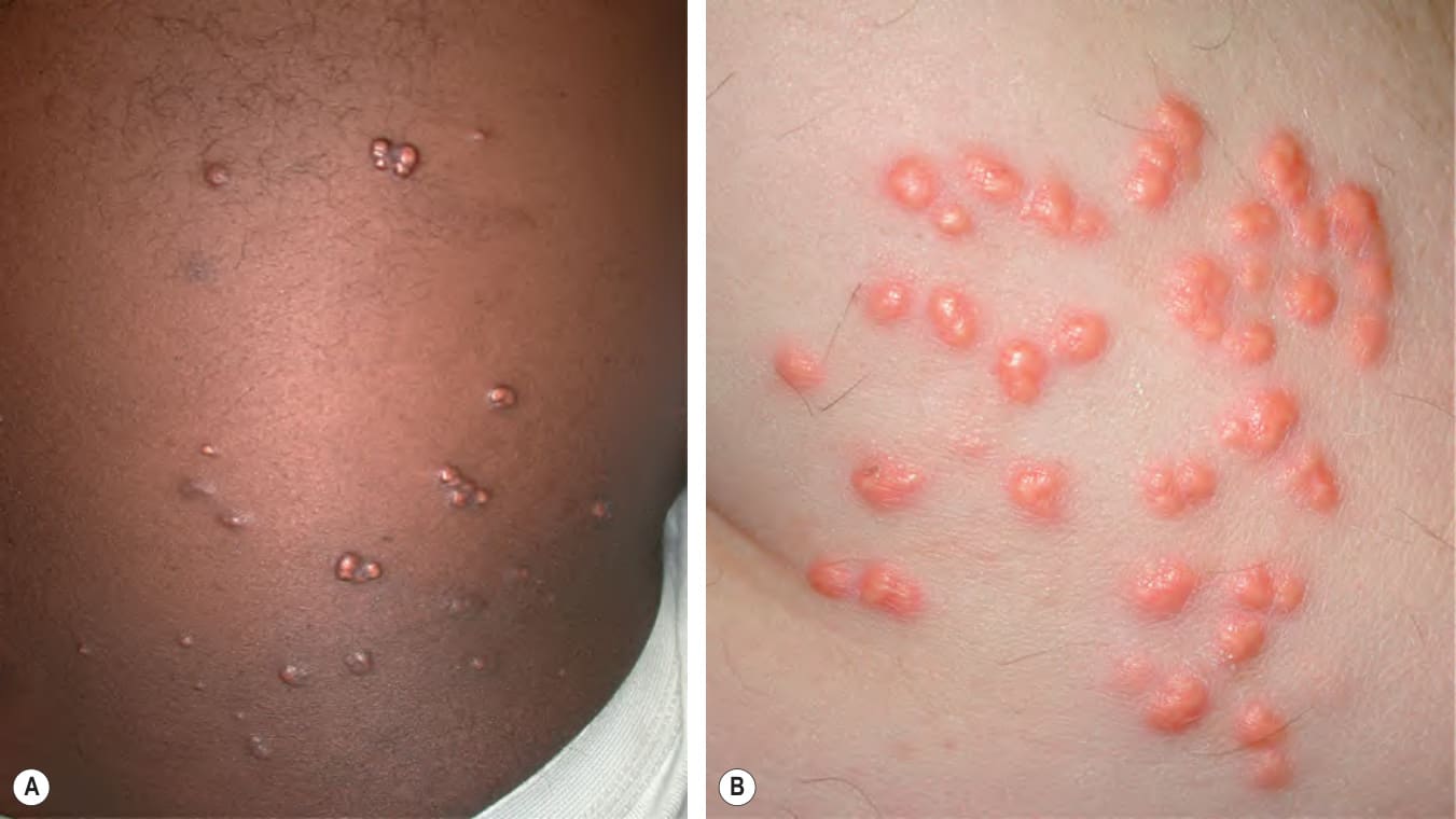
Fig. 92.3 Eruptive xanthomas.A, B
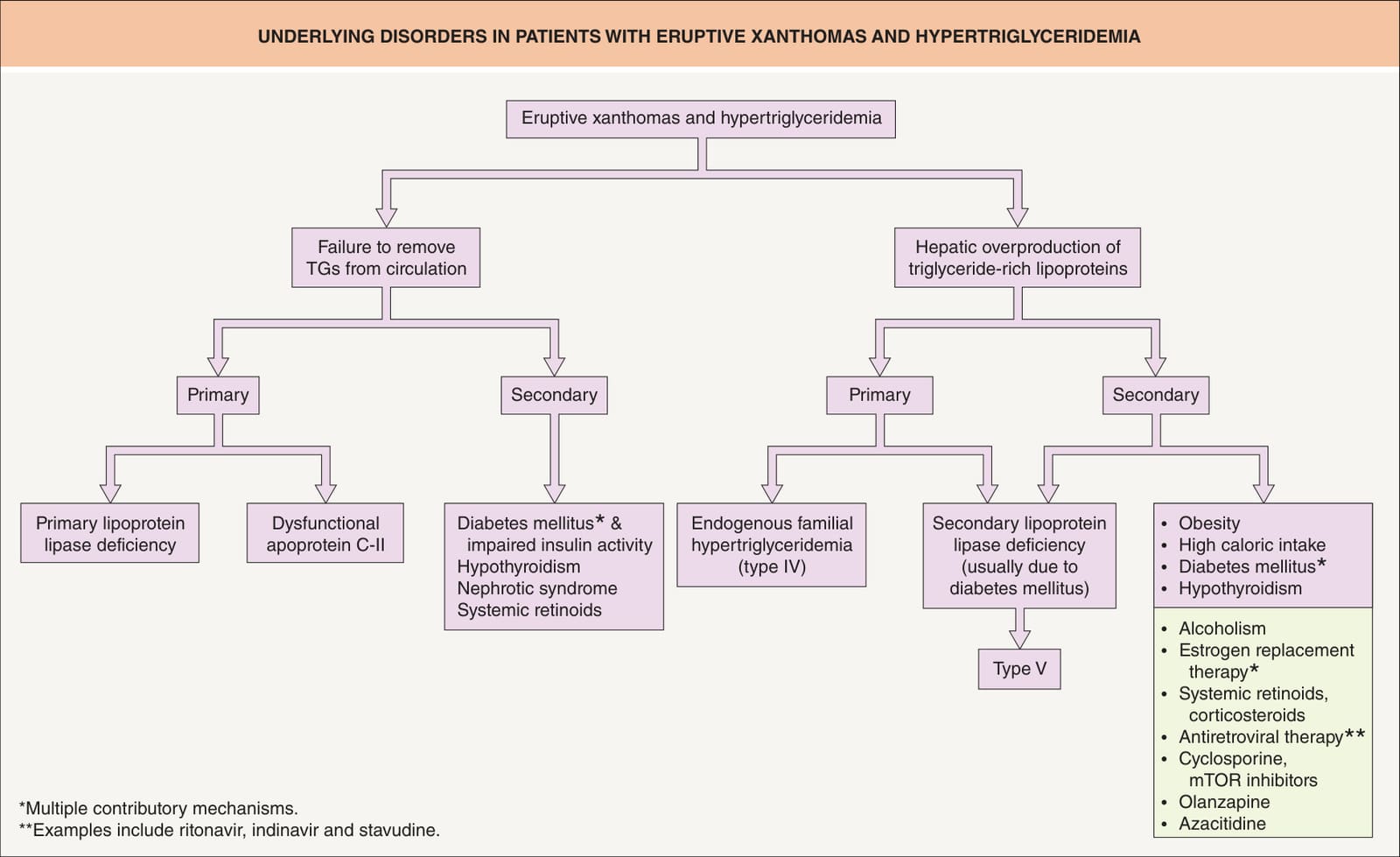
Fig. 92.4 Underlying disorders in patients with eruptive xanthomas and hypertriglyceridemia. mTOR, mammalian target of rapamycin; TGs, triglycerides.
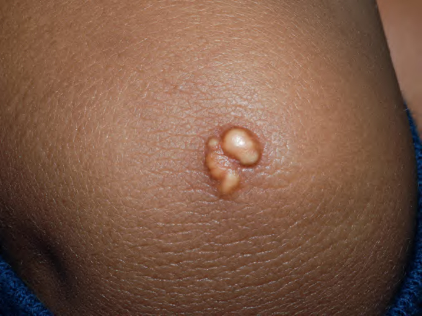
Fig. 92.5 Tuberoeruptive xanthomas on the elbow of a child with homozygous familial hypercholesterolemia. Note the yellowish hue. Courtesy Julie V. Schaffer, MD.
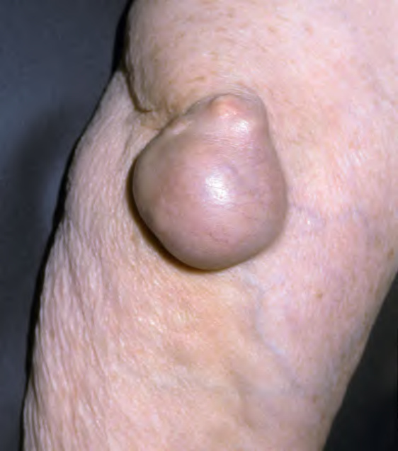
Fig. 92.6 Nodular tuberous xanthoma of the elbow.Courtesy Lorenzo Cerroni, MD.
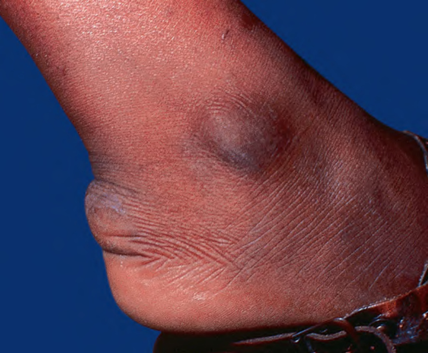
Fig. 92.7 Tendinous xanthoma. Linear swelling of the Achilles area, representing a tendinous xanthoma in a patient with dysbetalipoproteinemia.

Fig. 92.8 Tendinous xanthomas of the fingers in a patient with homozygous familial hypercholesterolemia.
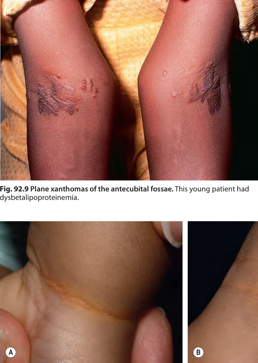
Fig. 92.9 Plane xanthomas of the antecubital fossae. This young patient had dysbetalipoproteinemia.
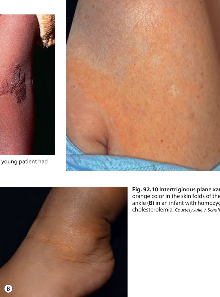
Fig. 92.10 Intertriginous plane xanthomas. Yellow– orange color in the skin folds of the wrist (A) and ankle (B) in an infant with homozygous familial hypercholesterolemia. Courtesy Julie V. Schaffer, MD.
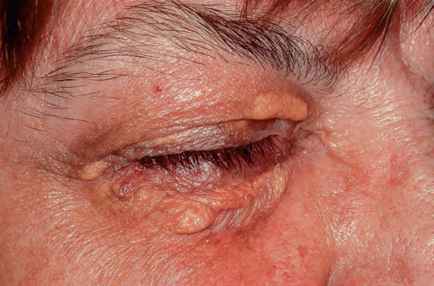
Fig. 92.11 Xanthelasma palpebrarum with typical yellowish hue.
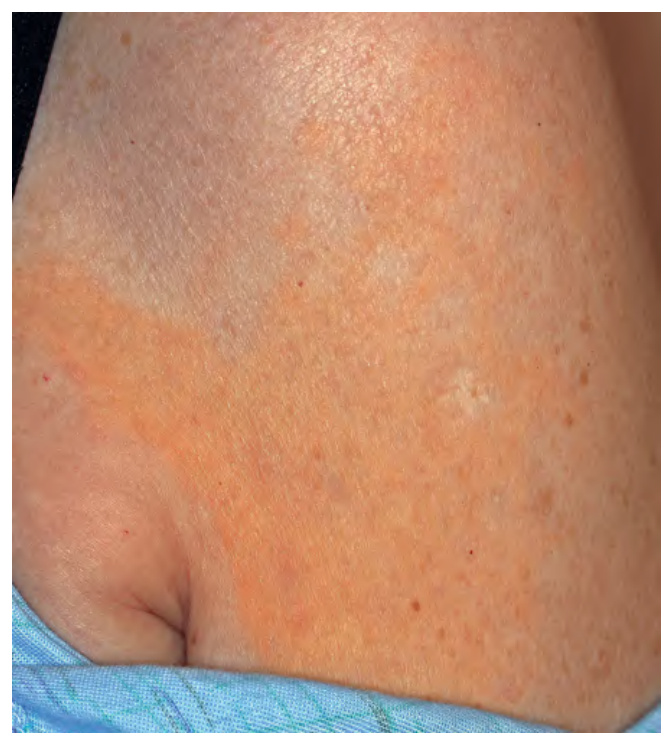
Fig. 92.12 Normolipemic plane xanthoma.
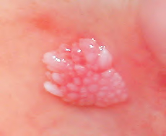
Fig. 92.13 Verruciform xanthoma of the oral mucosa.Courtesy Kishore Shetty, DDS.
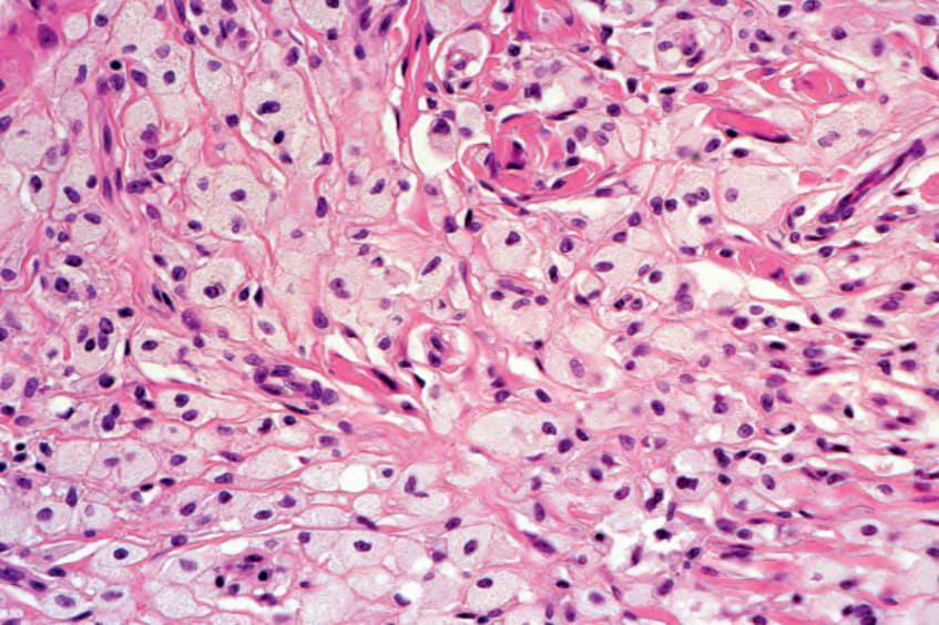
Fig. 92.14 Histology of a tuberous xanthoma. Foamy macrophages fill the dermis. Courtesy Lorenzo Cerroni, MD.
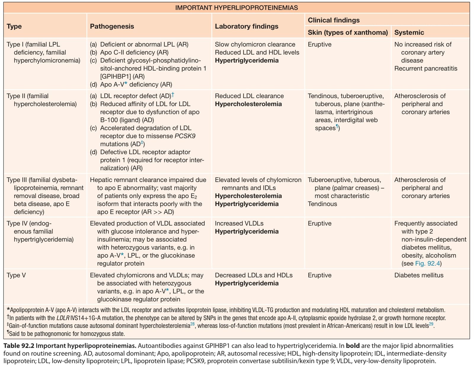
Table 92.2 Important hyperlipoproteinemias. Autoantibodies against GPIHBP1 can also lead to hypertriglyceridemia. In bold are the major lipid abnormalities found on routine screening. AD, autosomal dominant; Apo, apolipoprotein; AR, autosomal recessive; HDL, high-density lipoprotein; IDL, intermediate-density lipoprotein; LDL, low-density lipoprotein; LPL, lipoprotein lipase; PCSK9, proprotein convertase subtilisin/kexin type 9; VLDL, very-low-density lipoprotein.
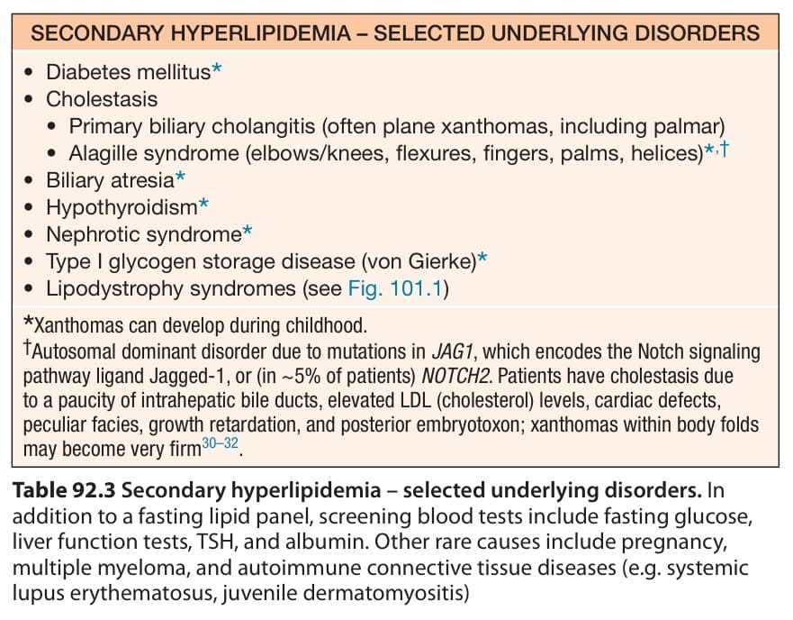
Table 92.3 Secondary hyperlipidemia – selected underlying disorders. In addition to a fasting lipid panel, screening blood tests include fasting glucose, liver function tests, TSH, and albumin. Other rare causes include pregnancy, multiple myeloma, and autoimmune connective tissue diseases (e.g. systemic lupus erythematosus, juvenile dermatomyositis)