PATHOLOGY
The characteristic histologic finding in xanthomas is the foam cell. Foam cells consist of macrophages that contain imbibed lipid within their cytoplasm (Fig. 92.14). All xanthomas contain dermal infiltrates of lipid, but they may vary in the degree of lipid content, the inflammatory infiltrate, the amount and location of the infiltrate, and the presence of extracellular lipid. The histologic appearance of xanthomas is somewhat altered by routine processing, as formalin fixation can remove deposits of lipid and leave artifactual clefting. Immunohistochemical staining for adipophilin can be performed to detect lipid accumulation. Adipophilin, also known as adipose differentiation-related protein, is a lipid droplet-associated protein.
Eruptive xanthomas contain lipid deposits in the reticular dermis. Of note, early in lesion development, foam cells are relatively small in number and size. The initial inflammatory infiltrate is mixed, containing both neutrophils and lymphocytes. As an increase in lipidization occurs, the appearance of the lesion becomes more typical of a xanthoma, but foam cells remain fewer in number compared with other types of xanthomas. Extracellular lipids are present in the dermis, which are seen as artifactual clefts filled with a wispy faint blue–gray material.
Tuberous xanthomas demonstrate large aggregates of foam cells in the dermis, often accompanied by fibrosis, but without a large number of inflammatory cells. Tendinous xanthomas have a similar histology, with foam cells that are even larger in size. Cholesterol esters are present in these lesions and can be seen with polarized microscopy.
Plane xanthomas such as xanthelasma can have a unique histologic appearance. The foam cells in this type of xanthoma are more super-ficial than in other types of xanthomas. Small aggregates of foam cells are often present in the superficial dermis. The lesions are non-inflammatory and have minimal fibrosis. With xanthelasma, there are often clues as to the location of the lesion, such as a thin epidermis, fine vellus hair follicles, and striated muscle fibers that are characteristic of the eyelid. In verruciform xanthomas, there is usually hyperkeratosis, acanthosis and papillomatosis, with foamy macrophages limited to the submucosa or dermal papillae.
A distinctive papular neutrophilic xanthoma, in which dermal foam cells are intermingled with neutrophils and prominent nuclear dust, has been described in HIV-infected patients with an IgA gammopathy. Xanthomatized neutrophilic dermatosis in the setting of myelodysplastic syndrome may represent a similar condition. Lastly, dermal xanthomatous cells observed in biopsies of cutaneous GVHD may be indicative of hepatic GVHD.
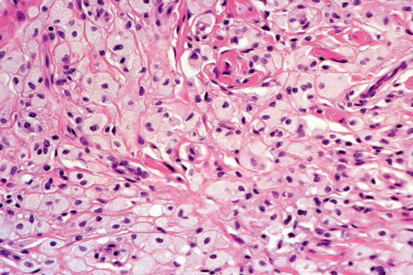
Fig. 92.14 Histology of a tuberous xanthoma. Foamy macrophages fill the dermis. Courtesy Lorenzo Cerroni, MD.
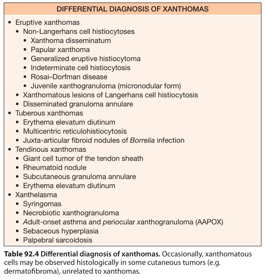
Table 92.4 Differential diagnosis of xanthomas. Occasionally, xanthomatous cells may be observed histologically in some cutaneous tumors (e.g. dermatofibroma), unrelated to xanthomas.