SARCOIDOSIS
Synonyms/subsets: Löfgren syndrome – erythema nodosum, hilar adenopathy, fever, arthritis Heerfordt syndrome – parotid gland enlargement, uveitis, fever, cranial nerve palsy Darier–Roussy disease – subcutaneous sarcoidosis
Key features
A systemic granulomatous disorder of unknown origin that most commonly involves the lungs
Cutaneous manifestations of sarcoidosis are seen in up to one-third of patients and may be the first clinical sign of the disease
Red–brown to violaceous papules and plaques appear most often on the face, in particular the nose, as well as the neck, upper back, extremities, and within scars and tattoos
Erythema nodosum is a nonspecific inflammatory skin finding that may be associated with an acute form of sarcoidosis which tends to remit
Histologically, sarcoidosis is characterized by non-caseating epithe- lioid granulomas, usually with a sparse or absent surrounding lymphocytic inflammation (i.e. “naked” granulomas)
Reactive granulomatous dermatitis (interstitial granulomatous dermatitis, palisaded neutrophilic and granulomatous dermatitis, and interstitial granulomatous drug eruption) represents an additional diagnostic consideration (see Table 93.4).
History
Sarcoidosis was described initially by Sir Jonathan Hutchinson in 1875 and cutaneous sarcoidosis (lupus pernio) by Besnier in 1889. Ten years later, Caesar Boeck coined the term “multiple benign sarkoid”, noting that the lesions resembled benign sarcomas.
Epidemiology
Sarcoidosis, which occurs in patients of all races and ages as well as both sexes, is characterized by a bimodal age distribution, with peaks between 25 and 35 years and then between 45 and 65 years in women. In the US, there is an increased incidence of sarcoidosis in African-Americans, ranging from 18 to 70 per 100 000. Middle-aged African-American women have the highest incidence (107 per 100 000), with African- American women having a lifetime risk of developing the disease of 2.7%. In comparison, the incidence in White individuals in the US ranges from 10 to 14 per 100 000. Sarcoidosis in African-Americans also tends to be more chronic and severe with a higher mortality rate than in other populations. Worldwide, the incidence of sarcoidosis is highest in Sweden (64 per 100 000) and the UK (20 per 100 000) and lowest in Spain and Japan (both 1.4 per 100 000). A greater number of patients with new-onset sarcoidosis are reported in the winter and spring.
Pathogenesis
Immune mechanisms
Sarcoidosis is a multisystem granulomatous disease characterized by hyperactivity of the cell-mediated immune system. Patients with a genetic susceptibility are exposed to a triggering antigen, leading to activation of macrophages and T cells, with subsequent granuloma formation. While classically considered a Th1-predominant immune response, the inflammatory cascade in sarcoidosis likely spans multiple pathways, including the innate immune system (via activation of pattern recognition receptors such as Toll-like receptors or NOD-like receptors) and the Th17 arm of the immune system. Specifically, CD4+ T helper cells differentiate into Th1 and Th17 effector cells that produce IFN-γ as well as Th17 cells that produce IL-17. This is triggered by antigen presentation by monocytes bearing MHC class II molecules, which initiates formation of epithelioid granulomas in a variety of tissue types. In the lung, an oligoclonal α/β T cell population has been described, suggesting that antigenic triggers of sarcoidosis favor a progressive accumulation and activation of specific T cell clones.
Increased production of Th1 cytokines, including interleukin (IL)-2, IL-12, IL-18, and interferon (IFN)-γ, as well as release of tumor necrosis factor (TNF) by macrophages and some CD8+ T cells, leads to persistent Th1 activity and persistent IFN-γ elevation. There is macrophage accumulation and hyperactivity, along with B cell stimulation and hypergammaglobulinemia. TNF and GM-CSF promote fusion of activated macrophages into the multinucleated cells seen within the granuloma.
Monocyte chemotactic factor (MCF), produced by activated T helper cells, attracts monocytes from the circulation into peripheral tissues. Compartmentalization of granuloma-forming T lymphocytes and monocytes within peripheral tissues leads to lymphopenia and decreased delayed-type hypersensitivity to common antigens (anergy), most pronounced during the initial stages of sarcoidosis. In addition, T regulatory cells may play a role in this anergic state. However, in some patients, there actually may be inadequate regulatory T cell function such that production of TNF or IFN-γ is not suppressed.
As macrophages differentiate into epithelioid cells and antigenpresenting cells, the phagolysosomal pathway plays an important role in sarcoidosis granuloma formation via promotion of mTORc1 (mechanistic target of rapamycin complex 1)/STAT3 signal transduction. In addition, IFN-γ stimulates the JAK (Janus kinase)/STAT pathway (see Fig. 128.11). With the discovery that this pathway plays a key role in
granuloma formation in a range of disorders including sarcoidosis, JAK inhibitors have become a therapeutic option.
The identity of the antigen responsible for the cascade of events leading to granuloma formation in patients with sarcoidosis remains uncertain. Some investigators have proposed an autoimmune etiology, while others have searched for exposures to inorganic dusts and particles (e.g. zirconium, talc) or an infectious cause. The latter is suggested by several observations, including the identification of microbial components such as mycobacterial DNA sequences in different sarcoidal tissues, the transmissibility of sarcoidal lesions via the Kveim reagent, and the observation that sarcoidal granulomas have developed in patients receiving organ transplants from donors with sarcoidosis. In a meta-analysis, mycobacterial nucleic acids were identified in ~25% of sarcoidal tissues. Also, the catalase-peroxidase (mKatG) protein from Mycobacterium tuberculosis has been proposed as a potential triggering antigen for granulomatous inflammation.
However, arguments against a purely infectious etiology include the observation that sarcoidosis patients do not develop fulminant infectious symptoms when placed on immunosuppressive agents. In addition, mycobacteria have not been cultured from sarcoidal tissues or from the sarcoid-like lesions that develop during antiretroviralinduced immune reconstitution. Occupational associations have been noted in healthcare workers, navy personnel on aircraft carriers, and firemen, with a marked increase in sarcoidosis-like illnesses in the first responders to the 9/11 World Trade Center terrorist attacks. In addition, exposure to beryllium can lead to berylliosis, a granulomatous disease that can closely mimic pulmonary sarcoidosis. Additional proposed triggers of sarcoidosis include viruses, Cutibacterium acnes, and an abnormal accumulation of serum amyloid A (SAA).
Over the past two decades, an increasing number of medications have been reported to induce cutaneous sarcoidosis and sarcoid-like granulomatous eruptions, including IFN, other systemic immunomodulators (e.g. TNF inhibitors > IL-17 inhibitors, IL-1 receptor antagonists), immune checkpoint inhibitors (e.g. ipilimumab, nivolumab, pembrolizumab), and targeted kinase inhibitors (e.g. vemurafenib), as has administration of the SARS-CoV-2 vaccine. Patients with lymphoma may also develop secondary sarcoidal reactions, most commonly in lymph nodes but occasionally in the skin.
With diascopy, a yellow– brown, “apple jelly” color is seen.
Sarcoidosis is a polygenic not a monogenic disease. A genetic component is supported by a twin study in which the monozygotic twins of patients with sarcoidosis had an 80-fold increased risk of having sarcoidosis. The largest case–control study to date demonstrated a familial relative risk of 4.7. The heritability of sarcoidosis has been estimated to range from 40% to 70%. The impact of HLA alleles varies significantly, depending upon disease subtype and racial group. Overall, HLA-DRB101 and DRB104 protect against sarcoidosis in some populations, while DRB103, DRB111, DRB112, DRB114, and DRB115 appear to confer risk; HLA-B8/DR3 may be associated with the development of Löfgren syndrome (see below). In African-Americans, HLA-DQB1 alleles may play a role, and patients with DRB103 may have a better prognosis. Specific susceptibility genes include TNF (variants associated with Löfgren syndrome) and IL23R (variants confer risk for Crohn disease and sarcoidosis). Whole genome sequencing of 41 members of a family with a high rate of sarcoidosis found variants within 34 candidate genes, one of which was JAK2 (see above).
While the etiologic agent clearly remains elusive, investigation of the immunopathogenesis of the disease (see above) has been aided by examining the cutaneous reaction produced by injection of a tissue suspension prepared from sarcoidal spleen. The suspension (Kveim– Siltzbach antigen) causes characteristic non-caseating granuloma formation in the skin of patients with sarcoidosis. Nowadays, however, this skin test is rarely performed.
Clinical Features
Up to a third of patients with systemic sarcoidosis develop skin lesions, which may be the first or only clinical manifestation of the disease.
Although cutaneous sarcoidosis most commonly presents as papules and plaques, often red–brown to violaceous in color (Figs. 93.2A–D), the skin manifestations are protean (Table 93.2). Sarcoidal lesions are fairly symmetric in distribution and favor the nose, especially the alae, and the periocular and perioral regions of the face, followed by the neck, upper trunk, and extremities. Involvement at sites of prior injuries, e.g. scars, tattoos, is a characteristic finding (Fig. 93.2E). Unusual variants include hypopigmented, ichthyosiform, micropapular, psoriasiform, and ulcerative (Fig. 93.3A,B; see Table 93.2).
Although many lesions are red–brown to violaceous in color, they can vary from yellow–brown to erythematous, especially in lightly pigmented skin. Upon diascopy, in which pressure induces blanching, the lesions are said to have the color of “apple jelly” (Fig. 93.4); this finding is usually easier to appreciate in lightly pigmented skin. Individual plaques can develop central clearing leading to an annular configuration or they can contain prominent telangiectasias (angiolupoid sarcoidosis). Dermoscopic features include small translucent yellow to orange globules, linear vessels, and central scar-like areas.
Additional variants of sarcoidosis of relevance to dermatologists include Löfgren syndrome, Darier–Roussy disease, and lupus pernio. Patients with Löfgren syndrome present acutely with fever, arthritis, hilar adenopathy, and erythema nodosum. They often require supportive measures and occasionally systemic corticosteroids, with spontaneous resolution occurring over 1–2 years. Patients with Darier– Roussy sarcoidosis present with painless, firm, subcutaneous nodules or plaques without epidermal changes. This variant represents sarcoidosis limited to the panniculus and, like other forms of cutaneous sarcoidosis, is often associated with systemic sarcoidosis. Lupus pernio is characterized by papulonodules and plaques, which are often on the nose and cheeks and have a violaceous color, sometimes along with scale;
there may be a beaded appearance along the nasal rim (see Fig. 93.3C). Recognition of lupus pernio is important because of its association with chronic sarcoidosis of the lungs (~75% of patients) and of the upper respiratory tract (~50% of patients), as well as its recalcitrant nature that often requires more aggressive therapy.
The most common and important nonspecific cutaneous manifestation of sarcoidosis is erythema nodosum (Fig. 93.5). In addition to its association with Löfgren syndrome, the presence of erythema nodosum portends a subacute, transient course of sarcoidosis that usually resolves spontaneously and typically does not require chronic immunosuppressive therapy. In general, there are no additional cutaneous manifestations.
Nail changes can be seen in sarcoidosis, including clubbing, subungual hyperkeratosis, and onycholysis. Oral sarcoidosis may affect the soft mucosa, gingival tissue, tongue, hard palate, and major salivary glands. Heerfordt syndrome (uveoparotid fever) includes parotid gland enlargement, uveitis, fever, and cranial nerve palsies, usually of the facial nerve.
Systemic manifestations of sarcoidosis are also protean. Lung disease occurs in ~90% of patients, ranging from alveolitis to granulomatous infiltration of the alveoli, blood vessels, bronchioles, pleura, and fibrous septa. The end stage of pulmonary sarcoidosis is fibrosis with bronchiolectasis and “honeycombing” of the lung parenchyma. Hilar and/or paratracheal lymphadenopathy, which is usually asymptomatic, occurs in 90% of patients. Additional systemic manifestations are outlined in Table 93.3.
The patient had received both ipilimumab and nivolumab. A search for internal involvement, e.g. lung, lymph nodes, is warranted. Courtesy Department of Dermatology, Medical University of Graz.
Childhood sarcoidosis is rare, and it usually presents with a triad of arthritis, uveitis, and cutaneous lesions, along with constitutional symptoms. Peripheral lymphadenopathy is frequently present. If sarcoidosis is being considered in a child, it is important to exclude Blau syndrome (see Ch. 45).
Pathology
The histopathologic hallmark of sarcoidosis is the presence of super-ficial and deep dermal epithelioid cell granulomas devoid of prominent infiltrates of lymphocytes or plasma cells (“naked tubercles”) (Fig. 93.6). Central caseation is usually absent, although fibrinoid deposition may be observed in up to 10% of cases. Multinucleated histiocytes (“giant cells”) are usually of the Langhans type, with nuclei arranged in a peripheral arc or circular fashion. The giant cells may contain eosinophilic stellate inclusions known as asteroid bodies (Fig. 93.7) or rounded laminated basophilic inclusions known as Schaumann bodies, although neither is specific or required for the diagnosis. Asteroid bodies represent engulfed collagen, whereas Schaumann bodies likely represent degenerating lysosomes. Notably, up to 25% of biopsies of sarcoidosis contain polarizable material; therefore its presence does not exclude the diagnosis. Rarely, the granulomatous infiltrate is arranged in a superficial (“lichenoid”) band. In vulvar sarcoidosis, transepidermal elimination of the granulomas may be seen.
A range exists within the histologic spectrum of sarcoidosis, from the characteristic tubercles with minimal or no surrounding lymphocytic inflammation to unusual cases with dense lymphocytic and plasmacytic infiltrates around and within the nodular histiocytic aggregates. Occasionally, these aggregates may extend into the subcutaneous fat, producing the clinical features of Darier–Roussy sarcoidosis.
Diagnosis and Differential Diagnosis
Sarcoidosis is a diagnosis of exclusion, both clinically and histologically. In order to establish the diagnosis, a supportive clinical history must be accompanied by the histologic presence of non-caseating granulomas in at least one organ system. Details of systemic manifestations are outlined in Table 93.3. An algorithm for initial and longitudinal evaluation has been proposed.
Serologically, elevated anti-nuclear antibody titers occur in ~30% of patients. The serum angiotensin-converting enzyme (ACE) level is elevated in ~60% of patients but is nonspecific for sarcoidosis; it has a false-positive incidence of 10%, making it a more useful test for monitoring disease progression in select patients where it is elevated, rather than for establishing the diagnosis. An ACE level >2–3 times the upper limit of normal is more suggestive of sarcoidosis but its clinical utility may still be limited. In sum, most sarcoidosis experts do not routinely order ACE levels.
Like syphilis, sarcoidosis is a great mimicker, and the clinical differential diagnosis depends upon the type of presenting clinical lesions. For example, a list of other disorders, in addition to sarcoidosis, that can present with annular lesions is provided in Table 19.1. Papules, nodules, and plaques may be nonspecific clinically, dictating a biopsy, and then the differential diagnosis rests upon the histopathologic findings. It is important to exclude the possibility of drug-induced cutaneous sarcoidosis (e.g. IFN-α for hepatitis C viral infection [use declining], TNF inhibitors, immune checkpoint inhibitors) (Fig. 93.8).
The histologic differential diagnosis is broad, and includes multiple infections that lead to granulomatous inflammation. Special stains for acid-fast and fungal organisms should be obtained. When clinically appropriate, tissue culture should be performed. Increasingly, PCR is being utilized to exclude infections, including mycobacterial. Both tuberculoid leprosy and lupus vulgaris are in the differential diagnosis and for the latter, a QuantiFERON®-TB Gold test may provide additional information. It is especially important to exclude infectious etiologies before initiating immunosuppressive agents.
Other histologic mimics include foreign body reactions to zirconium, beryllium, silica, tattoo ink, or soft tissue fillers. Special laboratory techniques, including histochemical, microincineration or spectrophotometric examinations, may be required to identify the causative agents (see Table 94.3). Specimens should be polarized to exclude birefringent foreign material as a causative agent, although up to 25% of known sarcoidal granulomas will have foreign material present within them. This finding is particularly common in lesions from the elbows and knees. Therefore, the dermatologic diagnoses of foreign body granuloma and sarcoidosis are not mutually exclusive.
In addition, granulomatous mycosis fungoides, Hodgkin disease, granulomatous rosacea, cutaneous Crohn disease, Blau syndrome (see Ch. 45), cheilitis granulomatosa, and the sarcoidal reaction to an underlying lymphoma can have histologic appearances similar to sarcoidosis. The histologic differences between sarcoidosis, granuloma annulare, annular elastolytic giant cell granuloma (AEGCG), necrobiosis lipoidica, rheumatoid nodule, and the reactive granulomatous dermatitides are outlined in Table 93.4.
Treatment
Corticosteroids, in either topical, intralesional, or systemic form, are the mainstay of therapy for systemic sarcoidosis (Table 93.5). Therapy is guided by organ involvement, disease severity and progression, and impact on patient quality of life. The typical oral prednisone dose for systemic disease is 0.5–1 mg/kg/day for 4–6 weeks, followed by a slow taper over months to years as dictated by the pulmonary disease, sarcoidosis of the upper respiratory tract, ocular disease, or other internal manifestations. The goal is alternate-day prednisone at the lowest possible dose that will maintain a remission. Cutaneous sarcoidosis may respond from the onset to lower doses of prednisone as well as every-other-day regimens. For limited cutaneous disease, topical or intralesional corticosteroids may suffice.
For treatment of more widespread cutaneous disease, alternative nonsteroidal systemic medications include hydroxychloroquine (200–400 mg/day), chloroquine (250–500 mg/day), minocycline or doxycycline (200 mg/day), and methotrexate (10–25 mg weekly). The use of thalidomide has declined, given recent negative studies, associated toxicities, and more effective alternative therapies. Improvement of both systemic and cutaneous sarcoidosis has been observed with TNF inhibitors, including infliximab and adalimumab. These agents may be particularly effective for chronic, recalcitrant sarcoidosis (e.g. lupus pernio), but notably TNF inhibitors occasionally can trigger sarcoidosis (most commonly etanercept). In selected patients, pentoxifylline, retinoids, and apremilast have been reported to be helpful. While additional immunosuppressives (e.g. leflunomide, mycophenolate mofetil, azathioprine) are more often used for extracutaneous sarcoidosis, they may have a role in some patients. A treatment algorithm, based upon evidence-based methodology, has been proposed. More recently, sustained responses have been observed with oral JAK inhibitors such as tofacitinib.
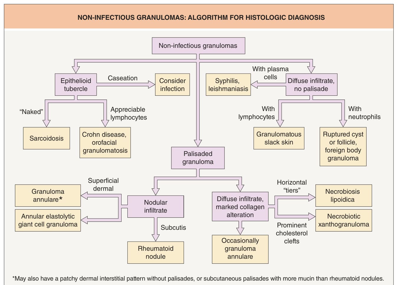
Fig. 93.1 Non-infectious granulomas: algorithm for histologic diagnosis.
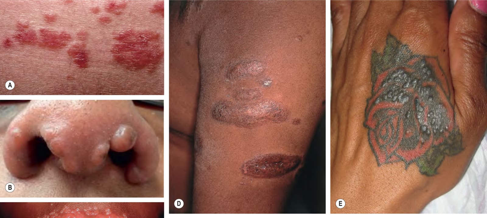
Fig. 93.2 Cutaneous sarcoidosis – papules and plaques.A Cutaneous sarcoidosis usually consists of papules and plaques with a typical reddish-brown to violet–brown color. B, C Papular lesions often favor the nose, lips, and perioral region. D Hyperpigmented plaques, some of which have scale. E Papules of cutaneous sarcoidosis arising within a tattoo; the differential diagnosis includes foreign body reaction. B, Courtesy Jonathan Leventhal, MD.
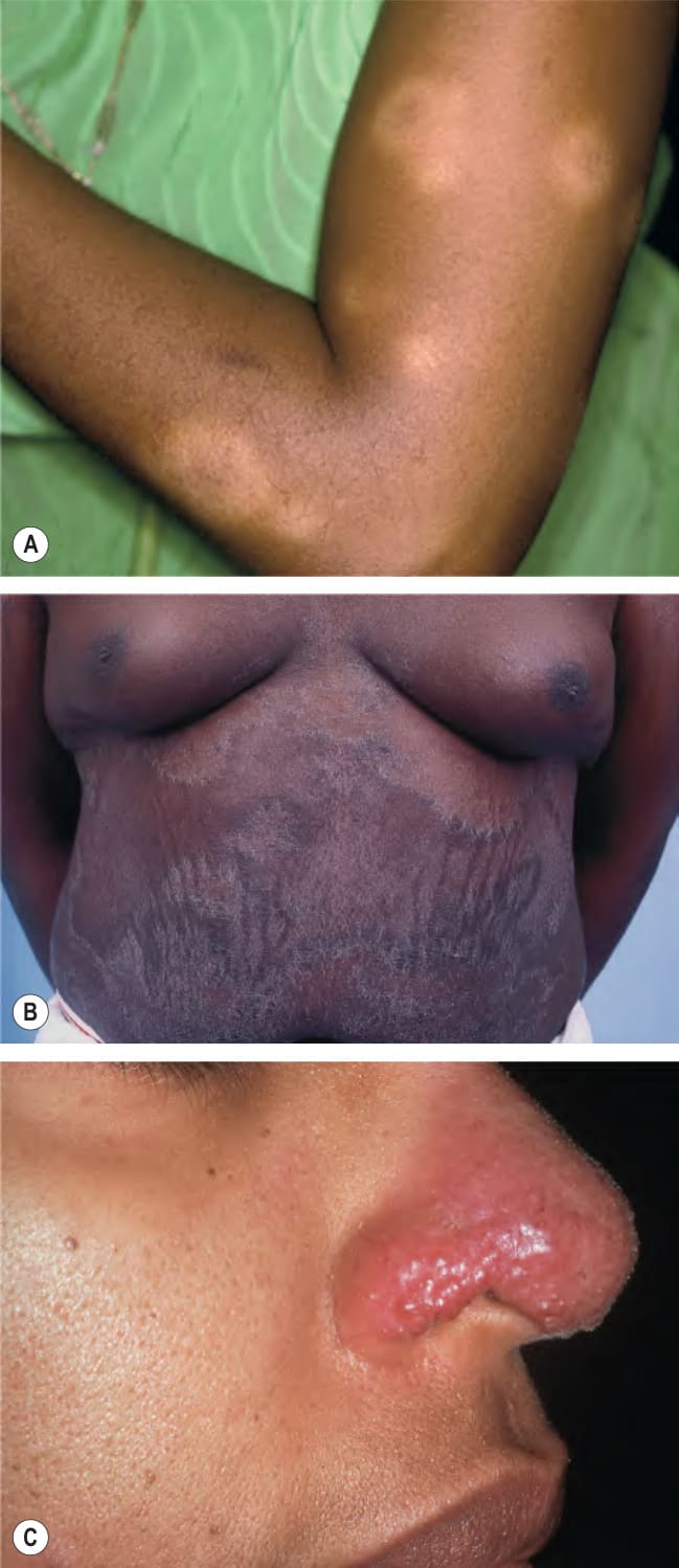
Fig. 93.3 Cutaneous sarcoidosis – clinical variants.A The hypopigmented variant is more noticeable in individuals with darkly pigmented skin. B Ichthyosiform presentation with obvious scale. C Coalescing violaceous papules on the nose in lupus pernio; note the notching of the nasal rim. A, Courtesy Louis A. Fragola, Jr, MD; B, Courtesy Jean L. Bolognia, MD.
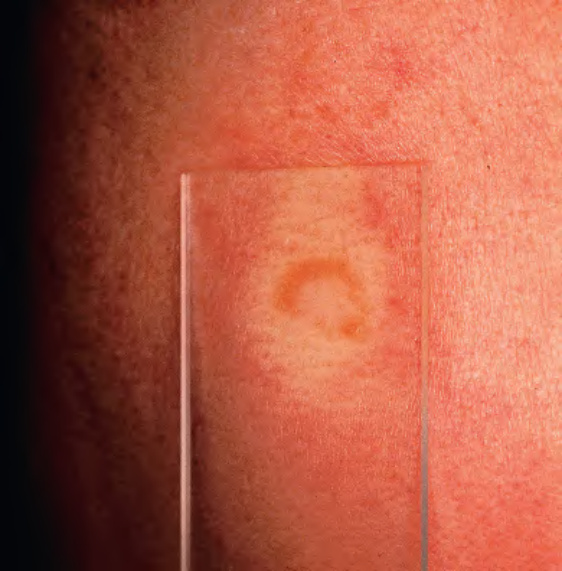
Fig. 93.4 Cutaneous sarcoidosis – diascopy.
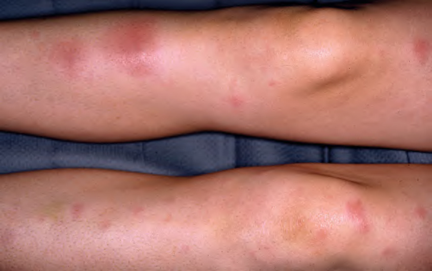
Fig. 93.5 Erythema nodosum in a patient with sarcoidosis. These patients often have hilar lymphadenopathy. Löfgren syndrome consists of erythema nodosum, hilar lymphadenopathy, fever, and arthritis. Courtesy Louis A. Fragola, Jr, MD.
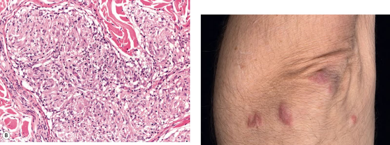
Fig. 93.6 Cutaneous sarcoidosis – histopathologic features.A Nodular aggregates of epithelioid histiocytes forming tubercles filling the dermis. B A sarcoidal tubercle with a sparse admixture of lymphocytes (“naked tubercle”). Courtesy Lorenzo Cerroni, MD.
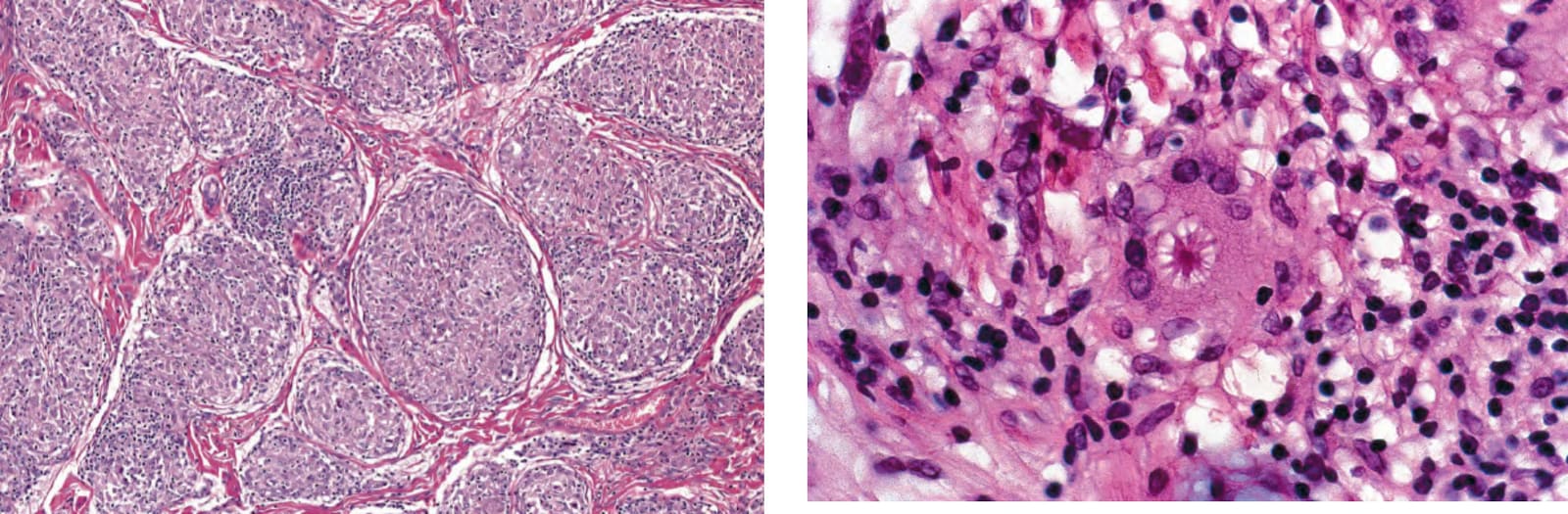
Fig. 93.7 Cutaneous sarcoidosis – histopathologic features. An asteroid body within the cytoplasm of a multinucleated epithelioid histiocyte (“giant cell”).
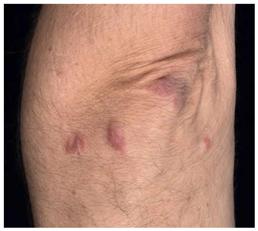
Fig. 93.8 Immune checkpoint inhibitor-induced cutaneous sarcoidosis.
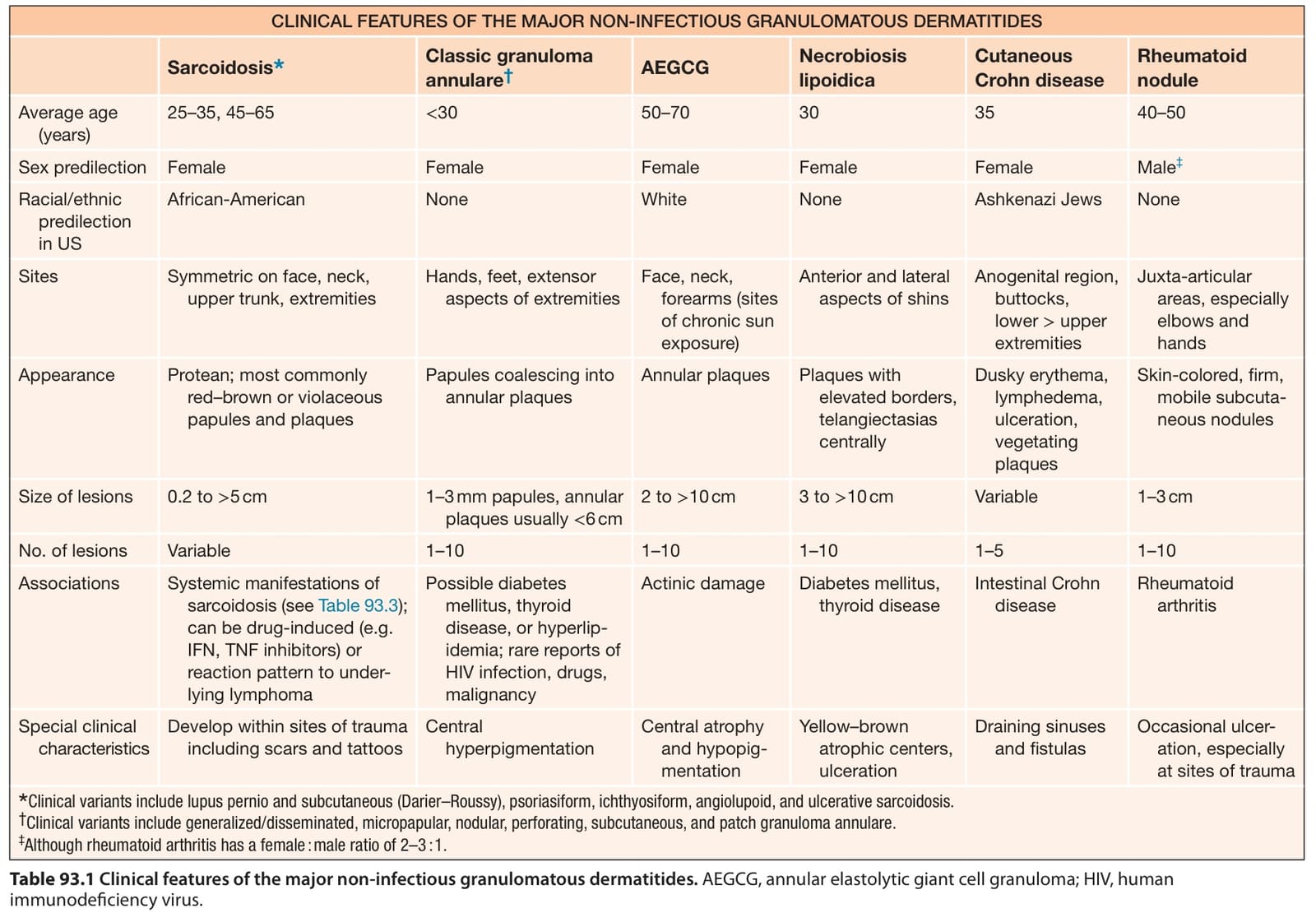
Table 93.1 Clinical features of the major non-infectious granulomatous dermatitides. AEGCG, annular elastolytic giant cell granuloma; HIV, human immunodeficiency virus.
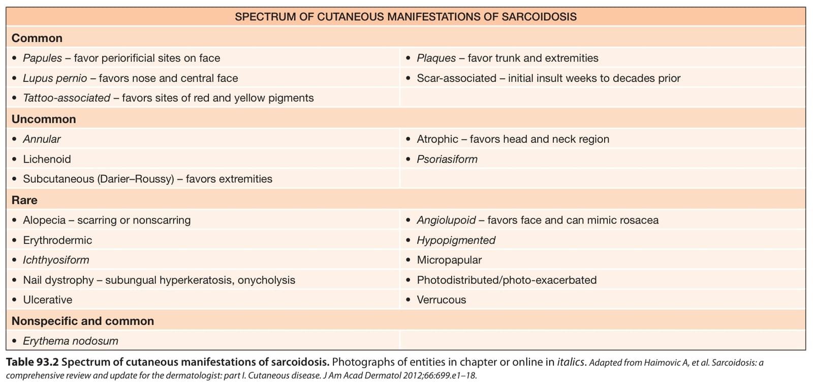
Table 93.2 Spectrum of cutaneous manifestations of sarcoidosis. Photographs of entities in chapter or online in italics. Adapted from Haimovic A, et al. Sarcoidosis: a comprehensive review and update for the dermatologist: part I. Cutaneous disease. J Am Acad Dermatol 2012;66:699.e1–18.
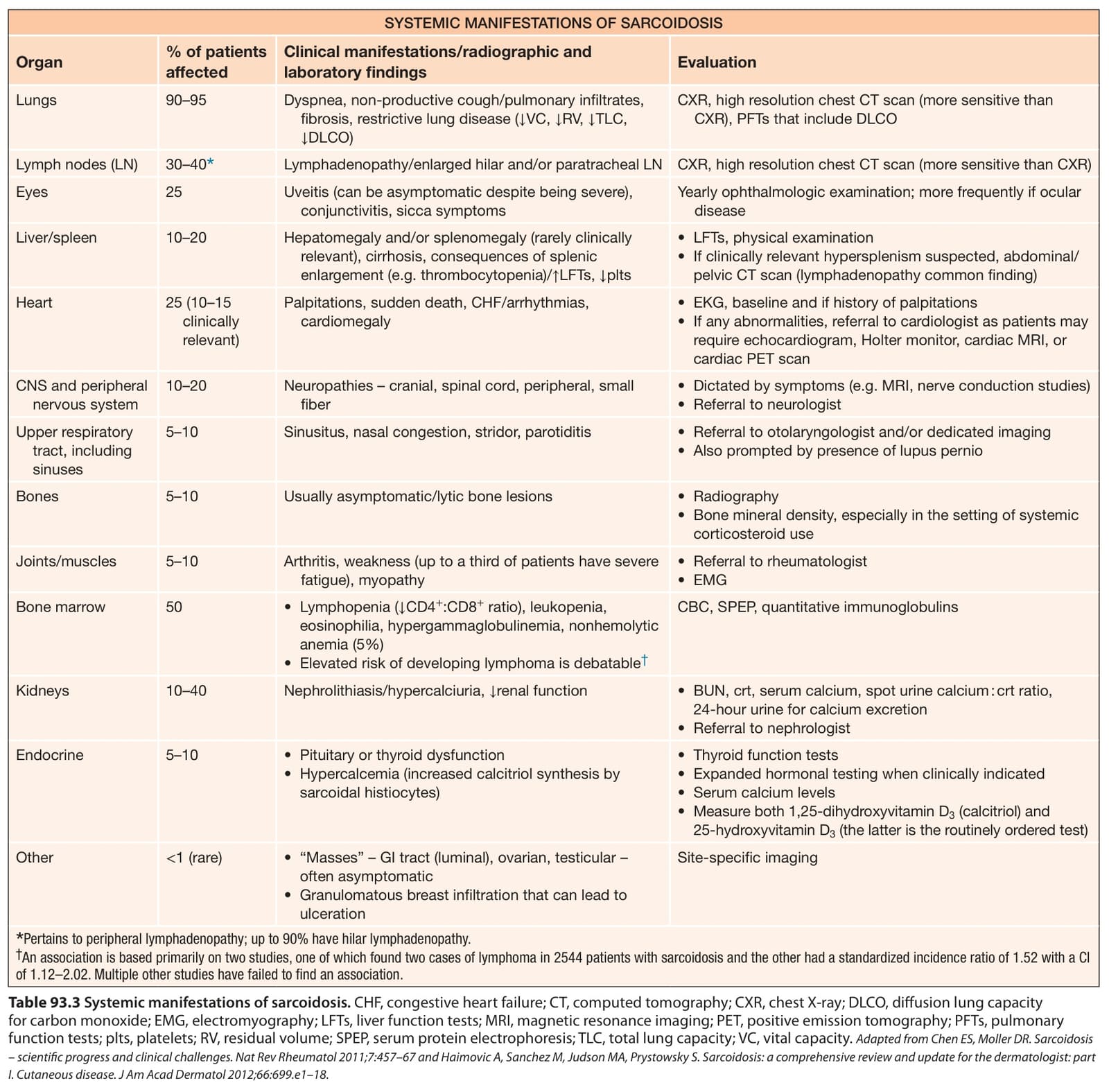
Table 93.3 Systemic manifestations of sarcoidosis. CHF, congestive heart failure; CT, computed tomography; CXR, chest X-ray; DLCO, diffusion lung capacity for carbon monoxide; EMG, electromyography; LFTs, liver function tests; MRI, magnetic resonance imaging; PET, positive emission tomography; PFTs, pulmonary function tests; plts, platelets; RV, residual volume; SPEP, serum protein electrophoresis; TLC, total lung capacity; VC, vital capacity. Adapted from Chen ES, Moller DR. Sarcoidosis – scientific progress and clinical challenges. Nat Rev Rheumatol 2011;7:457–67 and Haimovic A, Sanchez M, Judson MA, Prystowsky S. Sarcoidosis: a comprehensive review and update for the dermatologist: part I. Cutaneous disease. J Am Acad Dermatol 2012;66:699.e1–18.
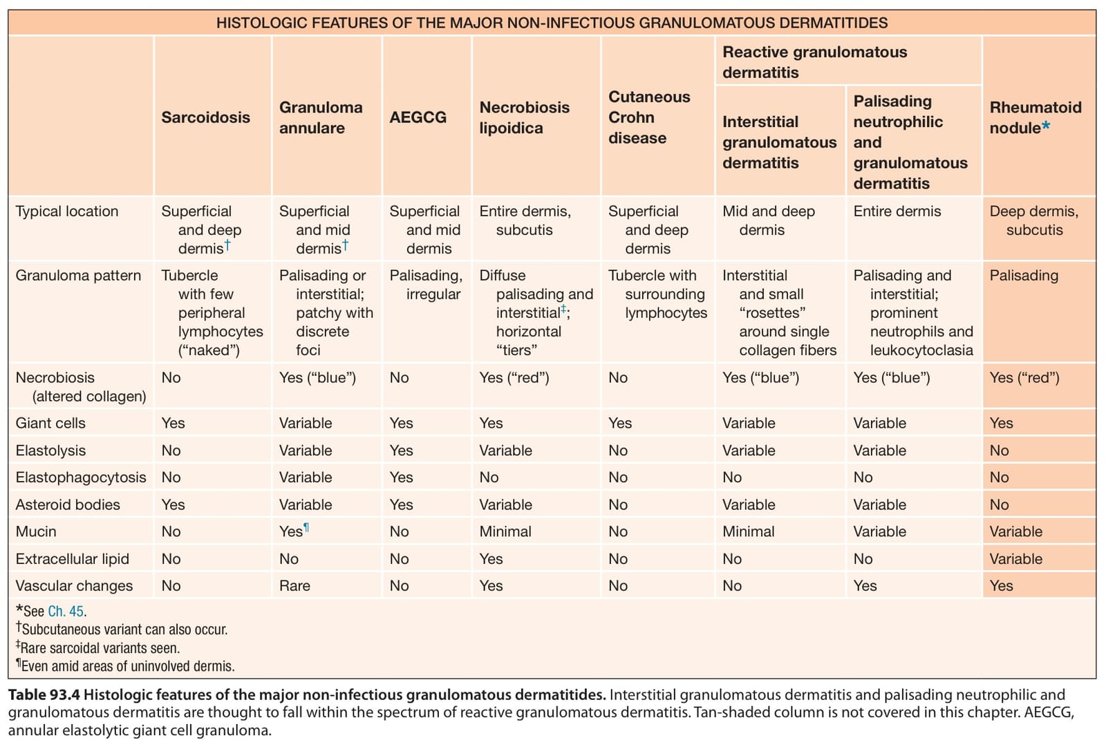
Table 93.4 Histologic features of the major non-infectious granulomatous dermatitides. Interstitial granulomatous dermatitis and palisading neutrophilic and granulomatous dermatitis are thought to fall within the spectrum of reactive granulomatous dermatitis. Tan-shaded column is not covered in this chapter. AEGCG, annular elastolytic giant cell granuloma.
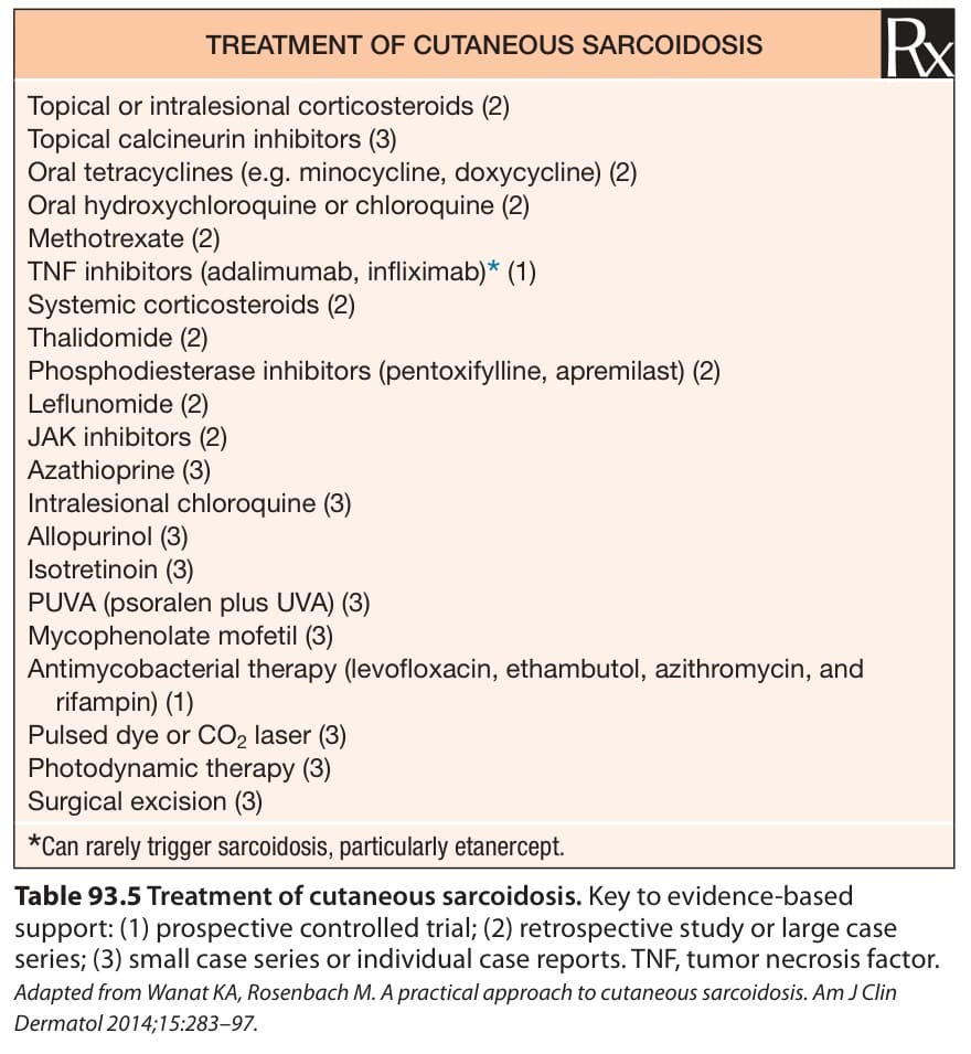
Table 93.5 Treatment of cutaneous sarcoidosis. Key to evidence-based support: (1) prospective controlled trial; (2) retrospective study or large case series; (3) small case series or individual case reports. TNF, tumor necrosis factor. Adapted from Wanat KA, Rosenbach M. A practical approach to cutaneous sarcoidosis. Am J Clin Dermatol 2014;15:283–97.