GRANULOMA ANNULARE
Synonyms: Pseudorheumatoid nodule – subcutaneous granuloma annulare variant Generalized granuloma annulare – disseminated granuloma annulare
Key features
Small grouped papules assuming an annular configuration, often in a symmetrical and acral distribution
Seen primarily in children and young adults
Clinical variants include localized, generalized, and subcutaneous forms
Possible associations with systemic diseases, including diabetes mellitus, hyperlipidemia, thyroid disease, and rarely HIV infection, remain controversial
Histologically, there is an infiltrative or palisading granulomatous dermatitis with focal degeneration of collagen and elastin and deposition of mucin
History
Calcott Fox first described “ringed eruption of the fingers” in 1895. Radcliffe-Crocker called this entity granuloma annulare (GA) in
1902. Some authors consider annular elastolytic giant cell granuloma (AEGCG) to be a variant of GA; in 1975, O’Brien first referred to this entity as actinic granuloma and then it was renamed AEGCG by Hanke et al. in 1979. Previous names included atypical (annular) necrobiosis lipoidica of the face and scalp, Miescher’s granuloma of the face, and possibly granuloma multiforme. Although controversy continues regarding the role of actinic damage in AEGCG and whether it is simply a variant of GA, in this chapter it will be considered a variant.
Epidemiology
Granuloma annulare typically presents during the first three to five decades of life. Subcutaneous GA occurs more commonly in children, whereas generalized GA is more often observed in older patients. While uncommon it is not a rare disease, representing 0.1%–0.4% of visits to dermatologists in several single-center studies. The female-to-male ratio is over 2 : 1, and GA is diagnosed more frequently in White patients. Compared to GA, AEGCG tends to occur in an older population.
Pathogenesis
The etiology of GA is unknown. Trauma, insect bite reactions, tuberculin skin testing, vaccination, UV exposure, Borrelia, and a number of viral infections have all been proposed as inciting factors. Based on the T cell subpopulations identified in GA lesions, a delayed-type hypersensitivity reaction to an unknown antigen has been postulated as the precipitating event. Morphologic similarities to other granulomatous processes suggest that GA is caused by a Th1 inflammatory reaction, with elevations in IL-2R-positive lymphocytes, IFN-γ- producing lymphocytes, and TNF production by macrophages. These lymphocytes release cytokines, including macrophage inhibitory factor, that cause monocytes to accumulate within the dermis and release lysosomal enzymes that can degrade connective tissue. In addition to inflammatory gene signatures associated with Th1 and innate immunity, when lesional skin was compared to non-lesional skin, there was upregulation of Th2 cytokines (IL-4, IL-31) and the JAK/STAT pathway (see Fig. 128.11). As with sarcoidosis (see above), this latter pathway is thought to play an important role in granuloma formation. As with other granulomatous disorders, elastophagocytosis may be observed in GA, but this most likely represents a secondary phenomenon.
Familial cases of GA have been reported, including cases in identical twins, and one small study suggested that an association may exist between generalized GA and HLA-Bw35. Vessel-based mixed inflammatory cell infiltrates with endothelial swelling are sometimes detected in routine histopathologic specimens, and direct immunofluorescence studies of GA may show vessel-based deposits of immunoreactants, but a role for these findings in the pathogenesis of this disease has not been established.
Granuloma annulare has also been described in association with various medications including systemic immunomodulators (e.g. TNF inhibitors, secukinimab, tocilizumab), antiepileptics (levetiracetam, topiramate), amlodipine, thalidomide, allopurinol, and paroxetine; the latter was in a photodistribution. The granulomatous eruptions seen with increasing frequency following administration of immune checkpoint inhibitors include GA as well as sarcoidosis.
With regard to AEGCG, its pathogenesis is not well understood but may be related to inflammation precipitated by actinic damage. Specifically, O’Brien postulated that “a state of auto-aggression develops in relation to the damaged [elastic] fibers”, in an attempt to repair or remodel damaged skin. Infiltrating T lymphocytes are predominantly of the helper subset, suggesting that the pathogenesis may involve a cellmediated immunologic response to a weakly antigenic determinant on altered elastotic fibers. However, destruction of elastic tissue has also been observed in classic GA and necrobiosis lipoidica, leading to the argument that this finding may represent a secondary event caused by the granulomatous inflammation rather than a precipitating one, and occasionally, a patient may have both AEGCG and necrobiosis lipoidica or sarcoidosis.
Clinical Features
GA is a benign, usually self-limited, cutaneous disease that presents as arciform to annular plaques on the dorsal hands or feet of young people (Fig. 93.9). However, there is a wide array of clinical presentations, from a papular form to lesions that are subcutaneous, macular (patch form), or have central perforation (Fig. 93.10). Distribution patterns also vary, from localized to occasionally photodistributed or generalized/disseminated (see Fig. 93.10C). In general, the distribution of GA is: 60% isolated to the hands and arms, 20% on the legs and feet, 7% on both upper and lower extremities, 5% on the trunk, and 5% on the trunk plus other areas. Facial lesions are rare. The plaques may be skin-colored, pink, or violaceous in color, and, upon close inspection, are found to be composed of individual small papules measuring a few millimeters in diameter. The lesions are usually asymptomatic.
In generalized/disseminated GA, the widespread, symmetrically distributed lesions can be annular plaques or discrete papules and they favor the trunk and/or extremities. This form is uncommon and has a later age of onset, poorer response to therapy, and an increased prevalence of the HLA-Bw35 allele. In one study of 100 patients with generalized GA, 45% had lipid abnormalities, including hypercholesterolemia, hypertriglyceridemia or both, while ~20% had diabetes mellitus. Thyroid disease has been reported in ~25% of patients with GA.
Deep dermal or subcutaneous GA manifests as large, painless, skin-colored nodules which may be mistaken for rheumatoid nodules, leading to the term pseudorheumatoid nodule (see Fig. 93.10E). It has a predilection for children <6 years of age. Typical locations include the palms, hands, anterior tibial surfaces and feet, as well as the buttocks, scalp, and rarely the eyelids. As many as 50% of individuals with deep GA lesions also have associated classic lesions.
In perforating GA, the small papules have central umbilication, scalecrust or focal ulceration, and they occur primarily on the dorsal hands and fingers (see Fig. 93.10A). This variant occurs in up to 5% of patients with GA, and histologically transepidermal elimination of degenerating collagen is seen. While umbilicated GA has been described, it probably represents a variant of perforating GA.
Patch GA is a distinct, unusual variant that is characterized by patches of erythema on the extremities and trunk. Symmetrical lesions on the dorsum of the feet often present as macular (i.e. “patch”) disease. Although an annular configuration may be absent, presence of the classic histopathologic findings of interstitial GA (see below) allow for the diagnosis to be made microscopically.
Patients with AEGCG have medium-sized to large annular plaques with a raised erythematous border (3–5 mm) and a slightly atrophic, hypopigmented center, distributed primarily in chronically sun-exposed areas (Fig. 93.11). The sites of predilection include the neck, face, upper trunk, and arms. Early on, skin-colored or pink papules may appear, either singly or in small groups, which then coalesce into annular plaques. Scale is rarely observed. In contrast to necrobiosis lipoidica, there is an absence of yellow color, telangiectasias, or associated alopecia. Individual lesions measure 1–10 cm in diameter, and the total number of lesions is generally fewer than
10. Patients are usually asymptomatic, although pruritus has been reported. A single plaque lasts for months to years, after which spontaneous remission may occur, leaving mottled dyspigmentation or normal-appearing skin. Conjunctival lesions with similar clinical and histologic features have also been described.
Rarely, GA has been reported as a possible paraneoplastic granulomatous reaction to solid organ tumors, Hodgkin disease, non-Hodgkin lymphoma, or granulomatous mycosis fungoides. In these patients, the clinical pattern is frequently atypical, with painful lesions in unusual locations, including the palms and soles. In a literature review and in a case–control study, a relationship between GA and malignancy was not identified; however, patients with atypical presentations may warrant a complete physical exam with focused, age-appropriate screening. While photodistributed GA has been reported, some of these patients may have had AEGCG.
Many reports either supporting or refuting the association of GA with diabetes mellitus have been published, but to date there have been no large-scale epidemiologic studies. In a retrospective study of 84 patients, 12% were found to have diabetes mellitus, and these patients were more likely to suffer from chronic relapsing GA than were non-diabetic patients. In a larger retrospective study of 1383 patients, diabetes mellitus was diagnosed in ~20% of patients with generalized GA, compared with ~10% of patients with localized GA. However, in a case–control study of 126 patients, an association was not identified.
An increased incidence of thyroid disease, in up to 25% of patients, has been described.
Classic GA and perforating GA may develop within herpes zoster scars (see Table 80.7) and following BCG vaccinations. Atypical variants of GA, including photodistributed, have been associated with HIV infection. Additional reported associations, based upon small studies, include hepatitis, post-vaccination, and post-SARS-CoV-2 infection. Rarely, based upon case reports, AEGCG has been observed in patients with lymphoma or leukemia.
Pathology
GA is a granulomatous dermatitis characterized by focal degeneration of collagen and elastic fibers, mucin deposition, and a perivascular and interstitial lymphohistiocytic infiltrate in the upper and mid dermis. The key to the histopathologic diagnosis of GA is the presence of mucin plus the identification of histiocytes in one of two patterns. The most common is the interstitial or infiltrative pattern, in which scattered histiocytes are distributed between collagen fibers (Fig. 93.12). Degeneration of collagen fibers is minimal, but granular, basophilic mucin deposition between collagen bundles can be highlighted with Alcian blue and colloidal iron stains. The second pattern is more obvious and is easier to diagnose. It consists of one to several palisading granulomas characterized by central connective tissue degeneration surrounded by a rim (“palisade”) of histiocytes and lymphocytes (Fig. 93.13). Mucin is abundant in the center of the palisaded granuloma, and fibrin, neutrophils, and nuclear dust may also be present. Rarely, a prominent (“pseudolymphomatous”) lymphoid infiltrate is seen.
Increased mucin can be detected in ~90% of biopsy specimens from GA lesions. The sensitivity of detection is increased by using at least two different mucin stains, such as colloidal iron and Alcian blue. Elastic tissue is reduced or absent within the histiocytic aggregates in ~20% of generalized GA and 35% of localized GA cases. Collagen
degeneration is more common in localized GA than in generalized GA, and elastophagocytosis is occasionally noted, but not as often as in AEGCG.
As noted previously, vascular changes in GA are variable, but include fibrin, C3, and IgM deposition in vessel walls (detected by direct immunofluorescence) and occlusion of vascular lumina. One study found that the presence of leukocytoclastic or granulomatous vasculitis or a thrombotic vasculopathy within the lesions of GA was predictive of associated systemic disease, and the possibility of palisading neutrophilic and granulomatous dermatitis as a reflection of disorders such as rheumatoid arthritis needs to be considered.
Subcutaneous GA is characteristically a septal panniculitis with palisaded granulomatous inflammation and it can closely mimic a rheumatoid nodule histologically. An interstitial pattern is not observed in this form of GA. In perforating GA, there is a superficial histiocytic infiltrate with transfollicular and/or transepidermal elimination of granulomatous inflammation and degenerated collagen.
In AEGCG, a non-palisading granulomatous infiltrate composed of histiocytes, foreign body-type multinucleated giant cells (with haphazardly arranged nuclei), and lymphocytes is present within the mid to upper dermis; there is an absence of altered collagen or mucin or lipid deposition. Elastic fibers are identified adjacent to and within the giant cells (elastophagocytosis; Fig. 93.14). Elastophagocytosis and asteroid bodies can be seen less frequently in other granulomatous disorders such as GA and necrobiosis lipoidica and therefore they are not specific for AEGCG. Elastic tissue stains, such as Verhoeff–van Gieson, show a characteristic complete absence of elastic fibers in the areas affected (or previously affected) by granulomatous inflammation. There is a sparse perivascular lymphocytic infiltrate, but no vascular changes.
Differential Diagnosis
GA is diagnosed based on its clinical and histopathologic features – there are no laboratory tests which aid in confirming the diagnosis.
When the diagnosis of deep GA versus rheumatoid nodule is in question, rheumatoid factor and anti-cyclic citrullinated peptide antibody levels can aid in identifying patients at risk for the latter. Rheumatoid nodules also demonstrate fibrin rather than mucin histologically.
The clinical differential diagnosis for conventional GA includes the annular entities listed in Table 19.1, including annular sarcoidosis and AEGCG. A clue to the latter is central hypopigmentation and/or atrophy. Arcuate and annular plaques of mycosis fungoides or borderline leprosy may also simulate GA, requiring biopsy for definitive diagnosis.
Papular GA can simulate flat warts, secondary syphilis, early eruptive xanthomas, and the non-X-histiocytoses (see Ch. 91). Subcutaneous GA can be clinically similar to rheumatoid nodules, the nodules of rheumatic fever, tendinous xanthomas, epithelioid sarcoma, subcutaneous sarcoidosis, idiopathic granulomatous mastitis, and deep granulomatous infections. Perforating GA must be differentiated from primary perforating disorders (see Ch. 96) and, occasionally, perforating calcinosis cutis, perforating gout, or molluscum contagiosum.
The clinical and histologic differences between GA, sarcoidosis, AEGCG, necrobiosis lipoidica, rheumatoid nodule, and reactive granulomatous dermatitides are outlined in Tables 93.1 and 93.4. The histopathologic differential diagnosis of GA also includes morphea, cutaneous T cell lymphoma, xanthomas, epithelioid sarcoma and, rarely, scleromyxedema. Lastly, the possibility of drug-induced GA needs to be considered.
Treatment
Given the self-limited and benign nature of GA, reassurance and clinical observation may be the treatment of choice for localized, asymptomatic disease. High-potency topical corticosteroids with or without occlusion and intralesional corticosteroid injections are the usual first-line local therapies. Cryosurgery, photodynamic therapy, pulsed dye laser, and topical tacrolimus may also be effective. For more extensive disease, systemic medications can be considered including: (1) anti-inflammatory drugs or immunomodulators (e.g. antimalarials, tetracycline-class antibiotics, pentoxifylline, niacinamide, dapsone, apremilast, retinoids); (2) phototherapy – PUVA, NB-UVB, or UVA1 (especially patch variant); and (3) immunosuppressants (TNF inhibitors, with adalimumab most often reported), while weighing the risk : benefit ratio (Table 93.6). In patients with photodistributed GA, oral medications such as dapsone are preferred over phototherapy. More recently, topical JAK inhibitors (e.g. compounded tofacitinib 2% ointment) have been used to treat GA as have oral JAK inhibitors.
Spontaneous resolution of GA occurs within 2 years in 50% of patients, but there is a 40% recurrence rate. The recurrent lesions tend to occur at the original sites, but clear more rapidly (80% within 2 years). The duration of untreated lesions has been reported to range from a few weeks to several decades.
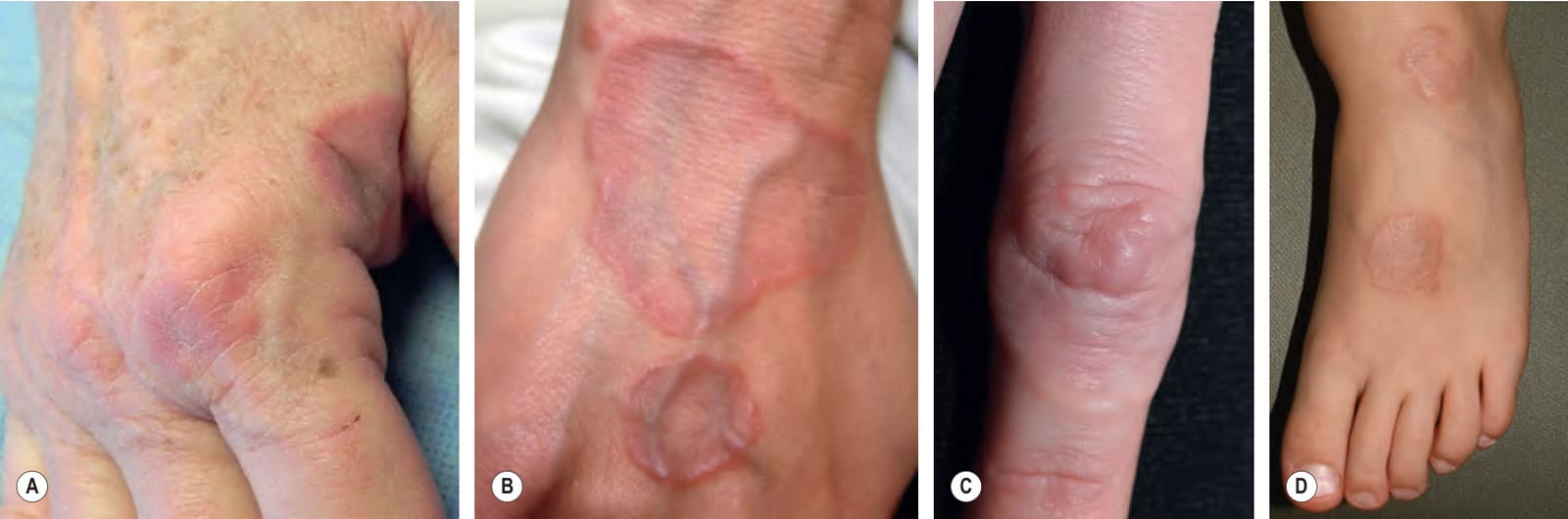
Fig. 93.9 Granuloma annulare – clinical features.A–D Annular pink to pink–brown plaques. The dorsal aspects of the hands and feet are a common location. Lesions can be skin-colored as well as pink (C). Grouped discrete papules may also be seen, with a more subtle annular or arciform configuration (A,C). C, Courtesy Julie V. Schaffer, MD; D, Courtesy Kalman Watsky, MD.
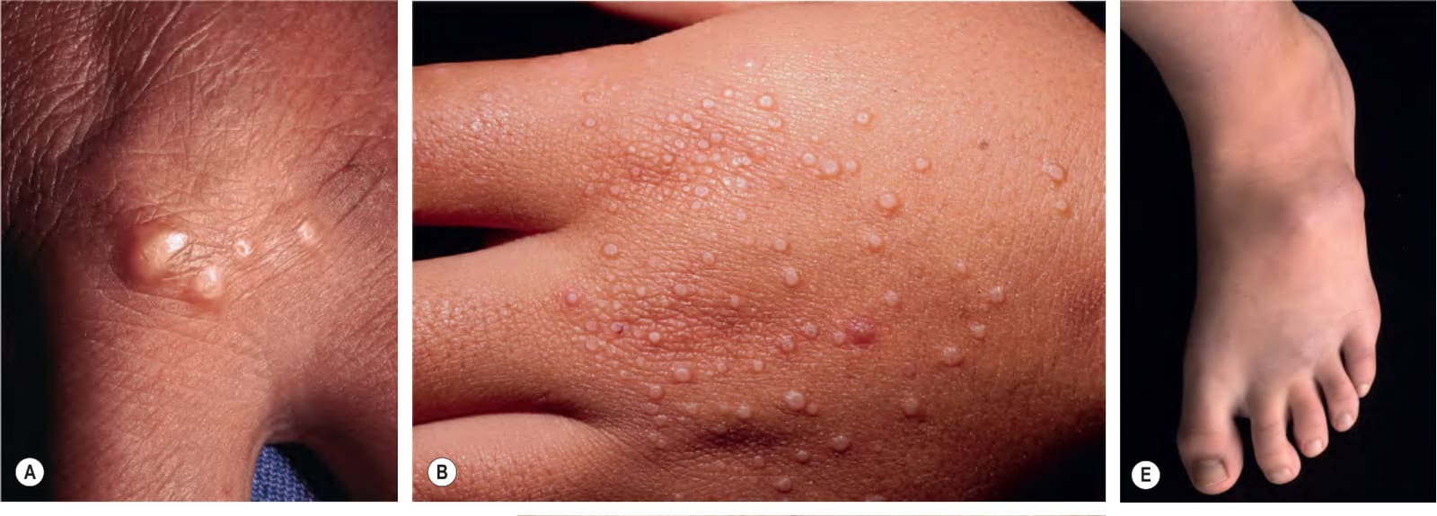
Fig. 93.10 Granuloma annulare (GA) – less common variants.A Perforating GA: papules characteristically have a central keratotic plug. B Micropapular GA: several of the lesions have a central dell. C Generalized/disseminated GA: discrete red–brown papules, many of which are in clusters. D Papular and nodular GA: the skin-colored nodules may be confused with rheumatoid nodules. E Skin-colored nodules of subcutaneous GA. A, Courtesy Ronald P. Rapini, MD; B, Courtesy Joyce Rico, MD; C, Courtesy Luis Requena, MD; D, Courtesy Kalman Watsky, MD; E, Courtesy Department of Dermatology, Medical University of Graz.
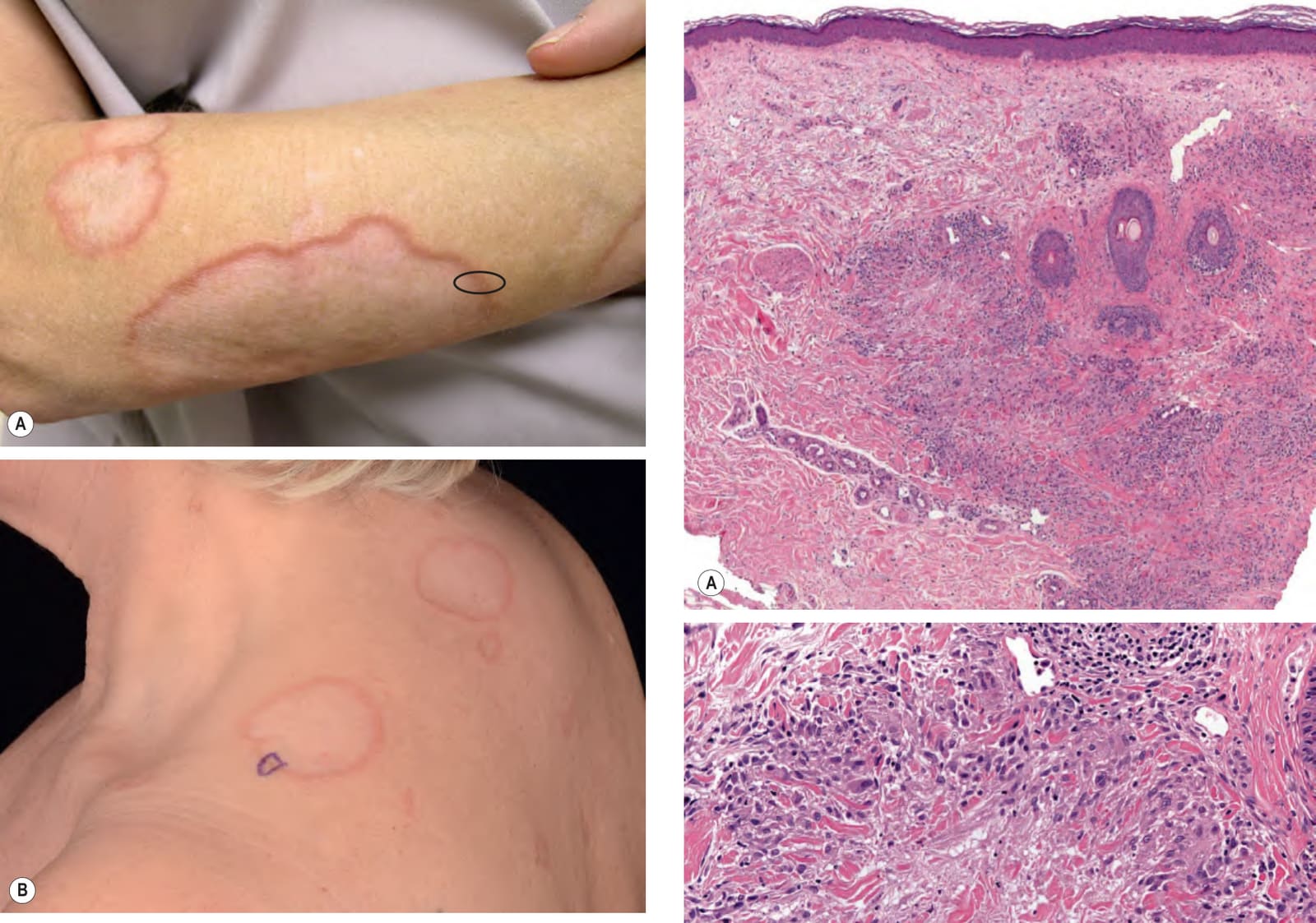
Fig. 93.11 Annular elastolytic giant cell granuloma – clinical features.A, B The inflammatory border resembles granuloma annulare but the central portion is hypopigmented and/or atrophic. A biopsy specimen that includes the areas outlined would contain the three characteristic histologic zones: absence of elastic fibers, granulomatous inflammation, and normal skin. Longitudinal sectioning of the surgical specimen is preferred. B, Courtesy Department of Dermatology, Medical University of Graz.
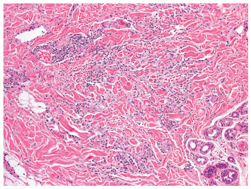
Fig. 93.12 Granuloma annulare – interstitial or infiltrative pattern. Infiltration of histiocytes between the dermal collagen fibers, with scant mucin. Courtesy Lorenzo Cerroni, MD.
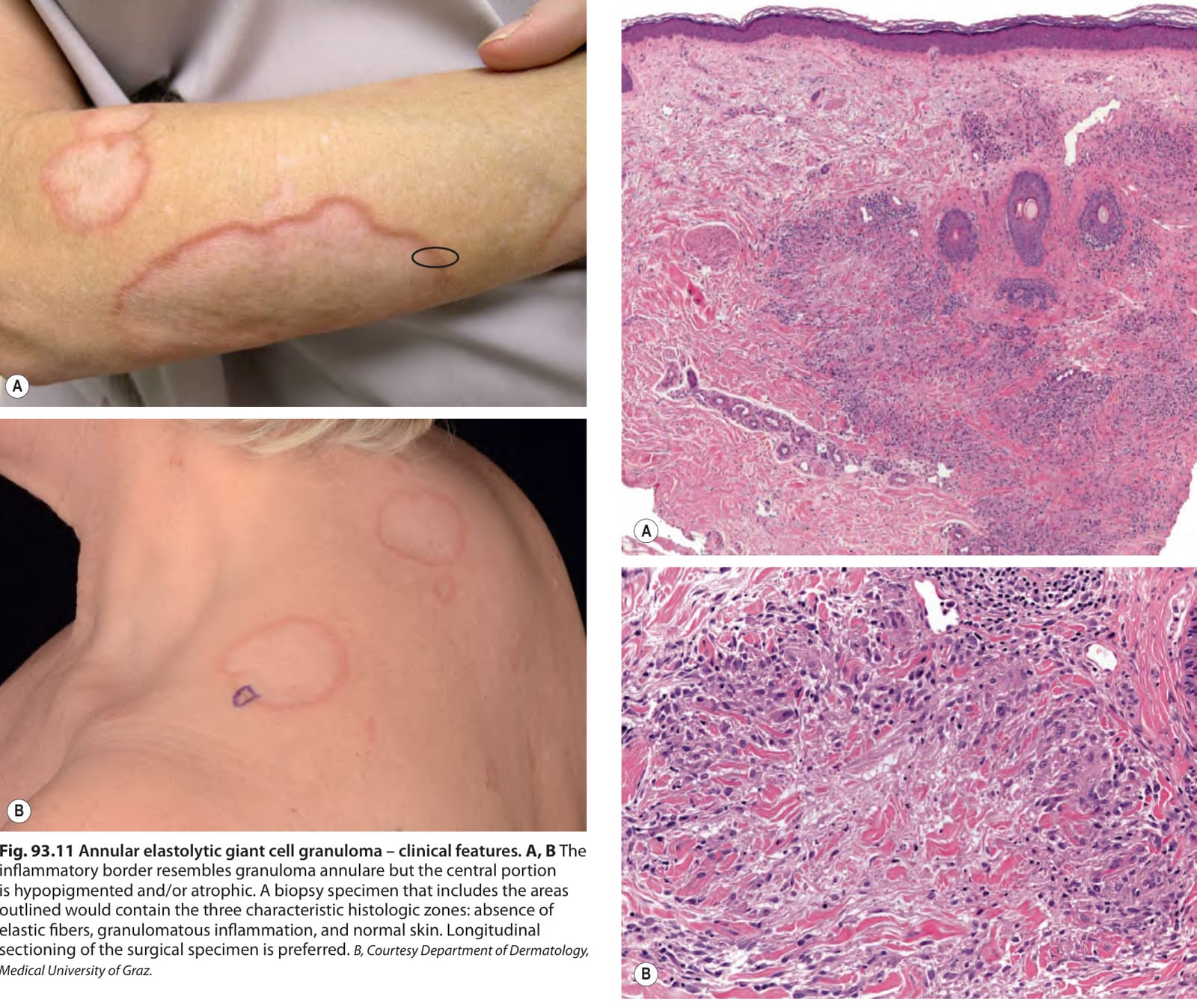
Fig. 93.13 Granuloma annulare – palisading granuloma pattern.A Multiple palisaded granulomas within the dermis. Degenerative collagen and mucin deposition are present in the central portion of the granulomas. B A palisaded granuloma with epithelioid histiocytes surrounding an anuclear dermis characterized by altered collagen and pallor due to deposition of acid mucopolysaccharides/glycosaminoglycans (mucin). Courtesy Lorenzo Cerroni, MD.
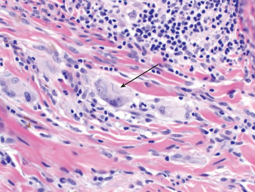
Fig. 93.14 Annular elastolytic giant cell granuloma – histopathological features. A dermal inflammatory infiltrate with several histiocytic giant cells. The presence of a fragmented elastic fiber within a giant cell (elastophagocytosis) is a characteristic finding (arrow).
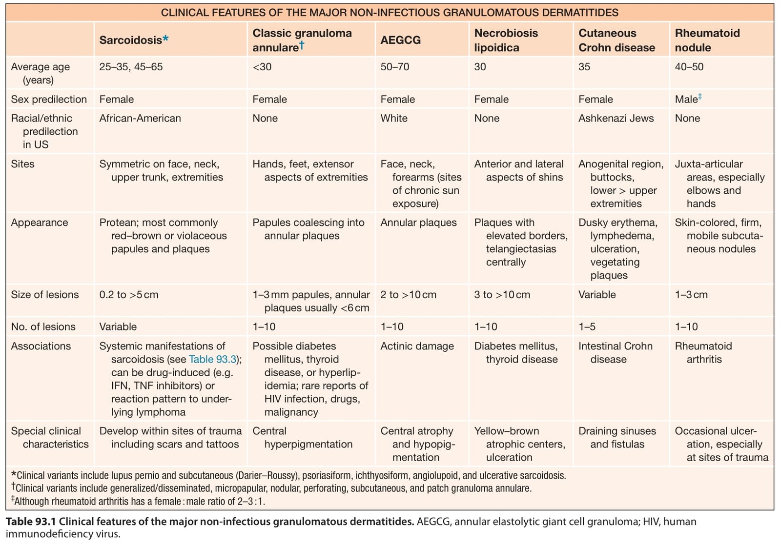
Table 93.1 Clinical features of the major non-infectious granulomatous dermatitides. AEGCG, annular elastolytic giant cell granuloma; HIV, human immunodeficiency virus.
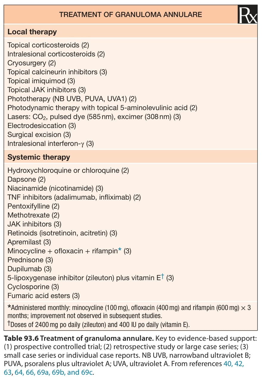
Table 93.6 Treatment of granuloma annulare. Key to evidence-based support: (1) prospective controlled trial; (2) retrospective study or large case series; (3) small case series or individual case reports. NB UVB, narrowband ultraviolet B; PUVA, psoralens plus ultraviolet A; UVA, ultraviolet A. From references 40, 42, 63, 64, 66, 69a, 69b, and 69c.