NECROBIOSIS LIPOIDICA
Synonym: Necrobiosis lipoidica diabeticorum (NLD)
Key features
Plaques with violaceous to red–brown, palpable peripheral rims and yellow–brown atrophic centers with telangiectasias
The most common site is the shins
Ulceration can occur following trauma
The proportion of patients with diabetes mellitus varies from 15% to 65%, but there is a strong association
Pathogenesis is unknown
Pathology shows a palisading granulomatous dermatitis with histiocytes and altered collagen that has a “layered” appearance and is often accompanied by perivascular plasma cells
History
First called “dermatitis atrophicans diabetica” by Oppenheim in 1929, the condition was renamed necrobiosis lipoidica diabeticorum by Urbach in 1932. The first non-diabetic patient with this condition was described in 1935 by Goldsmith, generating the more general term necrobiosis lipoidica (NL), which is currently the preferred term.
Epidemiology
NL typically occurs in young to middle-aged adults, with a female : male ratio that ranges from 3 : 1 to 8 : 1, depending on the study. Diabetic patients present at a younger age than those without diabetes, e.g. in a multicenter study, the mean ages were 45 years and 52 years, respectively. In a study of 171 patients with NL, diabetes mellitus, usually type 1, was present in ~65%; an additional 12%–15% had abnormal glucose tolerance tests. Also, over half of the patients with no evidence of diabetes and a normal glucose tolerance test reported a positive family history of glucose intolerance. However, a more recent retrospective study of 65 patients with NL seen in a dermatology outpatient clinic found that only 11% had type 1 or type 2 diabetes mellitus at the time of presentation. An additional 11% were later diagnosed with impaired glucose tolerance or diabetes.
Only 0.3% of patients with diabetes develop NL. However, there is no proven connection between a patient’s level of glycemic control and the likelihood of developing NL lesions. Diabetic patients with NL may or may not have a higher rate of diabetes-related complications such as peripheral neuropathy, retinopathy, and limited joint mobility, again depending upon the study. In retrospective reviews, thyroid dysfunction was noted in 15%–25% of patients with NL.
Pathogenesis
The cause of NL remains unknown. Immunologically mediated vascular disease has been suggested as the primary cause of the altered collagen seen in NL. This hypothesis is supported by the presence of immunoreactants deposited in vessel walls of lesional and uninvolved skin in patients with NL. Previous immunofluorescence studies had also found evidence for immune complex vasculitis in NL. In addition, it has been postulated that the microangiopathic vessel changes seen in diabetic patients could contribute to the development of collagen degeneration and subsequent dermal inflammation. A shared pathogenesis would provide an explanation for the high rate of complications such as retinopathy observed in some series of patients with NL.
When lesions favor the lower extremities of adults, venous hypertension as a contributing factor needs to be considered. Both venous insufficiency and hyperlipidemia have been proposed as triggers that incite an inflammatory cascade in some patients. Elevated plasma levels of fibronectin, factor VIII-related antigen, and α-macroglobulin have been detected in patients with NL, but the significance of these findings has not been determined. Theories regarding abnormally elevated platelet adhesion, increased thromboxane A production, and increased blood viscosity within NL lesions remain speculative.
Some investigators consider NL to be primarily a disease of collagen, with inflammation occurring as a secondary event. Anti-collagen antibodies have been detected in patients with both NL and GA, but no significant increase in antibody levels as compared with control patients has been demonstrated. The concentration of collagen is decreased in NL lesions, and electron microscopy reveals a loss of the cross-striations of collagen fibrils and significant variation in the diameter of individual fibrils. Fibroblasts cultured from NL lesions synthesize less collagen than their counterparts from unaffected skin. Although there are fewer collagen fibers overall, areas of fibrosis are seen. Overhydration of collagen due to hyperglycemia can lead to increased collagen cross-linking and stiffness, but it is unclear if this phenomenon occurs in NL.
From central Europe, there was a report of the detection of Borrelia spp. via focal floating microscopy in NL, but these findings need to be confirmed.
Clinical Features
Necrobiosis lipoidica is characterized by yellow–brown, atrophic, telangiectatic plaques with an elevated violaceous rim, most often in the pretibial region (Fig. 93.15). The plaques are usually multiple and bilateral. Lesions start as small, firm, red–brown papules that gradually enlarge and then develop central epidermal atrophy. Ulceration has been reported in 15%–35% of patients (see Fig. 105.20D), usually following minor trauma. Development of lesions secondary to Koebner phenomenon has also been reported. Less typical anatomic locations for NL include the upper extremities, face, and scalp, where the lesions may be more annular or serpiginous in configuration and are often less atrophic. As a result, they may resemble AEGCG clinically.
Decreased sensation to pinprick and fine touch, hypohidrosis, and partial alopecia can be observed within NL plaques. Decreased S100 staining within the cutaneous nerves of inflammatory plaques has been described, with degenerative neural changes possibly accounting for the loss of sensation. Of note, the cutaneous nerves in GA demonstrate normal S100 staining patterns. Although the lesions of NL are typically asymptomatic, some patients report pruritus, dysesthesia, or pain. Lesions that ulcerate after trauma or from transepidermal elimination of altered collagen and elastic fibers can be very painful. Rarely, squamous cell carcinoma has been reported to develop within lesions of NL. By dermoscopy, comma-shaped vessels and an irregular pattern of arborizing vessels are observed in early and more advanced lesions, respectively; whitish areas correspond to degenerated collagen and yellow to orange patches to granulomatous inflammation.
There are reports of patients with NL who also had GA or sarcoidosis. Possible versus random associations include systemic sclerosis, rheumatoid arthritis, and previous jejunoileal bypass surgery. The association of NL with diabetes mellitus and thyroid dysfunction are discussed in the Epidemiology section.
Pathology
In the palpable inflammatory border, a diffuse palisaded and interstitial granulomatous dermatitis that involves the entire dermis and extends into the subcutaneous fat septae is usually seen (Fig. 93.16). “Layered” tiers of granulomatous inflammation are composed of histiocytes surrounding horizontal tiers of degenerated collagen. The histiocytes are often multinucleated and the collagen fibers have irregular sizes and shapes. In areas of sclerosis, focal loss of elastic tissue may be seen. Extracellular lipid deposition can be demonstrated via adipophilin staining of routinely processed tissue sections or via oil red O staining of frozen tissue sections (latter rarely done nowadays). There is an associated superficial and deep perivascular infiltrate that is predominantly lymphocytic but often contains plasma cells and occasionally eosinophils. The epidermis is normal or atrophic.
In contrast to GA, there is no significant mucin deposition within the center of the palisading granulomas. Although early lesions of both NL and GA may have foci of leukocytoclasia, NL shows more prominent endothelial cell swelling, fibrosis, and hyalinization; the latter can lead to blood vessel wall thickening or even endarteritis obliterans. The vessel walls often contain a PAS-positive, diastase-resistant material suggesting neutral glycosaminoglycans.
Occasionally, NL lesions have less sclerotic collagen and more typical epithelioid cell granulomas rather than the horizontally tiered pattern of altered collagen and palisaded granulomatous inflammation described above. Vascular changes are rare, and this form of NL is felt to have a weaker association with diabetes mellitus than the classic lesions.
Differential Diagnosis
Clinically, the differential diagnosis of NL includes primarily GA, necrobiotic xanthogranuloma (NXG; see Ch. 91), sarcoidosis, diabetic dermopathy (see Ch. 53), and lipodermatosclerosis. Other entities in the differential diagnosis include additional forms of panniculitis (see Ch. 100), granulomatous infections (e.g. leprosy, dimorphic fungal infections, tertiary syphilis), morphea, lichen sclerosus, and sclerosing lipogranuloma.
Lesions of GA and sarcoidosis generally do not exhibit the same degree of atrophy, telangiectasias, or yellow–brown color as NL lesions, although prominent telangiectasias can be present in angiolupoid sarcoidosis and can occur following injections of triamcinolone. Ulcerated NL may be difficult to distinguish from ulcerative sarcoidosis or NXG.
Histopathologically, NL most commonly has to be distinguished from GA (see Table 93.4). NXG is also in the differential diagnosis, but there are abundant cholesterol clefts and larger zones of degenerated collagen. However, one case report did describe NL with prominent cholesterol clefts and transepithelial elimination of cholesterol crystals via hair follicles.
Treatment
No treatment for NL has proven to be effective in large, double-blind, placebo-controlled studies. In patients with diabetes mellitus, control of blood glucose levels may not have a significant effect on the course of NL. Spontaneous remission after an average of 8 to 12 years was observed in only 17% of 171 patients with NLD in one study.
First-line therapy consists of potent topical corticosteroids (including under occlusion) for early lesions and intralesional corticosteroids injected into the active borders of established lesions. Because the histologic changes can extend well into normal-appearing skin in clinically active NL lesions, some authors advocate the injection of intradermal corticosteroids into a rim of clinically normal skin around expanding plaques in an effort to halt disease progression. Careful monitoring for steroid-induced atrophy is important. Topical tacrolimus 0.1% ointment led to improvement of NL in uncontrolled case series. Topical GM-CSF or bovine collagen has been applied to ulcerations and tretinoin gel (0.025% BID) has been used to decrease lesional atrophy.
In addition to avoidance of trauma and local care of ulcers (see Ch. 145), therapies directed at increasing fibrinolysis, decreasing platelet aggregation, and/or decreasing thromboxane A synthesis have been employed in order to decrease the microangiopathy and vascular thrombosis. Pentoxifylline, stanozolol, inositol niacinate (inositol nicotinate), nicofuranose, and ticlopidine hydrochloride are all agents that have been tried in anecdotal reports or uncontrolled case series. The use of stanozolol is limited by its potential hepatotoxicity and ticlopidine can cause agranulocytosis. Controlled trials of aspirin versus placebo or aspirin plus dipyridamole versus placebo showed no significant differences compared to placebo. Perilesional injections of heparin have been reported in the Russian literature.
Based upon case series of patients, several systemic anti-inflammatory medications have been described as beneficial – antimalarials, niacinamide, mycophenolate mofetil, doxycycline, colchicine, methotrexate, thalidomide, TNF inhibitors, IL-12/23 inhibitors, and cyclosporine (severe ulcerations). As with sarcoidosis and GA, JAK inhibitors have emerged as a promising therapeutic option for selected patients. In addition, 5-week courses of systemic corticosteroids were found to be effective in a series of six patients with an average follow-up period of 7 months, but this can be associated with side effects such as elevation of serum glucose levels.
Multiple prospective, uncontrolled studies have demonstrated improvement in ~50% of patients with topical PUVA therapy. Lastly, there are case reports of beneficial effects from UVA1 phototherapy, pulse-dyed laser, fractional CO laser, or photodynamic therapy.
Surgical therapy may be necessary in patients with severe, refractory ulcerations. Excision to deep fascia or periosteum must be performed to minimize the chance of recurrence. Split-thickness skin grafting is performed following excision.
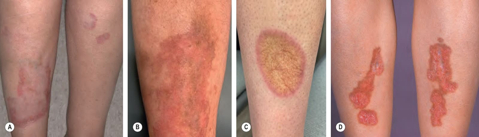
Fig. 93.15 Necrobiosis lipoidica – range of clinical features.A Annular and arciform pink–brown plaques on the shins with central atrophy. B Pink plaques on the shin with telangiectasias and peripheral hyperpigmentation; centrally there is atrophy. C Single yellow plaque with prominent telangiectasias and a dull pink rim. D Pink–yellow plaques with a brown rim on the shins; centrally there is atrophy, focal scarring, and telangiectasias. B, Courtesy Jeffrey Cohen, MD; C, Courtesy Aisha Sethi, MD.
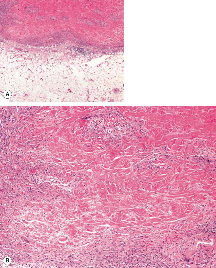
Fig. 93.16 Necrobiosis lipoidica – histopathologic features.A Multiple granulomas within the entire dermis extending into the subcutaneous fat. Note the layered tiers of granulomatous inflammation aligned parallel to the skin surface. B Palisaded granuloma composed of epithelioid histiocytes with altered collagen centrally. Courtesy Lorenzo Cerroni, MD.
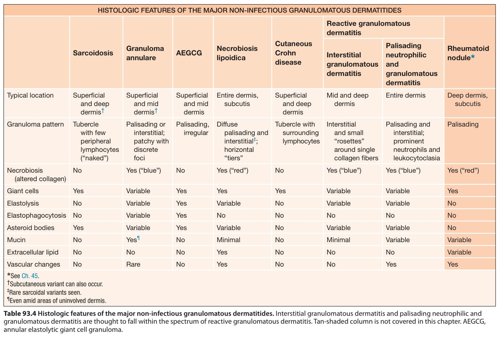
Table 93.4 Histologic features of the major non-infectious granulomatous dermatitides. Interstitial granulomatous dermatitis and palisading neutrophilic and granulomatous dermatitis are thought to fall within the spectrum of reactive granulomatous dermatitis. Tan-shaded column is not covered in this chapter. AEGCG, annular elastolytic giant cell granuloma.