CUTANEOUS CROHN DISEASE
Variants: Contiguous Crohn disease Distant/non-contiguous (“metastatic”) Crohn disease
Key features
Erythematous plaques, lymphedema, and knife-like ulcerations most commonly occur in the anogenital region
Cutaneous non-caseating granulomatous lesions that are not contiguous with intestinal Crohn disease have been referred to as “metastatic”
Approximately 20% of patients with cutaneous lesions have no preceding diagnosis of gastrointestinal Crohn disease
History
Crohn disease, first described in 1932, is characterized by segmental granulomatous inflammation of the intestinal tract and frequently involves cutaneous tissues as well. In 1965, cutaneous involvement was first described.
Epidemiology
Crohn disease usually begins between the second and fourth decades of life. Mucocutaneous findings occur in 20%–45% of patients (see Table 53.8). They can be subdivided into: (1) specific lesions including distant/non-contiguous cutaneous (“metastatic”) Crohn disease, contiguous perianal Crohn disease, and oral Crohn disease; (2) nonspecific or reactive dermatologic disorders, e.g. erythema nodosum, Sweet syndrome, pyoderma gangrenosum; (3) nutritional skin changes secondary to malabsorption; and (4) treatment-related side effects.
Cutaneous Crohn disease is relatively rare with <300 cases reported in the literature to date and <100 cases in children. Two-thirds of the patients are women, and the mean age of onset is 35 years. Approximately 20% of the reported cases had skin disease that preceded the diagnosis of intestinal Crohn disease by 3 months to 8 years.
Pathogenesis
Cutaneous Crohn disease occurs in association with or precedes intestinal Crohn disease, reflecting a multisystem disease. A number of genetic abnormalities which can lead to an exaggerated T cell response to certain commensal enteric bacteria and defective microbial clearance, including after breaks in the mucosal barrier or an alteration in the balance in gut flora (dysbiosis), have been linked to Crohn disease. The protein products of these genes have several functions, including regulation of NF-κB function (e.g. NOD2 [CARD15]) and autophagy (ATG16L1, IRGM). Patients with variants in TRAF3IP2, a psoriasis susceptibility gene, may have a greater risk of cutaneous involvement. Like psoriasis, Crohn disease is a predominantly Th1- and Th17-mediated disease, with elevated levels of IL-23 and IL-17 within involved tissues.
Clinical Features
Cutaneous Crohn disease may be either genital or extragenital, with genital involvement found in approximately two-thirds of children and one-half of adults. Labial, penile, or scrotal erythema and/or lymphedema are common presenting signs and can be dramatic (Fig. 93.17A–C,F). Linear or “knife-like” ulcerations that are within body folds (e.g. inguinal creases, perianal region, axillae) or involve the oral mucosa are relatively specific for mucocutaneous Crohn disease (see Figs. 53.28 and 72.25).
Perianal involvement, which may extend to the adjacent perineum, buttocks or abdomen, consists of sinus tracts, fissures, ulcers, and vegetating plaques (Fig. 93.17D,E). These findings occur in approximately one-third of patients with intestinal Crohn disease. Perianal skin tags, thought to be secondary to lymphedema, are observed in up to 40% of patients; as with other sites of lymphedema, granulomatous inflammation may be present as well. Sites of abdominal surgery, e.g. laparotomies, colostomies, ileostomies, may also develop granulomatous inflammation. For semantic purposes, the latter is considered to be “contiguous” rather than non-contiguous (“metastatic”) Crohn disease.
Oral lesions occur in 5%–20% of patients with Crohn disease, and these may consist of cobblestoning of the buccal mucosa, tiny gingival nodules, gingival hyperplasia, aphthae-like ulcers (Fig. 93.17G), linear, knife-like ulcerations, pyostomatitis vegetans (see Fig. 25.9A), angular cheilitis and ulceration, secondary orofacial granulomatosis (Fig. 93.17I), diffuse oral swelling, or indurated fissuring of the lower lip. Histologically, 90% of oral lesions associated with Crohn disease contain granulomas.
Non-genital disease presents with dusky erythematous plaques, often followed by the development of ulceration with undermined edges, draining sinuses and fistulas, and scarring (Fig. 93.17H). These lesions have been observed on the lower extremities and soles (40%), abdomen and trunk (25%), upper extremities and palms (15%), face and lips (10%), flexural areas (8%), and in a generalized distribution (4%). Other reactive cutaneous manifestations of Crohn disease include cutaneous arteritis (cutaneous polyarteritis nodosa), erythema nodosum, erythema multiforme, finger clubbing, cutaneous small vessel vasculitis, epidermolysis bullosa acquisita, vitiligo, palmar erythema, a pustular response to trauma (pathergy), Sweet syndrome, and pyoderma gangrenosum (see Ch. 53). Zinc deficiency may cause an acrodermatitis enteropathica-like syndrome in patients with severe Crohn disease.
Based upon a large retrospective series, cutaneous involvement may be indicative of colonic, rather than ileal, involvement. No consistent correlation exists between mucocutaneous disease activity and degree of gastrointestinal involvement. However, levels of fecal calprotectin can reflect the degree of intestinal inflammation.
Pathology
In both cutaneous and oral lesions of Crohn disease, small, nodular, non-caseating, epithelioid granulomas with surrounding lymphocytes are found in the superficial and deep dermis, sometimes extending into the subcutaneous fat. There are a few scattered multinucleated Langhans-type giant cells and a sparse perivascular lymphohistiocytic infiltrate; overlying ulceration may be present. Of note, the combination of lichenoid and granulomatous inflammation plus granulomatous perivasculitis can serve as a histopathologic clue. The walls of perianal sinuses and fistulas have similar granulomatous inflammation. However, the granulomatous foci may be subtle, requiring multiple sections to identify the granulomas, and clinicopathologic correlation as well as additional biopsies may be required.
Differential Diagnosis
Clinically, the differential diagnosis of cutaneous Crohn disease includes other granulomatous disorders such as cutaneous sarcoidosis, mycobacterial infections, deep fungal infections, and foreign body reactions. Other infections, such as actinomycosis or cellulitis, may mimic cutaneous Crohn disease. Ulcerated lesions may be misdiagnosed as pyoderma gangrenosum. In the case of genital swelling and ulcerations, one must consider granuloma inguinale, schistosomiasis, hidradenitis suppurativa, and chronic lymphedema due to obstruction.
The histopathologic picture of cutaneous Crohn disease can be indistinguishable from other granulomatous diseases with tuberculoid features, including lupus vulgaris. In the latter, central necrosis within tubercles is a helpful distinguishing clue. Differentiating cutaneous Crohn disease from sarcoidosis can also be challenging, although Crohn disease usually has denser collections of lymphocytes and granulomatous perivasculitis as well as the features described above. Foreign body granulomas and infectious granulomas should be considered in the histologic differential diagnosis. Special stains for acid-fast bacilli and fungal organisms should be obtained, and all skin biopsies should be polarized to look for foreign bodies. Pyoderma gangrenosum and cutaneous Crohn disease can sometimes have overlapping clinical and histopathologic features as neutrophilic inflammation may be present in both, but granulomas are absent in pyoderma gangrenosum.
Treatment
Cutaneous Crohn disease tends to be chronic, and its severity does not always correlate with the activity of the patient’s intestinal disease. For localized disease, topical or intralesional corticosteroids and/or topical calcineurin inhibitors may be utilized. Oral metronidazole (250 mg TID for at least 4 months) can be an effective treatment. For more extensive disease, use of systemic medications including corticosteroids, sulfasalazine, azathioprine, 6-mercaptopurine, and thalidomide may lead to improvement. By targeting the underlying immunopathogenesis, TNF inhibitors (e.g. infliximab, adalimumab) and ustekinumab have been reported to improve cutaneous disease. IL-17 inhibitors have been reported to exacerbate Crohn disease. Surgical excision of lesions is often complicated by wound dehiscence and disease recurrence, but can be considered for refractory disease.
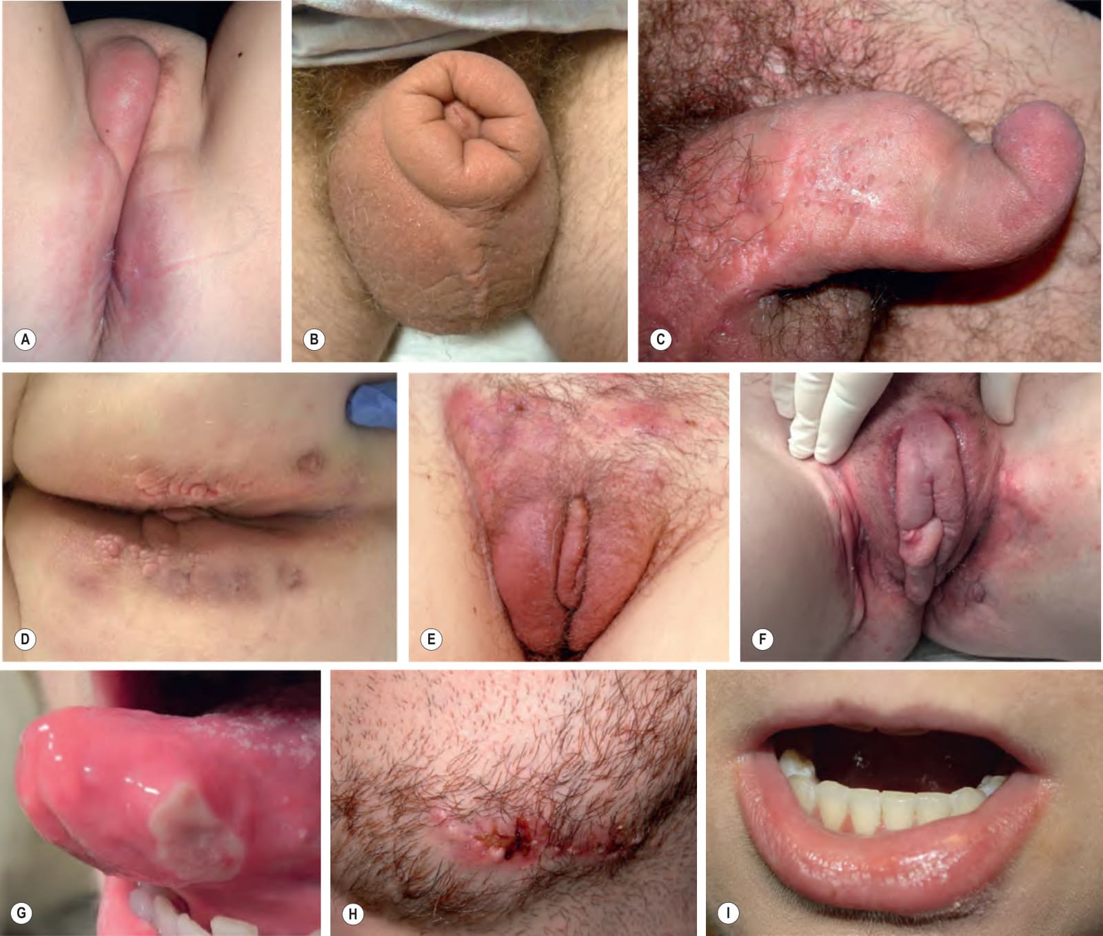
Fig. 93.17 Mucocutaneous Crohn disease.A Vulvar and perianal erythema and induration. Note the marked asymmetric vulvar swelling and perianal erosions. B Marked lymphedema of the prepuce with lymphedema and induration of the scrotum. C Inflammation and lymphedema leading to twisting of the penis along its long axis, referred to as a “saxophone penis”. D Perianal tags and indurated plaques. E Firm erythematous plaques of the mons pubis and labia majora and minora. There are also draining sinuses. F Symmetric erythematous plaques, lymphedema, and a “skin tag”; note the marked swelling of the labia minora. G Intraoral ulcer that resembles a major aphthous ulcer. H Ulcerated erythematous plaque of the mandible with draining sinuses. I Asymmetric enlargement of the lower vermilion lip due to granulomatous inflammation. A, Courtesy Julie V. Schaffer, MD; C, E, H, Courtesy Luis Requena, MD; D, Courtesy Mary Stone, MD; F, I, Courtesy Jeffrey P. Callen, MD, and Courtney R. Schadt, MD.