GENERAL CONSIDERATIONS
Definition
Literally, any material – living or non-living – introduced into the body is a “foreign body” and is treated by our immune system as “non-self” in order to elicit an appropriate response. Such a broad definition also includes infectious agents, but they are covered elsewhere (see Chs. 74–85). This chapter will focus on non-living materials that have been introduced into the dermis or subcutis. They represent inorganic compounds or organic material of high molecular weight (or their products) that resist degradation by the body’s inflammatory cells. Although granulomatous reaction patterns are most commonly observed, there is a broad range of clinicopathologic presentations. Foreign bodies reported to elicit skin reactions are listed in Table 94.1.
Routes of Entry
Accidental
Inoculation with foreign bodies may occur unintentionally during various activities such as gardening (wood splinters, cactus spines) or swimming and diving (coelenterate envenomation, sea urchin spines), or it may result accidentally, such as in a blast injury (silica particles) or motor vehicle accident.
Surgical procedures
Contamination of wounds by talc or starch powders used for lubrication of surgical gloves may incite a foreign body reaction. Surgical sutures also cause a foreign body response and this is routinely seen in surgical re-excisions (see Fig. 151.19). In addition, foreign body reactions have been observed in association with 2-octyl cyanoacrylate, a tissue adhesive used for small superficial wounds and lacerations, as well as a copolymer-containing lubricating agent used for liposuction.
Iatrogenic
Foreign material implanted into the skin for the purpose of tissue augmentation, e.g. injectable soft tissue fillers, can induce reactions in some individuals (Fig. 94.1). Paraffin is still being used illegally for tissue augmentation (see below), and a paraffin foreign body reaction can also occur after the application of paraffin-containing materials such as nasal packs.
Tattooing
Tattooing is the introduction of an exogenous pigment into the dermis, either deliberately or accidentally, resulting in permanent discoloration of the skin.
Topical application
When applied to the skin surface, antiperspirant and antipruritic preparations that contain zirconium oxide or zirconium lactate were noted to cause foreign body reactions. As a result, zirconium lactate was generally banned in 1978 (see below). These zirconium compounds have subsequently been replaced by aluminum–zirconium complexes and while the latter are less sensitizing, they may occasionally lead to foreign body reactions.
Self-inflicted
Self-administered intravenous and subcutaneous injections, a common practice among individuals with substance use disorder, may lead to the introduction of foreign material into the skin. An example is the injection of crushed talc-containing tablets.
Pathogenesis
The initial tissue response to most foreign substances involves an accumulation of neutrophils, which usually fail to properly deal with the foreign body. Its persistence attracts monocytes and local tissue macrophages that engulf the foreign material and become activated. Engulfed material may resist degradation and remain sequestered within the macrophages. Activated macrophages secrete a variety of specific biologically active substances, e.g. cytokines and chemokines, that attract additional macrophages and blood monocytes. The formation of a chronic granuloma represents an attempt by the body to sequester a persistent indigestible material. Individual macrophages may become larger (epithelioid histiocytes) or fuse to form multinucleated foreign body giant cells (Fig. 94.2). The infiltrate also contains T lymphocytes and fibroblasts. The pathogenesis of other patterns of reactions, e.g. lichenoid, pyogenic granuloma-like, pseudolymphomatous, remains speculative.
Clinical Features
The host response and, consequently, the clinical presentations of foreign body implantation are variable (Table 94.2 & 94.3). An acute inflammatory response occurs shortly after the entry of the foreign material. This may resolve, to be followed weeks, months, or even years later by a chronic inflammatory response. Although the latter may have a variety of clinical presentations, red to red–brown papules, nodules, and plaques (with or without ulceration) are most commonly observed. Over time, the lesions may become firmer due to fibrosis. The pattern
and arrangement of the cutaneous lesions will correspond to the route of inoculation, and observation of this patterning, in association with a relevant patient history, is crucial.
In addition to nodules and plaques, other forms of foreign body reaction include pyogenic granuloma-like lesions, lichenoid lesions, and a chronic draining fistula or wound.
Pathology
Apart from the acute reaction to the trauma that accompanies the introduction of the foreign body, a chronic reaction is the usual response. Although different patterns of chronic local tissue reactions have been described, including lichenoid, eczematous, and pseudolymphomatous, the most common is the granulomatous type of reaction
Formation of granulomas at bovine collagen injection sites occurs in ~1% of patients who have had two negative skin tests.
(see Table 94.3). Granulomas that develop as a reaction to foreign bodies are of two major types:
●allergic (immunologic)
●non-allergic (“foreign body”). The allergic type is characterized by the presence of collections of individual epithelioid histiocytes associated with variable numbers of lymphocytes and fewer multinucleated Langhans-type giant cells. In the non-immunologic type, the foreign body giant cell is the most conspicuous component of the infiltrate (see Fig. 94.2), which also contains histiocytes, lymphocytes, and other inflammatory cells.
A histologic decision as to whether a granuloma is of the foreign body type or of the allergic type is not always possible and different patterns may be seen in the same section. The foreign material inciting a reaction may be detected in conventional H&E-stained sections or may require special procedures for its identification (see Table 94.3).
Diagnosis
Foreign body reactions should be considered in the differential diagnosis of localized inflammatory nodules and plaques, especially when there is a persistent draining wound or sinus. Occasionally, these reactions present as pyogenic granulomas or localized lichenoid papules. Clinical morphology is usually not distinctive, but there may be a suggestive distribution pattern, and a thorough history is indispensable for arriving at the correct diagnosis.
Histopathologic examination can confirm the granulomatous nature of a lesion and occasionally foreign bodies may show distinctive
microscopic features in H&E-stained sections (Fig. 94.3). Because some foreign bodies are better visualized via polarized microscopy, this is routinely performed (Fig. 94.4). In addition to detecting radiolucent foreign bodies, high frequency ultrasound has been employed to assess the severity and depth of inflammatory reactions to tattoos and soft tissue fillers. CT, MRI, and PET-CT have also been used to evaluate foreign body reactions but can have limitations with regard to detection of small cutaneous foreign bodies.
Identification of the chemical nature of the foreign body requires sophisticated physicochemical procedures (e.g. energy-dispersive X-ray analysis, laser-induced breakdown spectroscopy) that are available in only a few research centers.
To summarize, accurate identification of a foreign body reaction depends on a high index of suspicion, thorough history-taking, and histologic examination, including polarized microscopy.
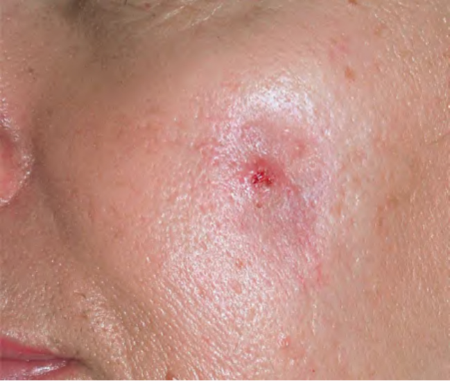
Fig. 94.1 Foreign body reaction at site of bovine collagen injection.
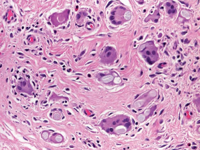
Fig. 94.2 Foreign body reaction to sutures. The foreign bodies have been engulfed primarily by foreign body-type giant cells; one Langhans-type giant cell is present at 12 o’clock. Courtesy Lorenzo Cerroni, MD.
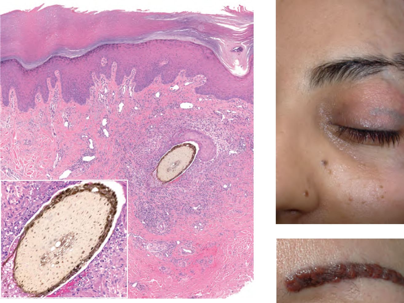
Fig. 94.3 Granulomatous foreign body reaction to implanted organic material in acral volar skin. A plant is the source of the foreign body and the latter is surrounded by a granulomatous reaction as well as neutrophils, lymphocytes, and cellular debris (inset). Superior to the foreign body is a portion of an epidermal cyst due to implantation of epidermal fragments into the dermis. Courtesy Lorenzo Cerroni, MD.
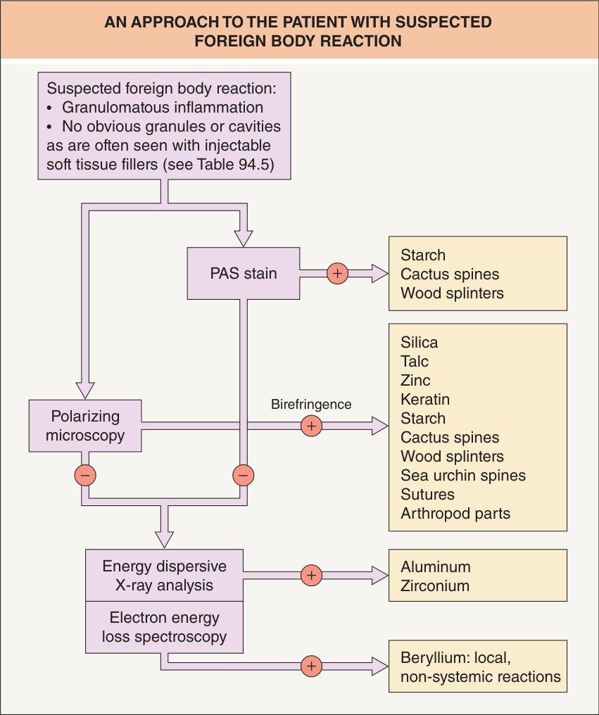
Fig. 94.4 An approach to the patient with suspected foreign body reaction.
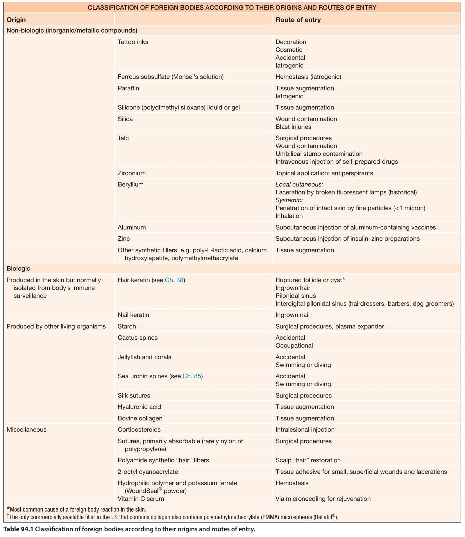
Table 94.1 Classification of foreign bodies according to their origins and routes of entry.
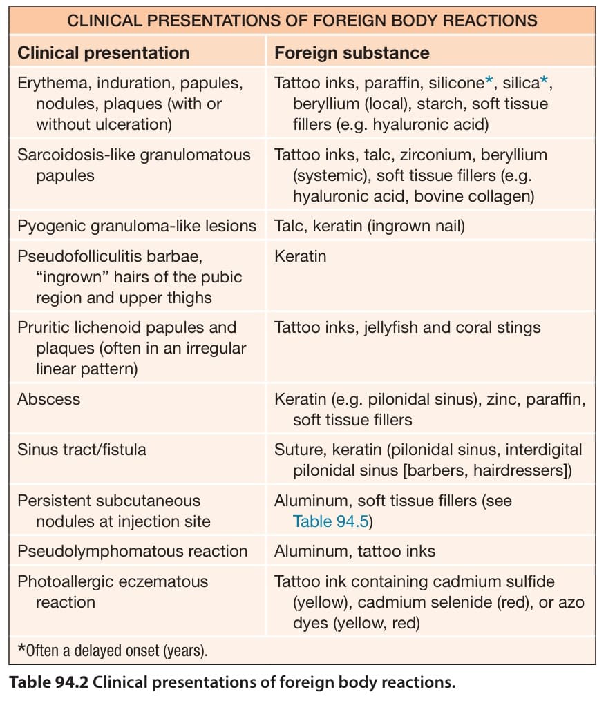
Table 94.2 Clinical presentations of foreign body reactions.
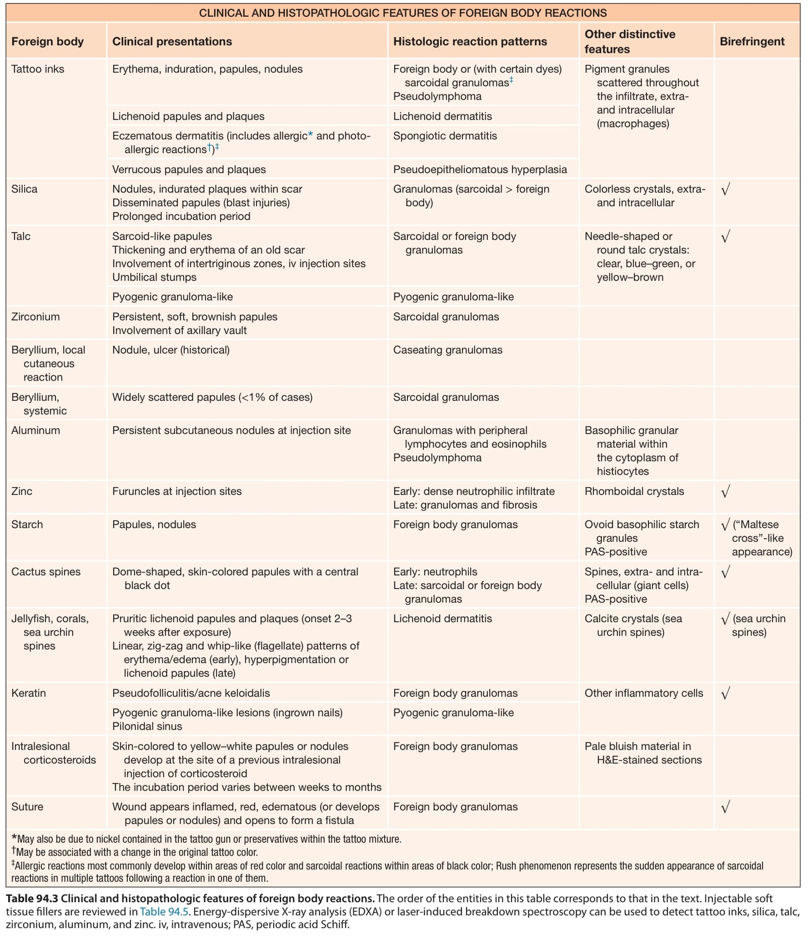
Table 94.3 Clinical and histopathologic features of foreign body reactions. The order of the entities in this table corresponds to that in the text. Injectable soft tissue fillers are reviewed in Table 94.5. Energy-dispersive X-ray analysis (EDXA) or laser-induced breakdown spectroscopy can be used to detect tattoo inks, silica, talc, zirconium, aluminum, and zinc. iv, intravenous; PAS, periodic acid Schiff.